3033
Hyperpolarized 13C 3D MRSI using Radial Multi-echo bSSFP and k-Space-based IDEAL1Division of Medical Physics, Department of Diagnostic and Interventional Radiology, University Medical Center Freiburg, Faculty of Medicine, University of Freiburg, Freiburg im Breisgau, Germany, 2Department of Nuclear Medicine, Technical University of Munich (TUM) School of Medicine, University Hospital rechts der Isar, Munich, Germany, 3German Cancer Consortium (DKTK), core center Heidelberg and German Cancer Research Center (DKFZ), Heidelberg, Germany, 4German Cancer Consortium (DKTK), partner site Freiburg and German Cancer Research Center (DKFZ), Heidelberg, Germany, 5NVision-Imaging Technologies GmbH, Ulm, Germany
Synopsis
Keywords: Hyperpolarized MR (Non-Gas), Hyperpolarized MR (Non-Gas), multi echo, bSSFP, radial readout
Motivation: 3D Metabolic imaging of hyperpolarized (HP) 13C agents demands specialized signal excitation and acquisition strategies.
Goal(s): This study introduces the combination of multi-echo bSSFP and radial readout with a spiral phyllotaxis pattern to rapidly image 13C-labeled contrast agents.
Approach: We implemented the novel sequence in a rodent in vivo experiment using HP [1-13C]pyruvate.
Results: Employing iterative signal decomposition and radial data reconstruction, we successfully captured the global signal dynamics with an unprecedented temporal resolution of 16 ms. Besides, we generated concentration maps for pyruvate, lactate, alanine, and calculated area-under-the-curve (AUC) metabolite ratio maps for Lac/Pyr and Ala/Pyr.
Impact: Hyperpolarized 13C 3D metabolic MRI is challenging due to the short-living magnetization. With 3D spiral phyllotaxis radial multi-echo bSSFP MRI and post-scan metabolite separation, large areas of interest can be acquired and reconstructed into signal time-curves and 3D metabolite maps.
Introduction
In 3D MRI with hyperpolarized (HP) 13C agents, specialized signal excitation and acquisition strategies are indispensable to fully harness the transient HP magnetization. Previously, a method combining the multi-echo balanced steady-state free precession (ME-bSSFP) sequence with an iterative decomposition approach, known as echo asymmetry and least-squares estimation (IDEAL), was introduced for efficient HP metabolic imaging and was even applied in initial clinical trials [1-3]. 3D radial acquisitions can be used for dynamic imaging of contrast agents, and a spiral phyllotaxis pattern for a radial k-space filling trajectory offers an efficient sampling strategy [4]. In this work, we present the successful implementation of a 3D ME-bSSFP radial sequence with spiral phyllotaxis sampling in a rodent in vivo experiment using HP [1-13C]pyruvate.Methods
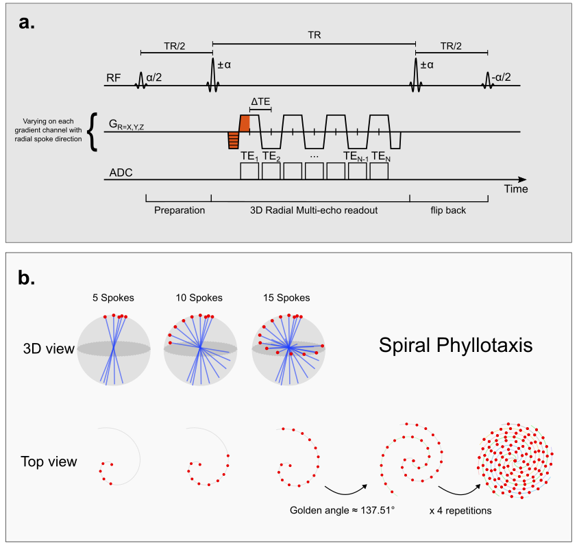
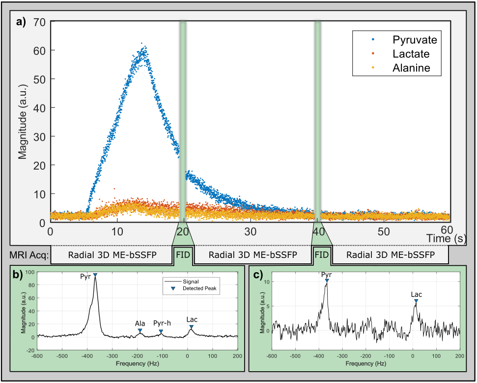
HP [1-13C]pyruvate (Concentration 80 mM, Injection dose 7.5 ml/kg body weight) was polarized by dissolution dynamic nuclear polarization (dDNP) using a HyperSense DNP Polarizer (Oxford Instruments). Upon tail-vein injection into healthy female Wistar rats (Charles River), HP 13C MRI was conducted on a 3T Biograph mMR (Siemens Healthineers) using a dual-tuned 1H/13C-transmit/receive volume coil (Rapid Biomedical). The 3D radial ME-bSSFP sequence (Figure 1a) used the following parameters: a = ±60° (after an initial a/2 preparation), TR = 16 ms, 5 echoes per TR, isotropic FoV = 290 mm, 300 radial readouts with 64 sampling points, leading to TA = 4.8 s per scan. The orientation angle between the readouts was incremented according to a spiral phyllotaxis pattern and each readout passes through the k-space center (Figure 1b) [4]. The sequence was dynamically repeated three times and non-localized spectroscopy (α = 10°, BW = 2000 Hz, Ns = 2048, TA = 1s) was interleaved to precisely determine the chemical shifts of all metabolites (Figure 2a). MATLAB was utilized for data processing. The multi-echo signals were decomposed using the IDEAL algorithm in k-space, followed by an in-house radial reconstruction script which transformed the initial radial k-space into Cartesian space with a matrix size of 64x64x64. The resulting spatial resolution of the image is isotropic 4.53 mm per voxel.Results
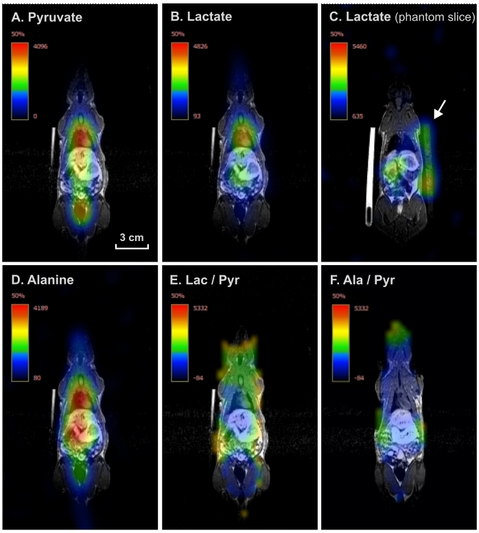
The peak resonance frequencies of [1-13C]pyruvate (-370 Hz), [1-13C]pyruvate-hydrate (-106 Hz), [1-13C]lactate (15 Hz), and [1-13C]alanine (-190 Hz) were accurately determined from the acquired spectrum (Figure 2b). The IDEAL algorithm applied to the k-space data effectively separated the metabolite amplitudes from the multiple echo contrasts per each radial orientation based on their peak frequencies. The metabolite-separated radial signals at the k-space center allowed us to dynamically monitor the global (non-localized) magnitude of each individual metabolite with an unprecedented temporal resolution of 16 ms, capturing the bolus of pyruvate and its conversion into lactate and alanine (Figure 2a). Following the reconstruction of the complete radial data, which involved concatenating the three ME-bSSFP acquisitions, 3D metabolite maps were generated and co-registered with 1H MRI. This process successfully localized the pyruvate, alanine, and lactate signals in the head, heart, and abdomen (Figure 3, A-D). Note that only the lactate map highlights the 13C-labelled, lactate reference phantom near the animal (Figure 3C). To further assess the metabolic conversion, voxel-wise division of the 3D metabolite maps yields area-under-the-curve (AUC) metabolite ratio maps. After masking the resulting Lac/Pyr and Ala/Pyr AUC-ratio maps to exclude regions with no measurable HP 13C signals, both AUC-ratio maps identified areas of high conversion, e.g., the animal's heart and head (Figure 3, E-F).Discussion and Conclusion
The integration of ME-bSSFP with center-through radial trajectory and spiral phyllotaxis golden-angle increments offers potential advantages over conventional Cartesian readouts. It allows for fast detection of HP 13C metabolites in large FoVs and is less sensitive to motion artifacts, which can be beneficial, for instance, for preclinical HP 13C MRSI with whole-animal or clinical HP 13C MRSI with extensive organ coverage. Furthermore, it features greater flexibility in data acquisition and reconstruction, opening up new possibilities: (a) Monitoring non-localized metabolic conversion with unprecedented temporal resolution; (b) Extracting metabolite maps and ratios with high SNR and spatial resolution from the complete dataset; (c) Reconstructing subsets of collected data in a sliding-window fashion to generate dynamic 3D maps with desired temporal resolution. (d) Offering flexibility for rapid acquisition and undersampling schemes. With improvements in metabolite-specific RF pulses, the decaying signals of downstream metabolic products from pyruvate may be better conserved.In conclusion, the successful acquisition, separation, and 3D reconstruction of HP 13C MRI signals acquired with a radial ME-bSSFP using a spiral phyllotaxis trajectory pattern was demonstrated. This method features great flexibility and holds promise for HP metabolic imaging with high SNR, and temporal-spatial resolution.
Acknowledgements
Research reported in this publication was funded by the German Federal Ministry of Education and Research (BMBF) in the program “Quantum Technologies – from Basic Research to Market” under the project “QuE-MRT” (contract number: 13N16448).References
[1] C. A. Müller et al., ‘Dynamic 2D and 3D mapping of hyperpolarized pyruvate to lactate conversion in vivo with efficient multi-echo balanced steady-state free precession at 3 T’, NMR in Biomedicine, vol. 33, no. 6, p. e4291, 2020.[2] R. Chowdhury et al., ‘Quantification of Prostate Cancer Metabolism Using 3D Multiecho bSSFP and Hyperpolarized [1-13C] Pyruvate: Metabolism Differs Between Tumors of the Same Gleason Grade’, Journal of Magnetic Resonance Imaging, vol. 57, no. 6, pp. 1865–1875, 2023.
[3] S. B. Reeder et al., ‘Iterative decomposition of water and fat with echo asymmetry and least-squares estimation (IDEAL): application with fast spin-echo imaging’, Magn Reson Med, vol. 54, no. 3, pp. 636–644, Sep. 2005.
[4] D. Piccini, A. Littmann, S. Nielles-Vallespin, and M. O. Zenge, ‘Spiral phyllotaxis: The natural way to construct a 3D radial trajectory in MRI’, Magnetic Resonance in Medicine, vol. 66, no. 4, pp. 1049–1056, 2011.
Figures