3032
A 3D Golden-Angle Radial Sequence for in vivo Hadamard-encoded 19F MRI in a Porcine Model1Division of Medical Physics, Department of Diagnostic and Interventional Radiology, University Medical Center Freiburg, Faculty of Medicine, University of Freiburg, Freiburg, Germany, 2Department of Cardiology, University Medical Center Freiburg, Faculty of Medicine, University of Freiburg, Freiburg, Germany
Synopsis
Keywords: Non-Proton, Non-Proton
Motivation: Perfluorooctyl bromide (PFOB) can be used to visualize the inflammatory reaction after myocardial infarction with 19F MRI, but effective methods for its imaging in large animals in vivo are still missing.
Goal(s): To develop a Hadamard-encoded sequence that removes the chemical shift artifacts of PFOB and increases the 19F SNR for application in large animal experiments.
Approach: A 3D center-out radial FLASH-sequence was developed and compared to a Cartesian FLASH-sequence in PFOB-phantoms. The radial sequence was tested in a pig after PFOB injection.
Results: Lowering TE increased the SNR by a factor of 2.4, which allowed to reconstruct 19F images of the animal.
Impact: Hadamard-encoded radial 19F MRI optimally uses the available multi-spectral information of PFOB which makes it the ideal candidate for monocyte tracking in large animals.
Introduction
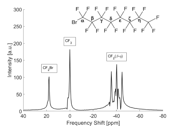
The spleen plays an important role during myocardial infarction (MI), since it functions as an emergency reservoir for monocytes, which are activated during the inflammatory process1. Since regulation of monocytes is important for MI-treatment, the egress of monocytes from the spleen needs to be monitored. Perfluorooctyl bromide (PFOB) is taken up by monocytes and can be used as a 19F MRI tracer. PFOB has been imaged in large animals2,3, however, to avoid chemical shift artifacts from the 3 main PFOB resonances spectrally selective excitation was applied which reduces the 19F MRI signal.To increase the SNR, a simultaneous excitation of all resonances can be performed, and the chemical shift artifacts from the 19F PFOB spectrum (Fig. 1) are corrected with spectral Hadamard-encoding. Therefore, the resonances of the spectrum are excited with varying 180° phase shifts, which allows to separate them during post-processing4. In these studies, conventional Cartesian k-space sampling has the disadvantage that the low readout bandwidth to maximize SNR increases TE. However, short TEs are important, because the complex CF2 multi-peak resonances of PFOB rapidly dephase in a few milliseconds. Therefore, we implemented a center-out radial sequence with the Hadamard encoding to minimize TE for 19F PFOB MRI.
Methods
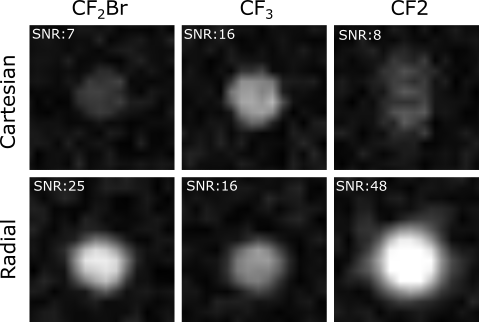
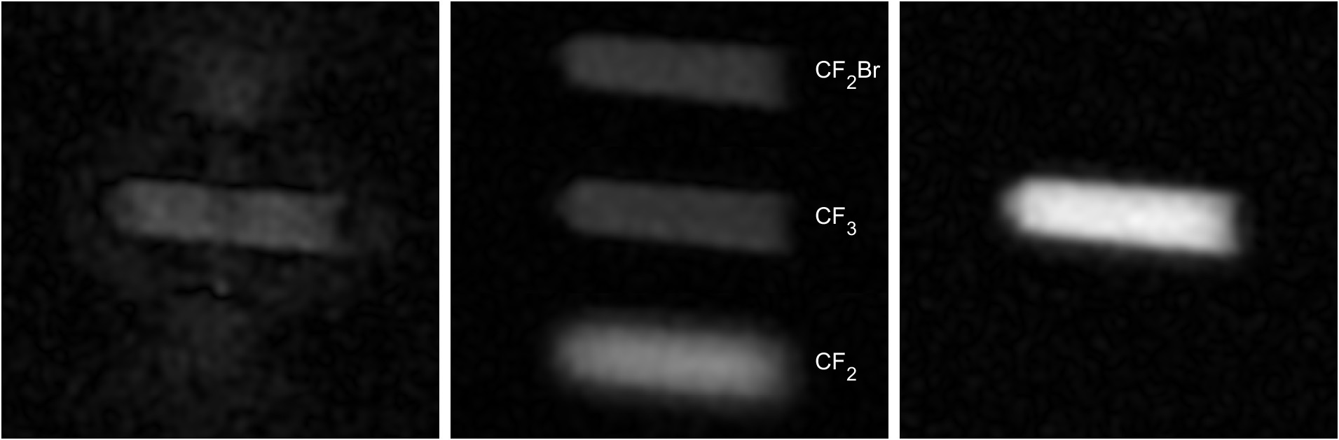
A 3D center-out radial FLASH sequence was developed using an interleaved spiral phyllotaxis pattern5 where consecutive radial spokes form evenly spaced spirals on the surface of a sphere. Hadamard-encoding was realized with 4 non-selective composite RF pulses with alternating phases for the 3 PFOB resonances. SNR comparisons with the original Cartesian FLASH sequence for Hadamard-encoding and the proposed radial sequence were performed on a phantom containing 10% PFOB in a fat water emulsion (Fig 2).Off-resonances in radial acquisitions lead to a uniform blurring (Fig. 3); thus, the Hadamard-decoded raw data of the individual resonances at a frequency shift δ had to be corrected individually by applying an inverse frequency shift in radial k-space:
$$S(k(\tau))=S_{meas}(k(\tau))*e^{-i\cdot\delta\cdot\gamma B_0\cdot\tau}$$
Here, $$$\tau$$$ is the elapsed time since the start of the ADC, $$$S_{meas}$$$ is the measured 19F signal and $$$S$$$ is the corrected signal. For PFOB, $$$\delta(CF_2Br)=18.1$$$ and $$$\delta(CF_2)$$$ were used assuming that the spectrum is centered at the CF3 resonance. From the corrected signals, 3D images were reconstructed of all resonances and combined to increase the SNR.
In vivo 1H and 19F images of a pig were acquired that had been injected with 300ml of a 40% PFOB emulsion3 2 days prior. All 19F measurements used an 8 channel transmit and 8 channel receive coil array that was specifically built for 19F MRI of large animals6. The Cartesian FLASH images of the phantom were acquired with TE=3.94ms, TR=10ms, BW=260Hz/px, (Δx)3=(5mm)3 in about 30 min. For the radial sequence phantom images were acquired with 25 spokes per spiral, 2584 interleaves, TE=1.7ms, TR=10ms, BW=390Hz/px, (Δx)3=(5mm)3 resulting in an acquisition time of 44:47 min:s, and in vivo images were acquired with 25 spokes per spiral, 1597 interleaves, TE=1.7ms, TR=10ms, BW=390Hz/px, (Δx)3=(5mm)3 resulting in an acquisition time of 27:41 min:s.
Results
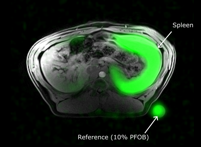
To compare the SNR despite the differences in acquisition time, the radial SNR values need to be divided by $$$\sqrt{1.5}\approx 1.22$$$. The resulting values are: SNR(CF2Br)=20, SNR(CF3)=13 and SNR(CF2)=39. SNR of Cartesian and radial acquisitions showed a significant increase in both the CF2Br and the CF2 signal by shortening of TE, whereas the SNR of the CF3 resonance slightly decreased. After signal combination, the total SNR increased by a factor of 2.4. The CF2 multi-peak appears blurred due to remaining chemical shift artifacts from the multiple resonances, which is also seen in the Cartesian data along the readout direction. The in vivo measurement in the pig clearly detected the 19F signals in the spleen with an SNR ≥ 50 (Fig.4).Discussion and Conclusion
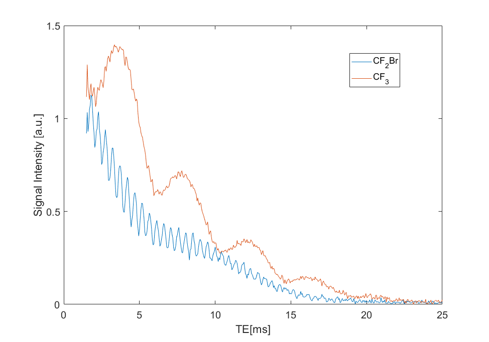
A significant increase in SNR for both the CF2Br and the CF2 resonance can be achieved by shortening of TE whereas the CF3 resonance decreased. FID measurements in a PFOB sample indicate that this is caused by additional dephasing effects (Fig. 5) which also depend on local field inhomogeneities. In summary, 19F PFOB MRI can be performed robustly in pigs with a radial Hadamard-encoded sequence, and monocyte accumulation in the spleen can be visualized in the 19F images.Acknowledgements
This study was funded by the Deutsche Forschungsgemeinschaft (DFG) under project #492563001 (BO 3025/17-1 and MA 7059/3-1), and it was part of the SFB1425, project #422681845.References
1. Senders ML, Meerwaldt AE, van Leent MMT, et al. Probing myeloid cell dynamics in ischemic heart disease by nanotracer hot spot imaging. Nat Nanotechnol. 2020;15(5):398-405. doi:10.1038/s41565-020-0642-4
2. Rothe M, Jahn A, Weiss K, et al. In vivo 19F MR inflammation imaging after myocardial infarction in a large animal model at 3 T. MAGMA. 2019;32(1):5-13. doi:10.1007/s10334-018-0714-8
3. Bönner F, Gastl M, Nienhaus F, et al. Regional analysis of inflammation and contractile function in reperfused acute myocardial infarction by in vivo 19F cardiovascular magnetic resonance in pigs. Basic Res Cardiol. 2022;117(1):21. doi:10.1007/s00395-022-00928-5
4. Bock M, Ilbey S, Maier A, Özen AC. Chemical Shift Artifact Correction for 19F MRI using Spectral Hadamard Encoding. In: Proc. Intl. Soc. Mag. Reson. Med. 30. London; 2022. doi:10.58530/2022/1008
5. Piccini D, Littmann A, Nielles-Vallespin S, Zenge MO. Spiral phyllotaxis: The natural way to construct a 3D radial trajectory in MRI. Magnetic Resonance in Medicine. 2011;66(4):1049-1056. doi:10.1002/mrm.22898
6. Özen AC, Spreter F, Schimpf W, et al. Scalable and modular 8-channel transmit and 8-channel flexible receive coil array for 19 F MRI of large animals. Magn Reson Med. 2023;89(3):1237-1250. doi:10.1002/mrm.29490
Figures