3025
A 3D Slicer Extension for Retrospective MP2RAGE Background Suppression1Radiology, Center for Magnetic Resonance Research, University of Minnesota, Minneapolis, MN, United States, 2Neuroscience, University of Minnesota, Minneapolis, MN, United States, 3Neurology, University of Minnesota, Minneapolis, MN, United States
Synopsis
Keywords: Data Processing, Data Processing
Motivation: MP2RAGE provides enhanced T1-weighted images but contains high-amplitude noise in areas of low signal. This can cause processing pipelines developed for traditional T1 images to fail. A “denoising” algorithm exists, but requires complex-valued image data which are not available retrospectively.
Goal(s): Provide an algorithm and easy-to-use interface for eliminating MP2RAGE background noise using only available scanner outputs.
Approach: We have developed a 3D Slicer extension for performing noise suppression with only the available MP2RAGE and inversion magnitude images.
Results: Our method generates background-suppressed and artifact-free images. The program was tested and optimized to be used with the HCP structural pipeline.
Impact: Here, we present a fast easy-to-use 3D Slicer extension for suppressing background noise in MP2RAGE images. It requires no extra phase data, enables users to reprocess already acquired images, and encourages the adoption of MP2RAGE as a primary T1-weighted acquisition.
Introduction
The MP2RAGE contrast1 generates excellent T1-weighted images and is useful for generating other derived contrasts (e.g. T1 maps, synthetic white matter-nulled T1s, and synthetic FGATIR2). However, it contains “salt-and-pepper” noise, rather than zero values, in areas of low or no signal. This noise can cause algorithms developed for traditional T1 images processing to fail. A “denoising” (more accurately, background suppression) method for eliminating such noise has been published3, but it relies on additional phase data which is typically not available. We have developed a method for retrospectively applying a similar noise suppression approach which relies only on available scanner output images.Methods
O’Brien et al.3 proposed reducing MP2RAGE background noise by applying a regularization factor $$$\beta$$$ which trades a small amount of bias field correction for noise suppression:$$ U=\frac{\Re(I_1^\ast I_2)-\beta}{|I_1|^2 + |I_2|^2 + 2\beta} \tag{1}$$
This relies on complex-valued images $$$I_1$$$ and $$$I_2$$$ from the first and second inversions of the MP2RAGE acquisition. However, typical scanner output consists of modified magnitude-only images $$$\left|\widetilde{I_1}\right|$$$ and$$$ \left|\widetilde{I_2}\right|$$$ and an un-suppressed (that is, $$$\beta=0$$$) image $$$U_0$$$. $$$\widetilde{I_1}$$$ and $$$\widetilde{I_2}$$$ may reflect additional processing, different coil combination methods, or other differences from the $$$I_1$$$ and $$$I_2$$$ values used to calculate $$$U_0$$$.
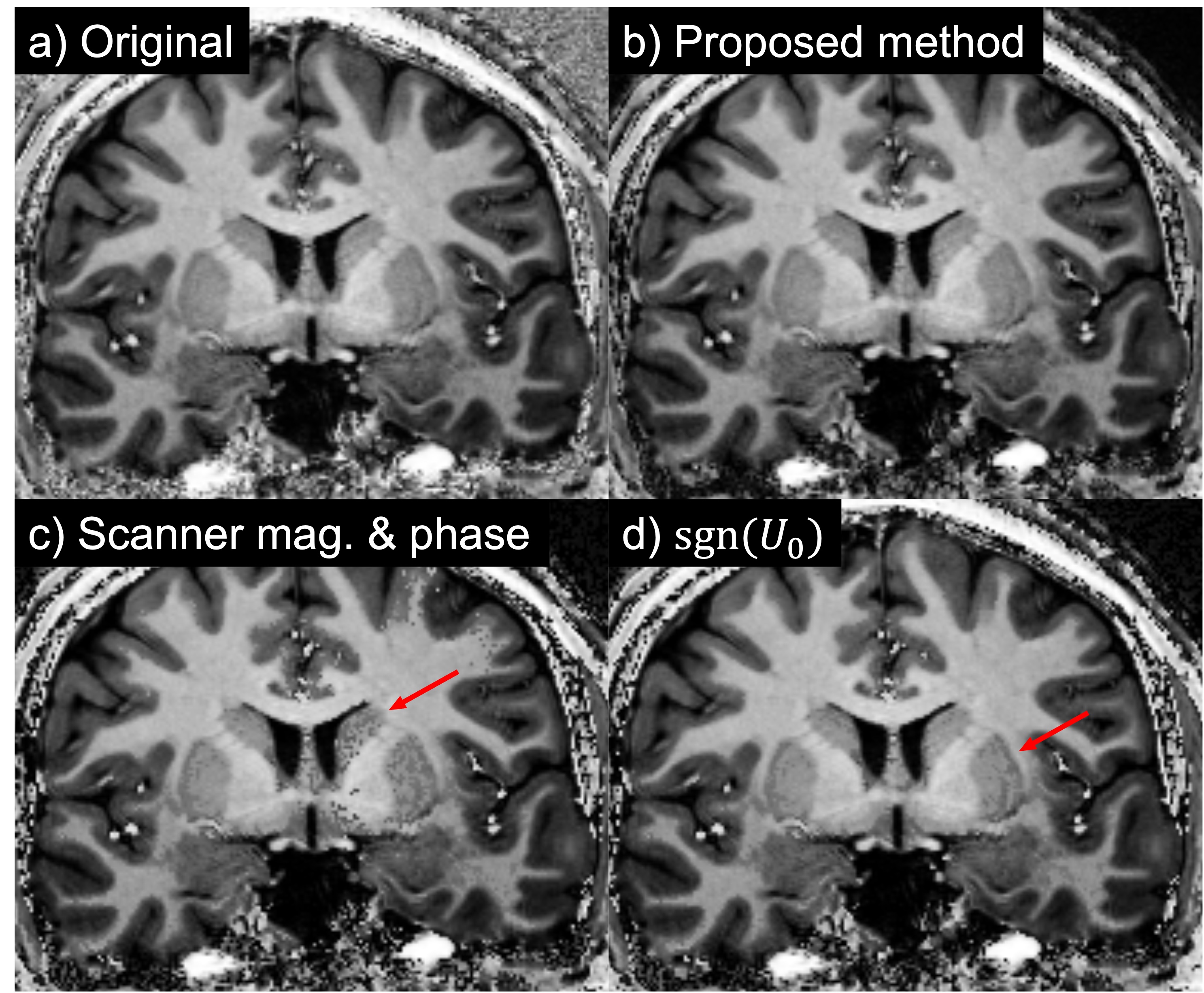
Phase images can also be obtained (e.g. through retrospective reconstruction), but these suffer from the same problems as the magnitude images. If we naively apply equation (1) to MP2RAGE output from a Siemens Terra 7T MRI scanner, phase instability between the 1st and 2nd inversions leads to obvious and unacceptable artifacts near large and rapid phase shifts (Figure 2(c)). A more robust approach is needed which can handle small magnitude and large phase differences between $$$\left(I_1\ ,\ I_2\right)$$$ and $$$\left(\widetilde{I_1},\ \widetilde{I_2}\right)$$$. The background suppression approach of O’Brien et al.3 therefore must be extended to function with only the typical scanner outputs $$$\left|\widetilde{I_1}\right|$$$, $$$\left|\widetilde{I_2}\right|$$$, and $$$U_0$$$.
Despite the challenges mentioned above, background suppression can still be applied retrospectively. First, assume without loss of generality that $$$I_2$$$ is real-valued and nonnegative (that is, $$$\angle I_2 = 0$$$). A signed version of the 1st inversion, $$$\widehat{I_1}$$$, can then be estimated by solving equation (1) with $$$\beta=0$$$:
$$\widehat{I_1} = \Re\left(\widetilde{I_1}\right) = \frac{U_0}{\widetilde{I_2}}\left( \left|\widetilde{I_1}\right|^2+ \left|\widetilde{I_2}\right|^2\right) \tag{2}$$
Substituting $$$\widehat{I_1}$$$ and $$$\widetilde{I_2}$$$ in equation (1), we can perform background suppression. Equation (2) may seem unnecessarily complicated: Why not use $$$\left|\ \widetilde{I_1}\right|$$$ and the sign of $$$U_0$$$ to determine $$$\widehat{I_1}$$$? That is, let
$$\widehat{I_1}=\text{sgn}\left(U_0\right)\widetilde{I_1} \tag{3}$$
This approach subtly fails because our available image $$$\widetilde{I_1}$$$ differs slightly from the $$$I_1$$$ image used to calculate U. This leads to visual artifacts around zero-crossings as shown in Figure 2(d).
We acquired MP2RAGE data on a Siemens Terra 7T scanner with TI1=780ms, TI2=2430ms, TE= 2.91ms, TR=6.3s, and flip angles of 8 and 10 degrees for the 1st and 2nd inversions, respectively. While images shown here are from a single 24-year-old female healthy control subject, we routinely acquire and process MP2RAGE data across a range of studies using this method.
Results and Discussion
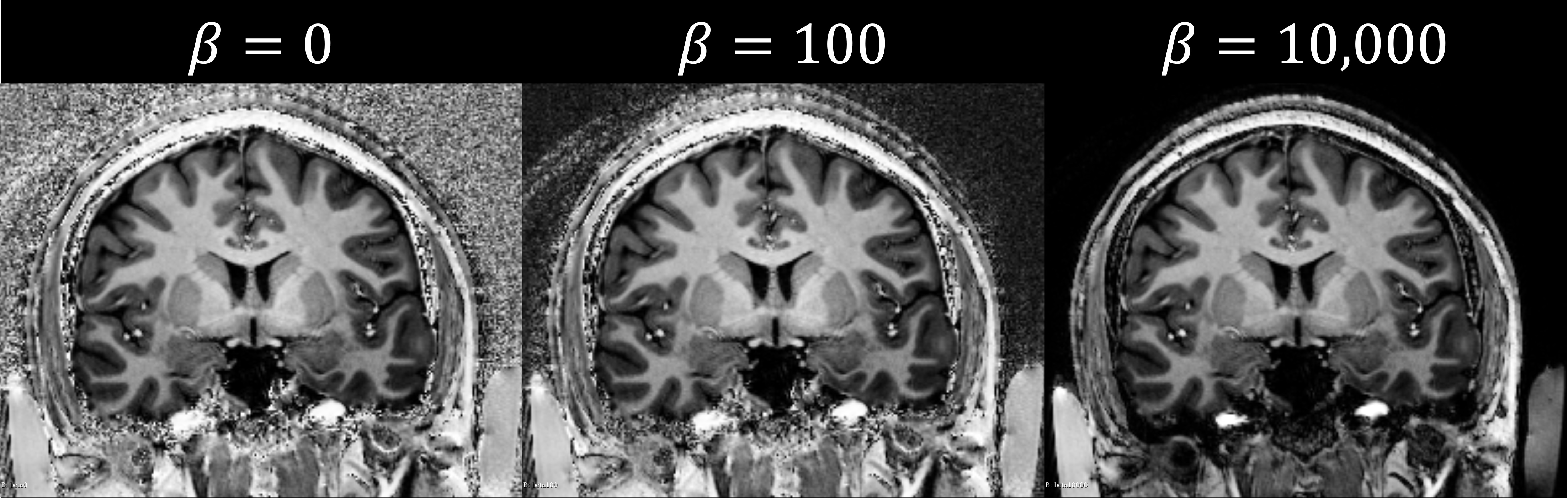
Figure 1 shows the result of applying MP2RAGE background suppression at values of 100 and 10,000. Note that higher beta values trade increased background suppression for decreased bias-field correction. While the Human Connectome Project (HCP)4 structural processing pipeline consistently failed on images, the proposed method yields outputs with consistent success.Figure 2 Compares the unprocessed $$$U_0$$$ image (a) against a variety of potential background suppression approaches described above. The proposed method, shown in (b), produces an artifact-free image, while generating complex-valued $$$\widetilde{I_1}$$$ and $$$\widetilde{I_2}$$$ from scanner phase outputs (c) or applying equation $$$\left(3\right)$$$ (d) both result in unacceptable artifacts.
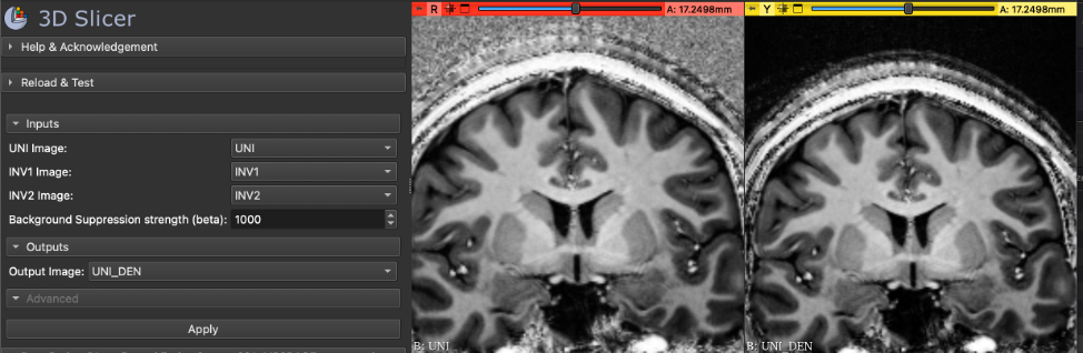
We have packaged the method described above as an easy-to-use 3D Slicer extension5 for Slicer version 5.4.0 and higher. Figure 3 shows a screenshot of the software user interface.
Conclusion
MP2RAGE has superior image contrast over MPRAGE for T1-weighted images, particularly at ultrahigh field. However, high-amplitude background noise presents a barrier to widespread replacement of MPRAGE. We have developed a method for suppressing this background noise and published it as an easy-to-use extension5 to the 3D Slicer6,7 software package. Our method relies only on default output images and can be applied retrospectively to already-acquired data.Acknowledgements
Research reported in this publication was supported by the Udall Center of the National Institutes of Health under award number P50 NS123109 and by the National Institutes of Health under award numbers S10 OD025256, P41 EB027061, P50 NS098753, R01NS081118, and R01 NS113746.
References
1. Marques JP, Kober T, Krueger G, Van Der Zwaag W, Van De Moortele PF, Gruetter R. MP2RAGE, a self bias-field corrected sequence for improved segmentation and T1-mapping at high field. NeuroImage. 2010;49(2):1271-1281. doi:10.1016/j.neuroimage.2009.10.002
2. Middlebrooks EH, Tao S, Zhou X, et al. Synthetic Inversion Image Generation using MP2RAGE T1 Mapping for Surgical Targeting in Deep Brain Stimulation and Lesioning. Ster Funct Neurosurg. 2023;101(5):326-331. doi:10.1159/000533259
3. O’Brien KR, Kober T, Hagmann P, et al. Robust T1-Weighted Structural Brain Imaging and Morphometry at 7T Using MP2RAGE. Margulies D, ed. PLoS ONE. 2014;9(6):e99676. doi:10.1371/journal.pone.0099676
4. Glasser MF, Sotiropoulos SN, Wilson JA, et al. The minimal preprocessing pipelines for the Human Connectome Project. NeuroImage. 2013;80:105-124. doi:10.1016/j.neuroimage.2013.04.127
5. Braun, Henry, Brenny, Samuel. MP2RAGE Background Suppression. https://github.com/harellab/SlicerMp2rageBackgroundSuppression
6. Kikinis R, Pieper SD, Vosburgh KG. 3D Slicer: A Platform for Subject-Specific Image Analysis, Visualization, and Clinical Support. In: Jolesz FA, ed. Intraoperative Imaging and Image-Guided Therapy. Springer New York; 2014:277-289. doi:10.1007/978-1-4614-7657-3_19
7. 3D Slicer. https://www.slicer.org/
Figures