3023
An Integrated Framework for Whole Brain Arteries Modeling on Compressed Sensing TOF-MRA at 7T1State Key Laboratory of Brain and Cognitive Science, Institute of Biophysics, Chinese Academy of Sciences, Beijing, China, University of Chinese Academy of Sciences, Beijing, China, The Innovation Center of Excellence on Brain Science, Chinese Academy of Sciences, Beijing, China, Beijing, China, 2University of Chinese Academy of Sciences, Beijing, China, Beijing, China, 3The Innovation Center of Excellence on Brain Science, Chinese Academy of Sciences, Beijing, China, 4The Innovation Center of Excellence on Brain Science, Chinese Academy of Sciences, Beijing, China, Beijing, China, 5Siemens Shenzhen Magnetic Resonance Ltd., Shenzhen, China., Beijing, China, 6Institute of Artificial Intelligence, Hefei Comprehensive National Science Center, Hefei, China, Hefei, China
Synopsis
Keywords: Segmentation, Brain
Motivation: Whole-brain vasculature modeling remains challenging due to background variations and diverse vessel sizes.
Goal(s): We pursued an integrated framework for the accurate segmentation and tracking of vasculature in the whole brain.
Approach: Small-patch CNN (SP-CNN) and centerline-Dice nnU-Net (CDNN) were utilized for multiscale segmenting of vessels. A random forest and soft-skeletonization were applied for tracking. A novel rotation algorithm was used to calculate diameters accurately.
Results:
We accurately reconstructed the arteries from compressed sensing time-of-flight MR angiography (CS-TOF-MRA) and geometric parameters of full-size arteries were obtained.
Impact: Our work enables the segmentation of full-size-arteries in CS-TOF-MRA. As this method integrates algorithms for higher accuracy and robustness, it will be beneficial for clinical diagnosis.
Introduction
As brain energy supply and neurological diseases are significantly related to the cerebral vascular network[1], accurate modeling of the cerebral vasculature is critical and aids in understanding cerebrovascular diseases and guiding treatment decisions[2]. TOF-MRA at 7T offers acceleration and noninvasive visualization of cerebral vasculatures[3], and compressed sensing has shown the potential to accelerate MR imaging[4]. However, there is a lack of algorithms for modeling vessels of whole brain due to background variations and diverse vessel sizes. This study presents an integrated framework to achieve full-size artery modeling: a combination of SP-CNN and CDNN was utilized for segmentation, and a random forest and soft-skeletonization were established for tracking. Our framework mitigates the limitations of different components while harnessing their individual strengths and ensuring precise and comprehensive vascular modeling. We demonstrate its effectiveness in advancing cerebrovascular imaging with ablation studies.Methods
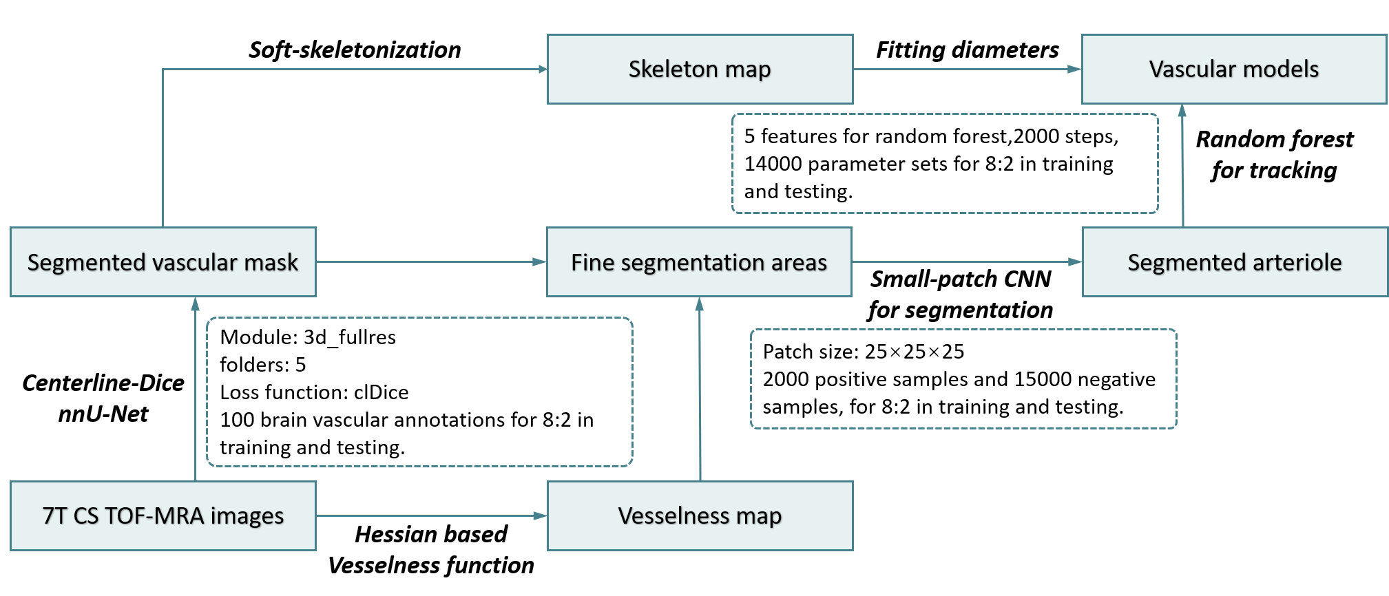
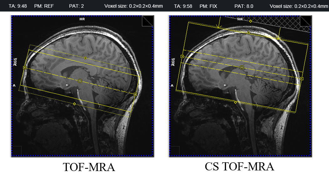
The workflow of our method was shown in Figure 1. The protocol of CS-TOF-MRA was resolution = 0.2 × 0.2 × 0.4 mm3, matrix = 864 × 960 × 272, TR = 148ms, TE = 5.29ms, FA = 24°, acceleration factor = 9, slab = 6. Unlike the TOF-MRA we previously used[5], this can cover the entire brain by utilizing CS-TOF with a similar scan time (Figure 2).We employed CDNN based on nnU-Net for segmentation of the cerebral vessels and optimized the loss function. To enable the equation of each-scale vessel preservation during the segmentation of the tubular objects, the dice was replaced by centerline-Dice (clDice), which is based on soft-skeletonization[6]. Regarding the loss function of clDice, we implemented two specific modifications. To increase the weight of small vessels, a dilation algorithm with the same scale was applied on the end of soft-skeleton algorithm. To maintain the geometric structure of the short vessels, we constrained the erosion on the normal plane during soft-skeletonization. The CDNN was trained on the CAS2023 dataset, which comprises 100 cases with reference annotations. The accuracy rate was 94%.
For accurate segmentation of small vessels like lenticulostriate arteries (LSA), after segmentation with CDNN, a vessel probability map was calculated by utilizing a Hessian-based vesselness function[7]. Voxels classified as background by CDNN, if associated with a high vesselness probability (>50%), were segmented using SP-CNN.
Finally, small-artery tracking was accomplished using the random forest model we previously proposed[5], while large artery modeling was performed through soft-skeletonization.
Results
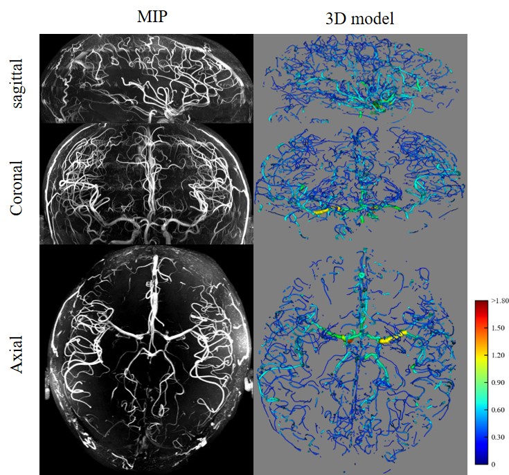
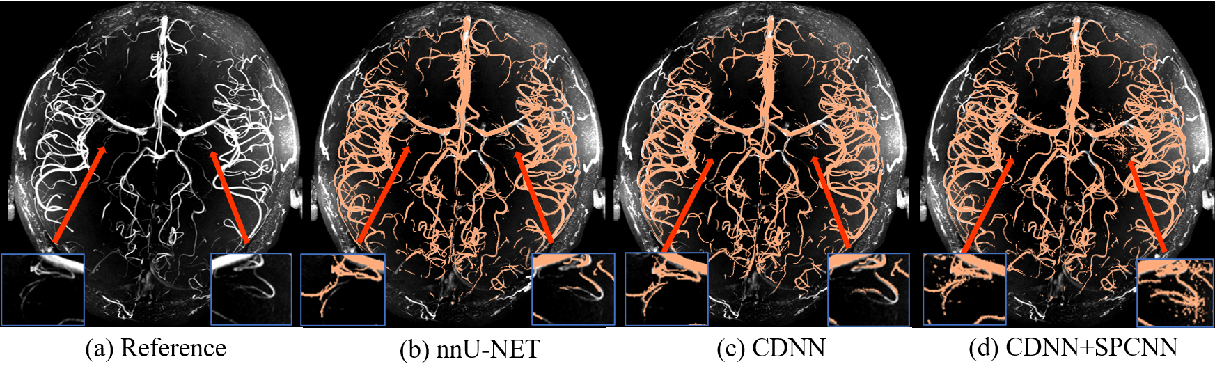
The 3D vascular model of whole brain of a healthy control was represented in Figure 3, in comparison with maximal intensity projections (MIP). The comparison of nnU-Net, CDNN and CDNN with SP-CNN was revealed in Figure 4.Despite the high accuracy of CDNN, the performance of small vessels like lenticulostriate arteries (LSA) was still poor, as illustrated in Figure 3b and Figure 3c.
Geometric parameters, such as lengths, diameters, curvatures, and amounts, can be evaluated automatically by the proposed method.
Discussion
In this study, we have proposed an integrated framework for full-size cerebral vascular modeling with optimized nnU-Net to CDNN utilizing the vesselness function as a threshold to determine whether to use SP-CNN. The theory of clDice is that extracting the skeleton and taking the intersection with the binary mask of the vessels enhances the weighting of smaller vessels[6]. Considering the variation in size within the whole-brain vasculature, because small vessels are more crucial, we dilated the vessel skeleton to increase the penalty for neglecting them. This is because even when large vessels are mistakenly counted as background, this error will be compensated for by the whole-brain threshold we used in the previous work5. In the soft-skeletonization algorithm, the erosion algorithm constrained in the normal plane effectively prevents a reduction in vessel length. Due to factors such as low signal-to-noise ratio, artifacts, and poor labeling quality, smaller vessels like perforating arteries are difficult to segment. However, using SP-CNN in the whole-brain interpolated image requires too much time. Therefore, the application of SP-CNN is determined by the vesselness function. As reported previously[5], SP-CNN exhibits a high true-positive rate that can effectively solve the drawback of nnU-Net's high false-positive rate. Our study is limited because of its high computational power consumption. Calculation of all geomatic parameters and complete 3D rendering requires 6–8 hours. This issue may be solved by using advanced hardware facilities.Conclusions
Our optimizations improved the ability of nnU-Net to segment multiscale vessels. We presented an integrated framework for segmenting and modeling full-size cerebral vasculature from 7T CS TOF-MRA images.Acknowledgements
This work was supported in part by National Natural Science Foundation of China (82271985, 81961128030), National Science and Technology Innovation 2030 Major Program (2022ZD0211900, 2022ZD0211901), Youth Innovation Promotion Association CAS (2022093) Natural Science Foundation of Beijing Municipality (7191003), Ministry of Science and Technology of China grant (2019YFA0707103), and National Nature Science Foundation of China grant (31730039)References
1. Pfeiffer, T., Y. Li, and D. Attwell, Diverse mechanisms regulating brain energy supply at the capillary level. Current Opinion in Neurobiology, 2021. 69: p. 41-50.
2. Wu, Y., et al., 3D Visualization of Whole Brain Vessels and Quantification of Vascular Pathology in a Chronic Hypoperfusion Model Causing White Matter Damage. Translational Stroke Research, 2023.
3. Ling, C., et al., Lenticulostriate arteries and basal ganglia changes in cerebral autosomal dominant arteriopathy with subcortical infarcts and leukoencephalopathy, a high-field MRI study. Frontiers in Neurology, 2019. 10: p. 870.
4. Ding, J., et al., Acceleration of brain TOF-MRA with compressed sensitivity encoding: a multicenter clinical study. American Journal of Neuroradiology, 2021. 42(7): p. 1208-1215.
5. Li, Z., et al., Small-patch CNN and random forest to model and quantify the lenticulostriate artery from 7T time-of-flight MR angiography.
6. Shit, S., et al. clDice-a novel topology-preserving loss function for tubular structure segmentation. in Proceedings of the IEEE/CVF Conference on Computer Vision and Pattern Recognition. 2021.
7. Straub, S., et al., A novel gradient echo data based vein segmentation algorithm and its application for the detection of regional cerebral differences in venous susceptibility. NeuroImage, 2022. 250: p. 118931.
Figures