3016
Exploring NORDIC denoising benefits in high-resolution Arterial Spin Labeling MRI1Krembil Brain Institute, University Health Network, Toronto, ON, Canada
Synopsis
Keywords: Data Processing, Perfusion, denoising
Motivation: NORDIC PCA denoising is a recent denoising technique that promises mitigation of thermal noise in MRI data, and its application in high-resolution ASL remains unexplored.
Goal(s): Assess the effectiveness of NORDIC denoising for improving the quality of high-resolution ASL perfusion signal.
Approach: NORDIC denoising was applied to ASL data in two different approaches. NORDIC1, on control and label separately, and NORDIC2 on the original ASL time course (control and label combined).
Results: NORDIC denoising results in a twofold increase in tSNR. The denoising effect is most effective when NORDIC is applied to ASL timeseries and in low SNR voxels.
Impact: ASL is often not utilized in clinical and cognitive neuroscience studies due to its low SNR. Thus, the improvement in tSNR afforded by NORDIC denoising paves the way for the implementation of high-resolution ASL in cutting-edge brain studies.
Introduction
Despite the recent advances in hardware, software, and analysis Arterial Spin Labelling (ASL) is still limited by its intrinsic low signal-to-noise ratio (SNR). For conventional low-resolution ASL scans (e.g., 3.4 × 3.4 × 4 mm3), spatial smoothing serves as an effective approach to reduce random noise (thermal noise). Another approach is the suppression of temporal noise in the control and label images using high-pass filtering1. Additionally, methods, such as Component-based noise correction (CompCor)2, global signal regression, wavelet denoising3, Total Generalized Variation (TGV)4, and machine learning methods, have all been explored for denoising5. NOise Reduction with DIstribution Corrected (NORDIC) PCA6 is a recent denoising approach, capable of distinguishing and ameliorating thermal noise components within MRI data whilst ideally not affecting the signal contribution. While initially developed to enhance high spatial resolution Diffusion MRI (dMRI), this method has also demonstrated significant utility in the fMRI field7, increasing signal stability of the time series and temporal SNR (tSNR). While applied to different fMRI contrasts, such as BOLD and VASO7,8,9, it has not been explored in ASL. In the present study, we aim to investigate the extent to which NORDIC denoising impacts high-resolution ASL data.Methods
Imaging was performed on 8 participants with a Siemens 3T Prisma MRI using the 64-channel coil. The acquisition consisted of a 3D GRASE pCASL with a resolution of 2mm isotropic voxels, 66 slices, post-labeling delay/labeling duration=1800/1800 ms, TE/TR=16.80/4000 ms, and 12 repetitions. Additional sequence details can be found in 10. We ran the NORDIC denoising algorithm V.1.111 on magnitude data. No separate noise scan, additional smoothing, or temporal filtering was used. We employed two different NORDIC approaches, (1) NORDIC denoising applied on control and label images separately (NORDIC1), and (2) to the full ASL time series (NORDIC2) wherein control and label are interleaved. Next, Perfusion-weighted images (PWI) were calculated following control-label subtraction. GM and WM matter masks were extracted from the mean perfusion image using SPM12 segmentation and were used for statistical analysis.We compared both NORDIC approaches with the standard perfusion data. We evaluated mean PWI and perfusion tSNR and repeated measures ANOVA with pairwise comparison (Bonferroni) was used to assess statistical differences. Perfusion tSNR was calculated as the ratio of the mean perfusion images divided by its standard deviation. To evaluate the amount of gain in tSNR, we calculated a voxel-wise linear regression on the tSNR maps and a two-sample paired t-test to assess differences.
Results
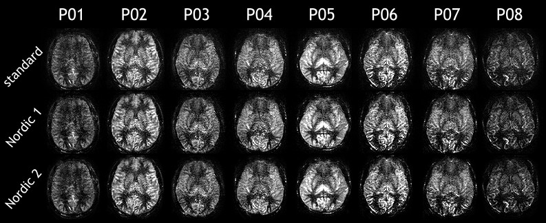
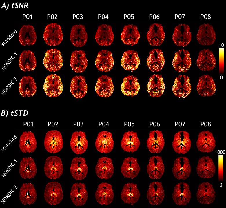
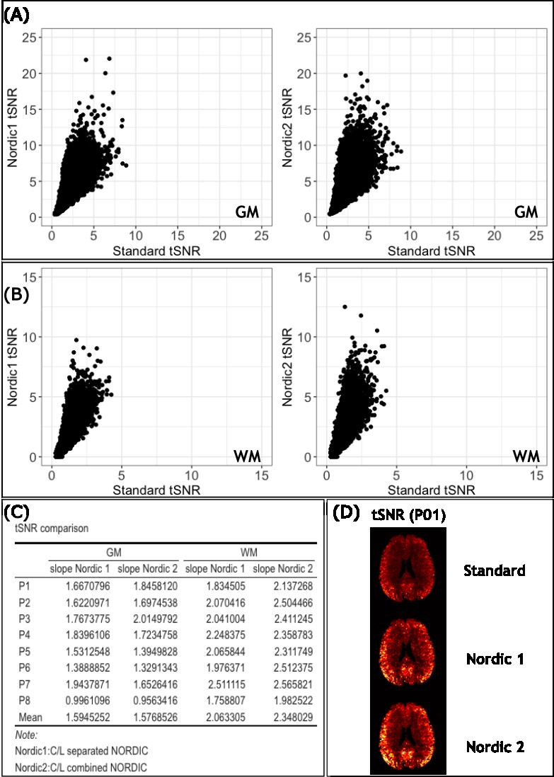
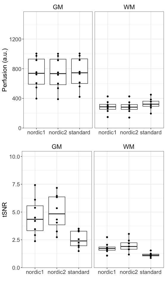
Figure 1 shows a single slice of PWI from all participants and conditions. The effect of NORDIC is noticeable in WM regions, yielding lower perfusion-weighting (NORDIC1=287±82, NORDIC2=284±83) than standard (325±75.5). In Figure 2A, we show tSNR maps from all participants and conditions, and Figure 2B shows the standard deviation perfusion maps (denominator of tSNR maps), we observed a smaller variability for NORDIC than standard perfusion images. We calculated the tSNR gain by computing linear regression, see Figure 3(A)-(B). The slopes, representing the gain, from all participants, are depicted in Panel(C). For GM, there was no significant difference between NORDIC1&2 (NORDIC1 mean slope=1.60, NORDIC2 mean slope=1.58), whereas for WM the slopes were significantly different (p=0.001), with NORDIC2 yielding higher slopes than NORDIC1 (NORDIC1 mean slope=2.06, NORDIC2 mean slope=2.35).In Figure 4, we observed significant differences in both GM and WM mean perfusion, with stronger differences in WM. For GM, pairwise comparison between standard and NORDIC2 was statistically significant (p=0.03). For WM, both NORDIC approaches reached a statistical significance level (p<0.0001) when compared with standard data. For tSNR analysis, we observed a significant improvement after using NORDIC (p<0.05 for both NORDIC approaches and regions).
Discussion
Our findings show that NORDIC denoising can enhance ASL tSNR by approximately two-fold. Regarding the mean perfusion maps, the denoising effect is stronger in the WM than in the GM perfusion. This is an interesting outcome since WM perfusion using ASL is typically unreliable and noisier due to longer transit times and much shorter blood flow values compared to GM12,13. In addition, smaller variability (higher precision) of the perfusion values is observed, possibly increasing the inter-subject reliability of ASL for clinical studies. Applying NORDIC on the ASL time series (NORDIC2) in contrast to denoise control and label separately further improved the tSNR.Conclusion
We presented the results of the usage of NORDIC denoising in ASL images, we showed that applying NORDIC to the full ASL time series (NORDIC2) provides higher tSNR gains and lower perfusion in WM.Acknowledgements
No acknowledgement found.References
1. Wang Z. Improving Cerebral Blood Flow Quantification for Arterial Spin Labeled Perfusion MRI by Removing Residual Motion Artifacts and Global Signal Fluctuations. Magn Reson Imaging. 2012;30(10):1409-1415. doi:10.1016/j.mri.2012.05.004
2. Behzadi Y, Restom K, Liau J, Liu TT. A component based noise correction method (CompCor) for BOLD and perfusion based fMRI. NeuroImage. 2007;37(1):90-101. doi:10.1016/j.neuroimage.2007.04.042
3. Bibic A, Knutsson L, Ståhlberg F, Wirestam R. Denoising of arterial spin labeling data: wavelet-domain filtering compared with Gaussian smoothing. Magn Reson Mater Phys Biol Med. 2010;23(3):125-137. doi:10.1007/s10334-010-0209-8
4. Spann SM, Shao X, Wang DJJ, et al. Robust single-shot acquisition of high resolution whole brain ASL images by combining time-dependent 2D CAPIRINHA sampling with spatio-temporal TGV reconstruction. NeuroImage. 2020;206:116337. doi:10.1016/j.neuroimage.2019.116337
5. Wang Z. Arterial Spin Labeling Perfusion MRI Signal Processing Through Traditional Methods and Machine Learning. Investig Magn Reson Imaging. 2022;26(4):220-228. doi:10.13104/imri.2022.26.4.220
6. Moeller S, Pisharady PK, Ramanna S, et al. NOise reduction with DIstribution Corrected (NORDIC) PCA in dMRI with complex-valued parameter-free locally low-rank processing. NeuroImage. 2021;226:117539. doi:10.1016/j.neuroimage.2020.117539
7. Vizioli L, Moeller S, Dowdle L, et al. Lowering the thermal noise barrier in functional brain mapping with magnetic resonance imaging. Nat Commun. 2021;12(1):5181. doi:10.1038/s41467-021-25431-8
8. de Oliveira ÍAF, Siero JCW, Dumoulin SO, van der Zwaag W. Improved Selectivity in 7 T Digit Mapping Using VASO-CBV. Brain Topogr. December 2022. doi:10.1007/s10548-022-00932-x
9. Knudsen L, Bailey CJ, Blicher JU, Yang Y, Zhang P, Lund TE. Improved sensitivity and microvascular weighting of 3T laminar fMRI with GE-BOLD using NORDIC and phase regression. NeuroImage. 2023;271:120011. doi:10.1016/j.neuroimage.2023.120011
10. Kashyap S, Oliveira ÍAF, Uludağ K. Feasibility of High-Resolution Perfusion Imaging Using Arterial Spin Labelling MRI at 3 Tesla. Neuroscience; 2023. doi:10.1101/2023.08.02.551576
11. https://github.com/SteenMoeller/NORDIC_Raw
12. van Osch MJP, Teeuwisse WM, van Walderveen MAA, Hendrikse J, Kies DA, van Buchem MA. Can arterial spin labeling detect white matter perfusion signal? Magn Reson Med. 2009;62(1):165-173. doi:10.1002/mrm.22002
13. Wu WC, Lin SC, Wang DJ, Chen KL, Li YD. Measurement of Cerebral White Matter Perfusion Using Pseudocontinuous Arterial Spin Labeling 3T Magnetic Resonance Imaging – an Experimental and Theoretical Investigation of Feasibility. PLoS ONE. 2013;8(12):e82679. doi:10.1371/journal.pone.0082679
Figures