3008
Comparing Myocardial Strain in Healthy Individuals of Different Ages: An Assessment of Two Vendor Software Tools1Central Hospital of Dalian University of Technology, Dalian, China, 2Department of Graduate School, Dalian Medical University, Dalian, China, 3GE HealthCare MR Research, Beijing, China
Synopsis
Keywords: Software Tools, Data Analysis, Data Processing, Software Tools, Heart
Motivation: There is no uniform standard for the value of myocardial strain of different software vendors.
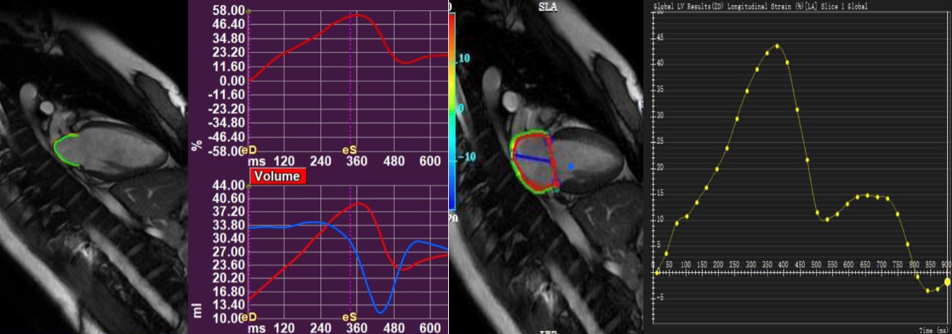
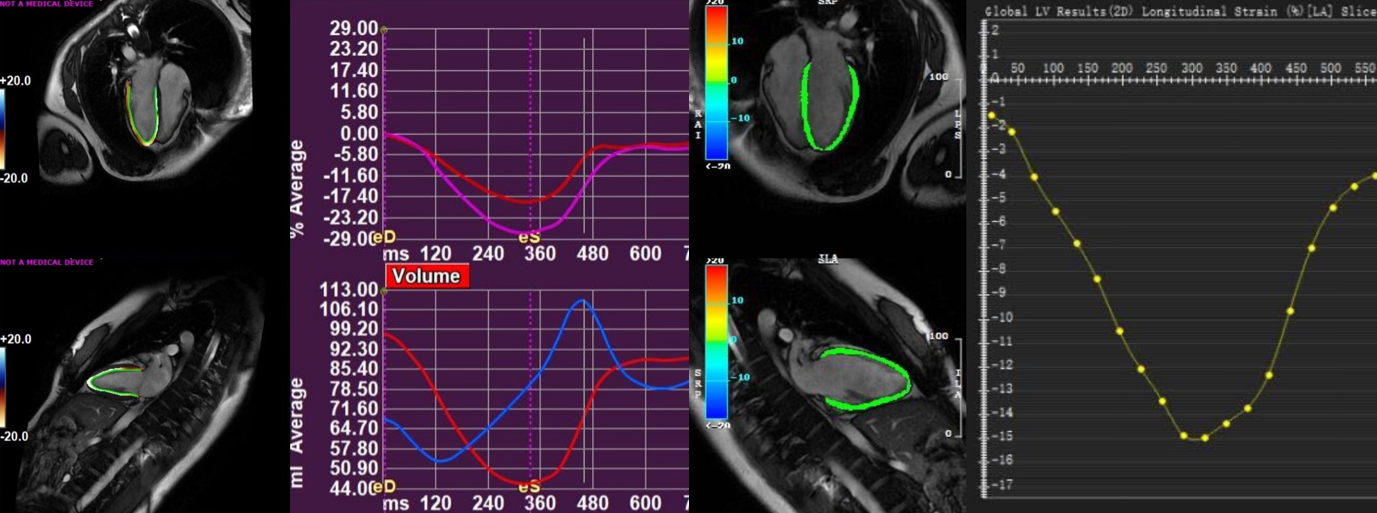
Goal(s): The study utilizes cardiac magnetic resonance feature tracking technique to assess the differences in myocardial strain parameters among healthy individuals of different age groups while employing two distinct vendor software tools.
Approach: The study utilizes cardiac magnetic resonance feature tracking technique to assess myocardial strain parameters while employing two distinct vendor software tools.
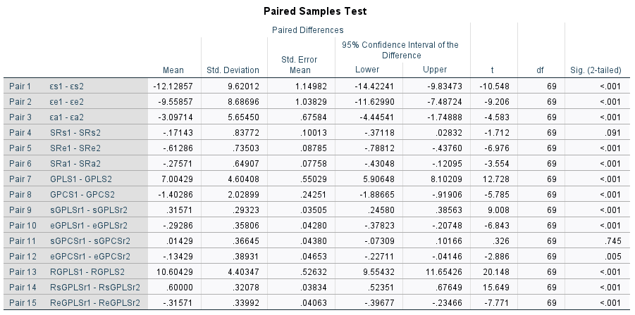
Results: The findings indicate variations in strain parameters across healthy individuals of varing age groups based on measurements obtained from CVI42 and Medis. Significant disparities in certain parameters are observed between the two suppliers.
Impact: The normal reference values of myocardial strain from different suppliers can not be unified, and large samples and multi-center studies are needed in the future.
Introduction
Cardiac magnetic resonance feature tracking technology offers a non-invasive method to assess myocardial mechanics. It enables a quantitative and accurate analysis of global and regional myocardial deformation. CMR-FT myocardial strain holds substantial potential for early diagnosis, treatment, and prognostic evaluation of various cardiovascular diseases. However, the absence of standardized reference values for myocardial strain measurement among different suppliers poses a significant obstacle to its clinical application. Additionally, it’s important to recognize that myocardial strain undergoes physiological changes with age. This study aims to conduct a longitudinal investigation into the variations in myocardial strain among healthy individuals of different age groups and a cross-sectional examination of the differences in myocardial strain between CVI42 and Medis suppliers in the same cohort of healthy individuals.Methods
Seventy healthy Chinese individuals, comprising 30 males and 40 females, meeting the inclusion criteria and displaying normal cardiac magnetic resonance results, were enrolled and divided into three groups based on age: 24 participants in group 1 (18-45 years), 24 participants in group 2 (46-60 years) and 22 participants in group 3 (≥61 years). Film sequences of standard 2ch,4ch, and short-axis views of the heart were obtained by balanced steady-state free procession sequences. Two physicians independently measured strain and strain rate in the left atrium, left ventricle, and right ventricle using CVI42 and Medis, with cross-repeated measurements. Statistics were performed using one-way ANOVA, Friedman's analysis, paired t-test and Wilcox test. Intra- and inter-observer group consistency tests were conducted.Results
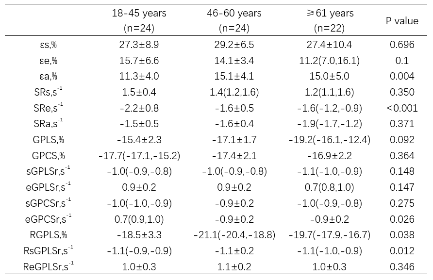
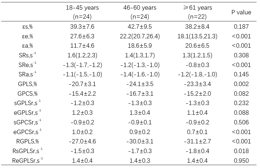
1) Myocardial strain in the 3 groups:Based on CVI42 measurements, significant differences were found in εa, SRe, sGPLSr, eGPCSr, RGPLS, and RsGPLSr. Notably, εa differed between Group 1 and Group 2 (P=0.003), and εa and SRe differed significantly when comparing Group 1 and Group 3 (both P<0.01). Based on Medis measurements, signnificant differences were observed in εe, εa, SRe, GPLS, eGPCSr, RGPLS, and RsGPLSr. Group 1 and Group 2 exhibited significant differences in εa, GPLS, and RGPLS (P<0.01 for all), and Group 1 and Group 3 showed significant differences in εe, εa, GPLS, eGPCSr, and RGPLS (P<0.01 for all). 2)Comparison between CVI42 and Medis measurements: εs (27.9±8.6% vs. 40.1±8.6%), εe (14.0±5.6% vs. 23.6±7.7%), εa (13.7±4.6% vs. 16.8±6.8%), GPLS (-15.6±4.5% vs. -22.6±3.6%), GPCS (-17.1±1.9% vs. -15.7±2.5%), sGPLSr (-0.9±0.2s-1 vs. -1.2±0.3 s-1), eGPLSr (0.9±0.2 s-1 vs. 1.2±0.4 s-1),RGPLS (-18.7±3.1% vs. -29.3±3.9%), RsGPLSr (-1.0±0.2 s-1 vs. -1.6±0.3 s-1), and ReGPLSr (1.0±0.3 s-1 vs. 1.3±0.4 s-1) all exhibited statistically significant differences (P<0.01 for all). Myocardial strain and strain rate measured by CVI42 and Medis demonstrated good intra- and inter-observer reproducibility.Conclusion
The most significant physiological changes in atrial strain occur at different ages, and it’s evident that the normal reference values for myocardial strain cannot be standardized across different providers. Addressing this issue may require advancements in technology and the incorporation of artificial intelligence in the future.Acknowledgements
This work is supported by Central Hospital of Dalian University of Technology.References
1、Pathan F, Zainal Abidin HA, Vo QH, Zhou H, D'Angelo T, Elen E, Negishi K,Puntmann VO, Marwick TH, Nagel E. Left atrial strain: a multi-modality, multi-vendor comparison study. Eur Heart J Cardiovasc Imaging. 2021 Jan1;22(1):102-110. doi: 10.1093/ehjci/jez303. PMID: 31848575.2、Truong VT, Palmer C, Wolking S, Sheets B, Young M, Ngo TNM, Taylor M, NaguehSF, Zareba KM, Raman S, Mazur W. Normal left atrial strain and strain rate usingcardiac magnetic resonance feature tracking in healthy volunteers. Eur Heart JCardiovasc Imaging. 2020 Apr 1;21(4):446-453. doi: 10.1093/ehjci/jez157. PMID:31504357.3、Gao Y, Zhang Z, Li G, Zhou S, Lou M, Zhao Z, Zhao J, Li K, Pohost GM.Reference Values for Left Atrial Strain and Strain Rate Based on a Large Sampleof Healthy Chinese Adults: An MR-Feature Tracking Study. J Magn Reson Imaging.2021 Dec;54(6):1784-1793. doi: 10.1002/jmri.27768. Epub 2021 Jun 15. PMID:34131972.4、Cau R, Bassareo P, Suri JS, Pontone G, Saba L. The emerging role of atrialstrain assessed by cardiac MRI in different cardiovascular settings: an up-to-date review. Eur Radiol. 2022 Jul;32(7):4384-4394. doi:10.1007/s00330-022-08598-6. Epub 2022 Apr 22. PMID: 35451607; PMCID: PMC9213357.5、Xu J, Yang W, Zhao S, Lu M. State-of-the-art myocardial strain by CMR featuretracking: clinical applications and future perspectives. Eur Radiol. 2022Aug;32(8):5424-5435. doi: 10.1007/s00330-022-08629-2. Epub 2022 Feb 24. PMID:35201410.6、Erley J, Tanacli R, Genovese D, Tapaskar N, Rashedi N, Bucius P, Kawaji K,Karagodin I, Lang RM, Kelle S, Mor-Avi V, Patel AR. Myocardial strain analysisof the right ventricle: comparison of different cardiovascular magneticresonance and echocardiographic techniques. J Cardiovasc Magn Reson. 2020 Jul23;22(1):51. doi: 10.1186/s12968-020-00647-7. PMID: 32698811; PMCID: PMC7376701.7、Lange T, Schuster A. Quantification of Myocardial Deformation Applying CMR-Feature-Tracking-All About the Left Ventricle. Curr Heart Fail Rep. 2021Aug;18(4):225-239. doi: 10.1007/s11897-021-00515-0. Epub 2021 May 1. PMID:33931818; PMCID: PMC8342400.Figures