2993
Quantification of high-resolution magic-angle spinning (HR-MAS) NMR spectroscopy in cerebral organoids1University of Toronto, Toronto, ON, Canada, 2Sunnybrook Research Institute, Toronto, ON, Canada, 3NMR Solutions, Helsinki, Finland
Synopsis
Keywords: Data Processing, Spectroscopy, Organoids
Motivation: 12
Goal(s): 2
Approach: 2
Results: 2
Impact: 2
Introduction
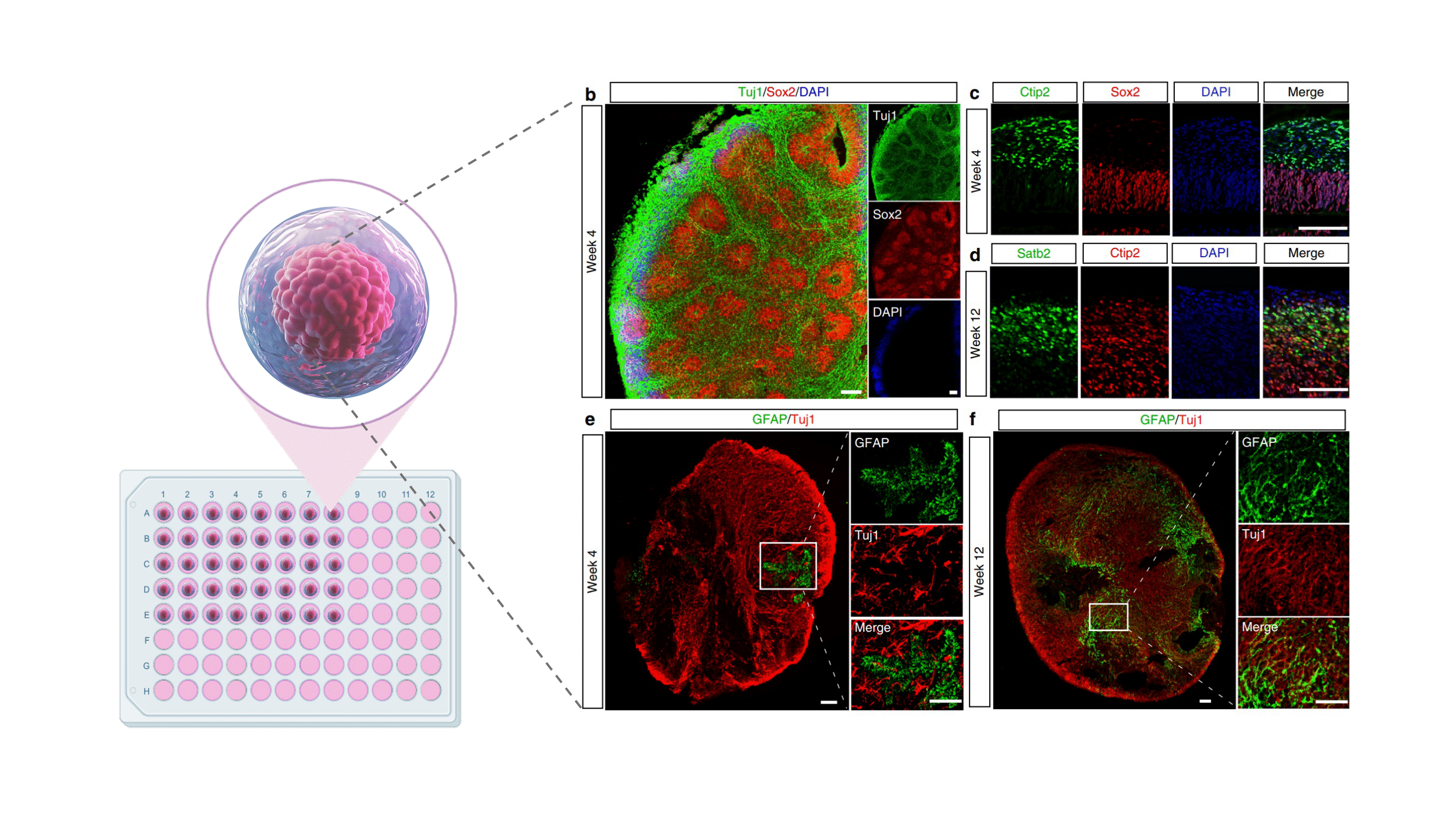
Cerebral organoids (COs), derived from pluripotent stem cells, are three-dimensional self-organising clusters of cultured brain tissue resembling the human brain’s genetics and phenotype1–4. Establishing an in vitro neurophysiology model requires novel methodologies to assess their similarity to the brain and translate therapeutics from COs to the clinic. NMR Spectroscopy (NMRS) offers non-invasive, high-reproducibility, non-destructive analysis, providing quantification of metabolites involved in different cellular and metabolic pathways5–8. However, NMRS faces challenges in obtaining high-quality data from COs, including magnetic field homogeneity and dipolar coupling effects, acquisition times, sample concentration, and biomolecular weight6,7,9,10. These limitations result in broad linewidths, non-flat baselines, overlapping peaks, and a low signal-to-noise ratio, complicating further analysis of COs neurochemical profile. While pre-processing is essential for noise reduction and spectrum correction7,9, using approaches such as high-resolution magic-angle spinning (HR-MAS) NMR can improve the signal quality from small-volume, low-concentration samples11,12. HR-MAS NMR is a powerful tool to study COs as fast spinning reduces linewidth, facilitating metabolite identification. However, the quantification of HR-MAS NMR spectra from complex metabolite mixtures such as brain tissue remains complicated11,12. Analysis of in vivo brain MR spectra is routinely performed using linear combination modelling software such as LCModel, ABfit, FSL-MRS and OSPREY. These tools perform well even in the presence of minor basis set imperfections, due to the relatively high degree of line-broadening observed in vivo. In contrast, the very narrow linewidths observed In HR-MAS spectra result in poor performance of standard linear combination modelling tools if there are relatively minor errors in the basis set parameters, such as the chemical shifts and J-coupling constants. Therefore, a methodology with flexibility to adjust the coupling and chemical shifts of basis spectra is needed for accurate linear combination modelling of HR-MAS spectra of complex mixtures6,10,15. In this work, we explore the use of a new software tool for analysis of HR-MAS NMR spectra for improved metabolite identification and quantification in COs.Methods
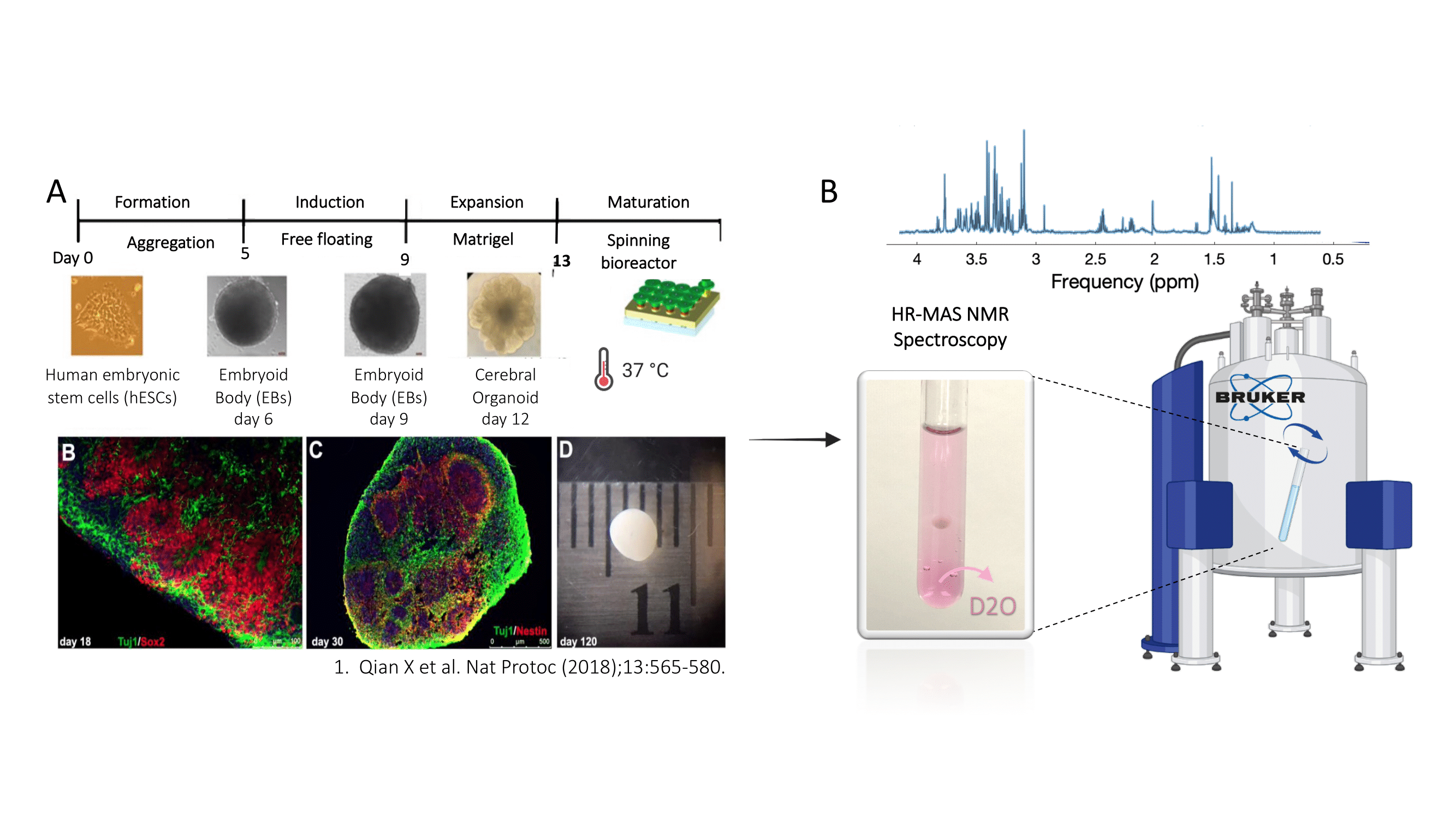
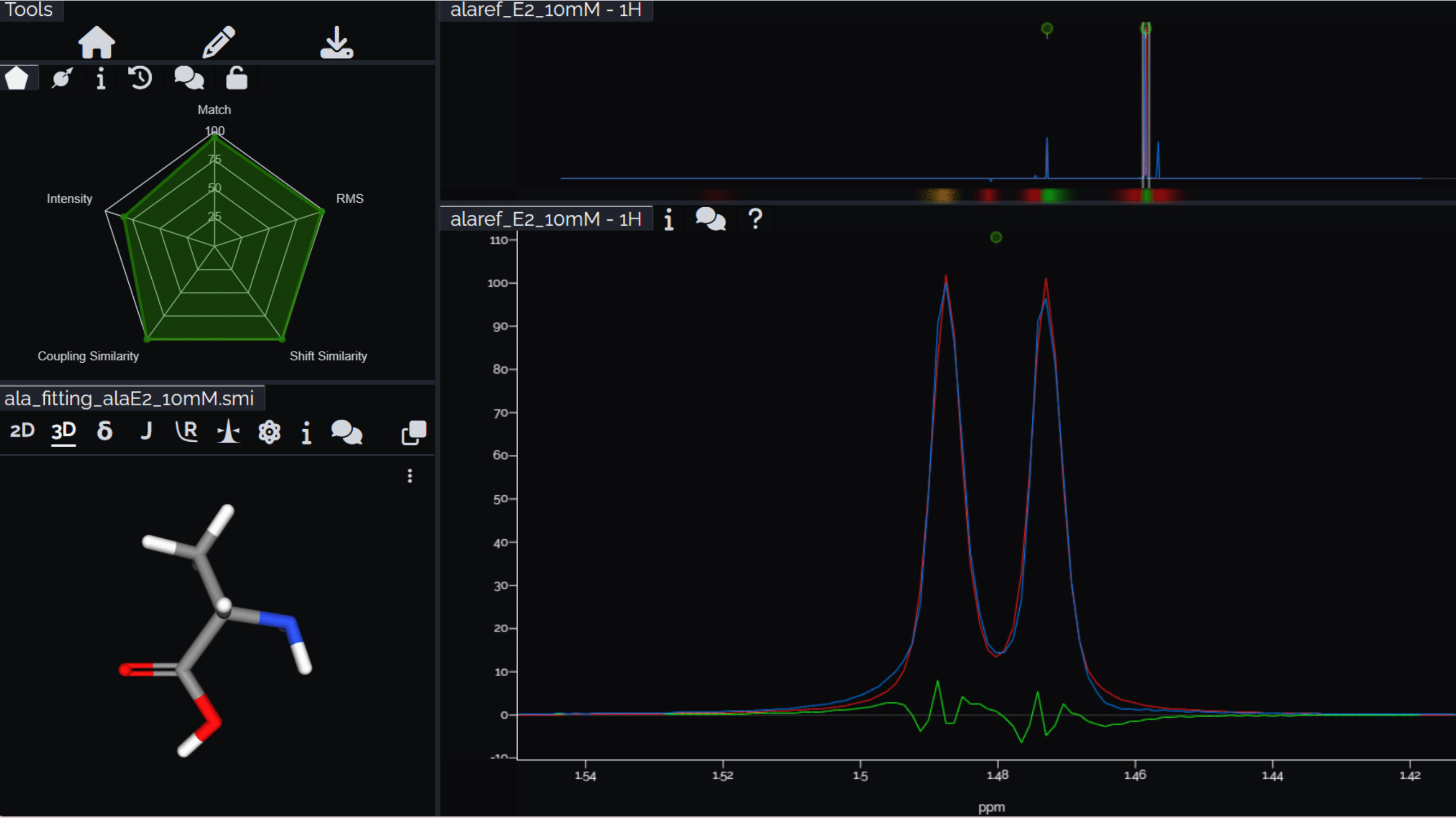
Here, we propose metabolite identification and quantification in 500 MHz HR-MAS NMR spectra from ~100-day-old COs derived from Pluripotent Stem Cells, adjusting chemical shifts and J-coupling constants for a consistent fitting model. After pre-processing data in FIDA (Matlab v.R2023b)16 and Topspin v.4.3.0 (Bruker) to visualize and correct phase and baseline, we employed Cosmic Truth (CT; ct.nmrsolutions.io) from NMR solutions, a quantum mechanical spectral analysis tool for 1D-1H NMR spectroscopy, to extract NMR parameters, including chemical shifts and coupling constants from COs NMR spectra with high order effects and overlapping signals. Acquired HR-MAS spectra were loaded into CT, and initial basis functions were loaded using the simplified molecular input line entries (SMILEs) of metabolites of interest, followed by semi-automatic adjustment of coupling and chemical shifts to achieve spectral similarity with the spin system entries. For metabolite quantification, a reference scan was performed in a phantom containing a 20mM solution of alanine. Metabolite concentrations were then estimated by measuring their signal intensity relative to the alanine reference signal.Results
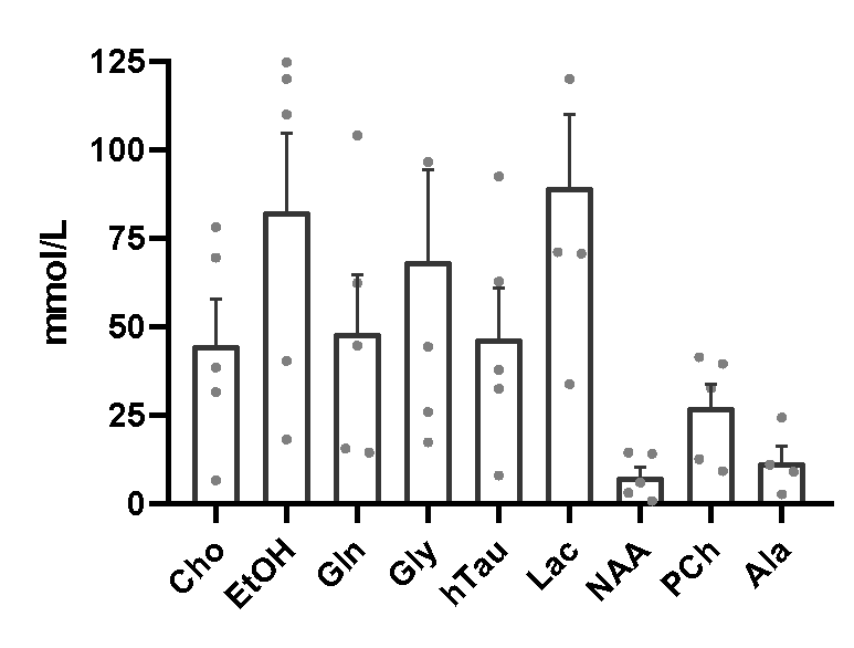
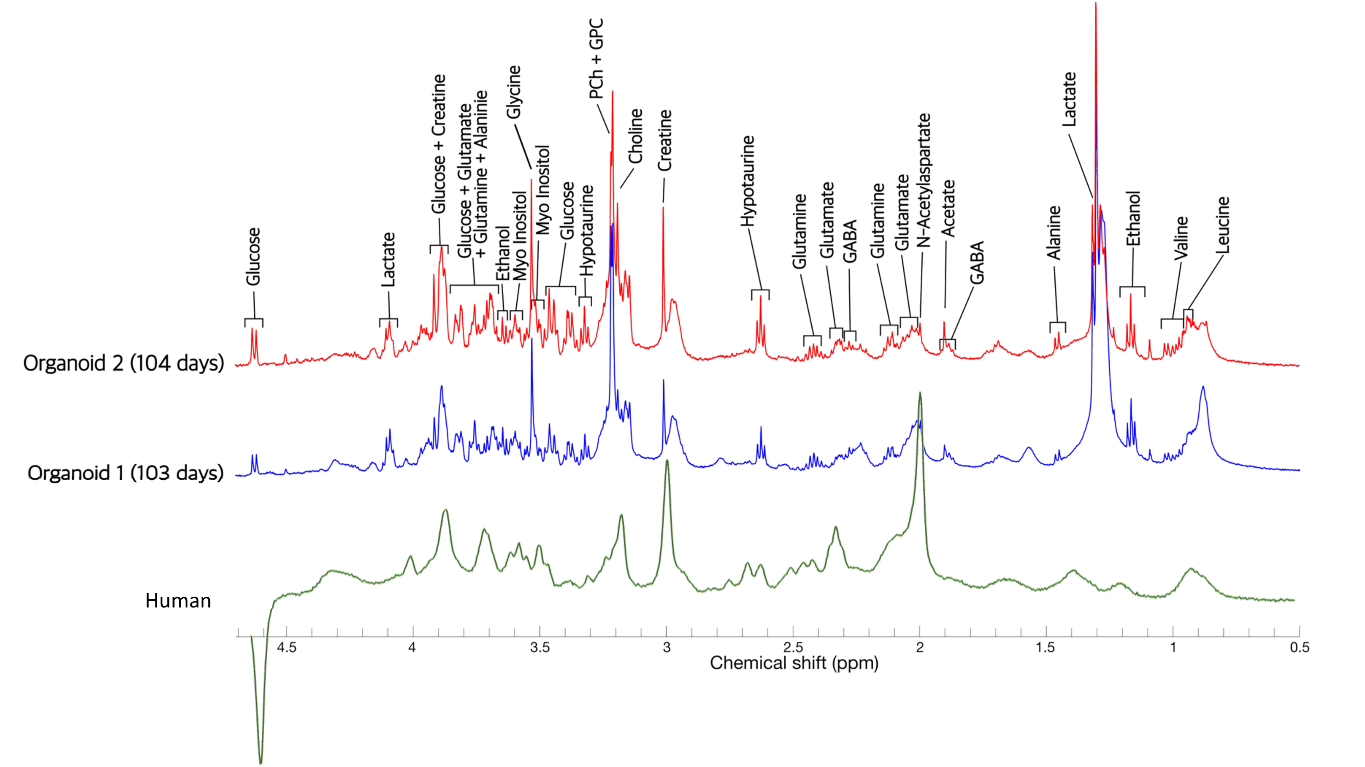
We identified and quantified 17 metabolites in HR-MAS NMR spectra of ~100 day-old COs, revealing important distinctions from the human brain. NAA is notably absent in COs, which may be due to early maturation stages or the absence of crucial neuronal support cell interactions, like microglia and oligodendrocytes. In contrast, COs exhibit elevated levels of lactate, ethanol, choline, phosphocreatine, glycine, and glucose. The high concentration of lactate may stem from inner-core hypoxia within COs, suggesting potential anaerobic glycolysis in this cerebral platform. Future research will focus on metabolic pathways activated in later COs maturation stages and compare genetic expression with HR-MAS NMR quantification.Conclusions
This study proposes an analysis method for the identification and quantification of metabolites in cerebral organoids, which account for flexible parameters, such as chemical shifts, and coupling constants. It addresses many of the shortcomings of conventional linear combination modelling approaches for the analysis of high-resolution NMR data. In addition, this method may extend its application to quantify metabolites and small molecules in different human brain platforms, going beyond cerebral organoids.Acknowledgements
This work is supported by the Canadian Institutes for Health Research (JN, AS and CS, Grant #: PJT-183715).References
1. Chiaradia, I., and Lancaster, M.A. (2020). Brain organoids for the study of human neurobiology at the interface of in vitro and in vivo. Nat Neurosci 23, 1496–1508. 10.1038/s41593-020-00730-3.
2. Di Lullo, E., and Kriegstein, A.R. (2017). The use of brain organoids to investigate neural development and disease. Nat Rev Neurosci 18, 573–584. 10.1038/nrn.2017.107.
3. Eichmüller, O.L., and Knoblich, J.A. (2022). Human cerebral organoids — a new tool for clinical neurology research. Nat Rev Neurol 18, 661–680. 10.1038/s41582-022-00723-9.
4. Frontiers | In vitro Models of Neurodegenerative Diseases https://www.frontiersin.org/articles/10.3389/fcell.2020.00328/full.
5. Alderson, T.R., and Kay, L.E. (2021). NMR spectroscopy captures the essential role of dynamics in regulating biomolecular function. Cell 184, 577–595. 10.1016/j.cell.2020.12.034.
6. Emwas, A.-H., Roy, R., McKay, R.T., Tenori, L., Saccenti, E., Gowda, G.A.N., Raftery, D., Alahmari, F., Jaremko, L., Jaremko, M., et al. (2019). NMR Spectroscopy for Metabolomics Research. Metabolites 9, 123. 10.3390/metabo9070123.
7. Emwas, A.-H., Saccenti, E., Gao, X., McKay, R.T., dos Santos, V.A.P.M., Roy, R., and Wishart, D.S. (2018). Recommended strategies for spectral processing and post-processing of 1D 1H-NMR data of biofluids with a particular focus on urine. Metabolomics 14, 31. 10.1007/s11306-018-1321-4.
8. Kumar, S., Villanueva, M., and Zuklic, J. (2016). Quantitative Determination of Acetaminophen by q-NMR. Comparison of Analysis with TopSpin software. MestReNova software, and ERETIC Method. GSU Research Day.
9. Wishart, D.S. (2019). NMR metabolomics: A look ahead. Journal of Magnetic Resonance 306, 155–161. 10.1016/j.jmr.2019.07.013.
10. Lin, A., Andronesi, O., Bogner, W., Choi, I.-Y., Coello, E., Cudalbu, C., Juchem, C., Kemp, G.J., Kreis, R., Krššák, M., et al. (2021). Minimum Reporting Standards for in vivo Magnetic Resonance Spectroscopy (MRSinMRS): Experts’ consensus recommendations. NMR in Biomedicine 34, e4484. 10.1002/nbm.4484.
11. Vonhof, E.V., Piotto, M., Holmes, E., Lindon, J.C., Nicholson, J.K., and Li, J.V. (2020). Improved Spatial Resolution of Metabolites in Tissue Biopsies Using High-Resolution Magic-Angle-Spinning Slice Localization NMR Spectroscopy. Anal. Chem. 92, 11516–11519. 10.1021/acs.analchem.0c02377.
12. Hassan, Q., Dutta Majumdar, R., Wu, B., Lane, D., Tabatabaei-Anraki, M., Soong, R., Simpson, M.J., and Simpson, A.J. (2019). Improvements in lipid suppression for 1H NMR-based metabolomics: Applications to solution-state and HR-MAS NMR in natural and in vivo samples. Magnetic Resonance in Chemistry 57, 69–81. 10.1002/mrc.4814.
13. Oeltzschner, G. Osprey Documentation. (2018) RainCloudPlots tutorials and codebase (Version v1.1). Zenodo. http://doi.org/10.5281/zenodo.3368186
14. Izquierdo-Garcia, J., Villa Valverde, P., Kyriazis, A., Puerto-Nevado, L., Pérez-Rial, S., Rodriguez, I., Hernandez, N., and Ruiz-Cabello, J. (2011). Descriptive Review of Current NMR-Based Metabolomic Data Analysis Packages. Progress in nuclear magnetic resonance spectroscopy 59, 263–270. 10.1016/j.pnmrs.2011.02.001.
15. Liu, Y., Navarro-Vázquez, A., Gil, R.R., Griesinger, C., Martin, G.E., and Williamson, R.T. (2019). Application of anisotropic NMR parameters to the confirmation of molecular structure. Nat Protoc 14, 217–247. 10.1038/s41596-018-0091-9.
16. FID-A – Advanced MR spectroscopy processing and simulation – Open Source Imaging https://www.opensourceimaging.org/project/fid-a-advanced-processing-and-simulation-of-mr-spectroscopy/.
Figures