2978
Enhanced approach for three-dimensional myelin-weighted imaging using a novel flow suppression technique in the ViSTa1Department of Biomedical Engineering, Hankuk University of Foreign Studies, Yongin-si, Korea, Republic of, 2Imaging institute, Cleveland Clinic Foundation, Cleveland, OH, United States
Synopsis
Keywords: Multiple Sclerosis, Multiple Sclerosis, Myelin Water Imaging
Motivation: ViSTa offers improved myelin-weighted image. However, the presence of flow artifacts was seen when scanning a subject with a high flow speed.
Goal(s): This study aimed to construct a novel 3D ViSTa sequence by implementing a series of flow saturation pulses.
Approach: Three flow saturation pulses were placed as evenly as possible between the 1st inversion pulse and the excitation pulse to effectively mitigate the presence of flow with various speeds.
Results: The results from the proposed ViSTa sequence reveal a significant reduction in flow artifacts.
When we conduct MS patient scans, lesions have a significantly diminished signal level, providing a distinct demarcation.
Impact: The proposed method provides improved whole-brain covered 3D myelin-weighted images in a clinically reasonable scan time (< 7 min). Moreover, it shows good sensitivity to MS lesions. These features make the proposed method appealing for clinical neuroimaging applications.
Introduction
Myelin density measurement in the brain has important clinical and fundamental scientific implications, particularly with rising interest in the development of remyelination therapies for MS. Myelin water imaging (MWI) has been proposed as a biomarker, offering sensitivity and specificity in the detection and quantification of myelin[1].Our group proposed a ViSTa[2] imaging technique to acquire a short T2* signal without using multi-exponential fitting. ViSTa was utilized to capture the signal of the short T2* component of the signal, associated with myelin water by leveraging the T1 relaxation disparity between different water pools[3]. ViSTa offers substantially enhanced image quality when compared to conventional approaches.
In a previous study[2] on healthy controls, we used saturation pulses to suppress flow artifacts. However, we subsequently observed flow artifacts in a patient, presumably because disease-associated stenosis of arteries leads to high flow speed that exceeds the capabilities of the saturation pulseIn order to acquire a reliable ViSTaMWI-map, it is crucial to implement a flow suppression technique that effectively functions across a large range of flow velocities. This study aimed to construct a novel 3D ViSTa sequence by implementing a series of flow saturation pulses.
Methods
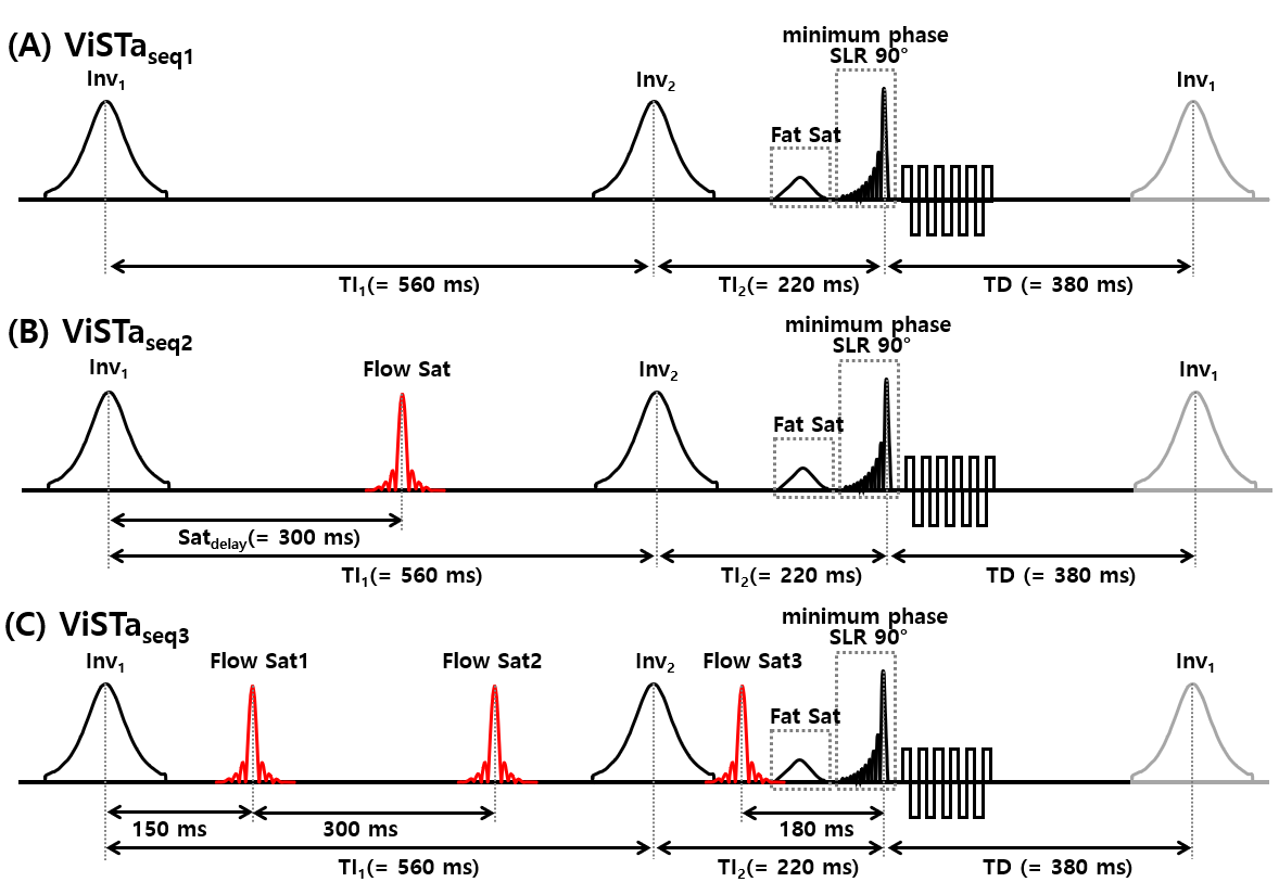
Data were collected from a healthy control and an MS patient (IRB approved) at 3T (Prisma, Siemens).As shown in Fig. 1, the diagram depicts three variations of the 3D ViSTa sequence: (A) without a flow saturation pulse, (B) with one flow saturation pulse, and (C) the proposed 3D ViSTa sequence. In the ViSTaseq2 sequence, which was used previously, the flow saturation pulse (flip angle = 180°, duration = 3.84 ms, and TBW = 8) was located between the two inversion RF pulses (300 ms after the peak of the first inversion pulse). In the proposed ViSTa sequence (ViSTaseq3), three flow saturation pulses were placed as evenly as possible between the 1st inversion pulse and the excitation pulse to mitigate the presence of flow with various speeds effectively. The flow saturation band covered 11 cm thickness in the lower head and neck areas and was positioned below an imaging volume with a 5 mm gap.
To quantify apparent myelin water fraction (aMWF), a proton density (PD) weighted GRE scan that had the same readout as the 3D ViSTa sequence was obtained with TR = 75 ms and flip angle = 5° (scan time = 30 sec). The ViSTaMWI-map was derived by dividing the ViSTa data by the PD-weighted GRE data and multiplying the result by a scaling factor. The utilization of a scaling factor was employed in order to account for the influence of T1 and T2* weighting in both the ViSTa and GRE datasets.
Results
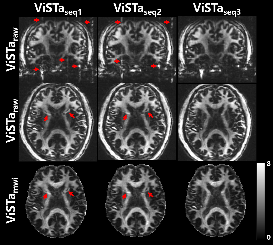
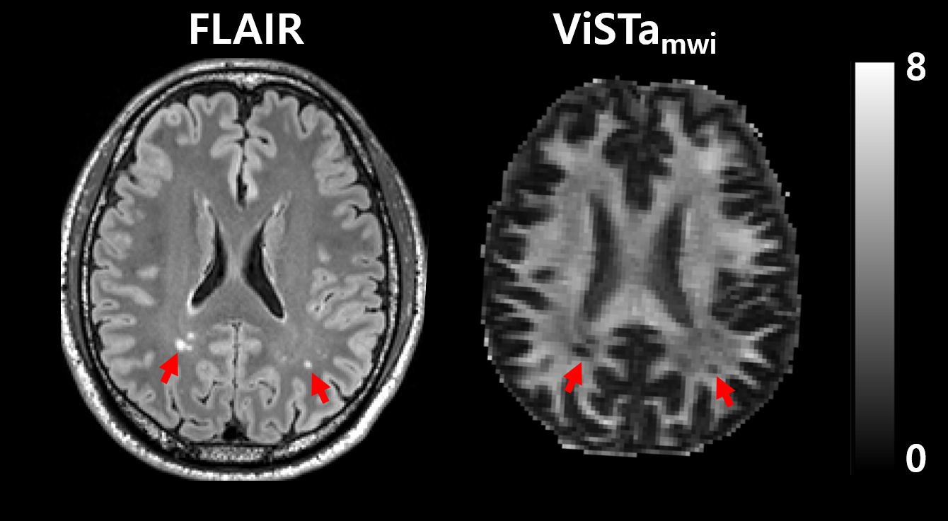
Figure 2 represents unnormalized 3D ViSTa images (ViSTaraw) and normalized ViSTaMWI-maps from three different ViSTa sequences. Upon comparing the images, it is evident that all the images exhibit a comparable signal distribution and tissue contrast. Results from ViSTaseq1 show the presence of significant flow artifacts. Despite utilizing a flow saturation RF in ViSTaseq2, the flow artifact is seen. Whereas the results from ViSTaseq3 reveal a significant reduction in flow artifacts.Figure 3 illustrates results from a patient diagnosed with MS. MS lesions demonstrated hyperintensities on the FLAIR. Corresponding regions on the ViSTaMWI-map also demonstrate diminished signal levels, suggesting that these are demyelinated lesions. Flow artifacts originating from arterial sources were not observed.
Discussion and Conclusion
This work demonstrated an enhanced approach for reducing inflow artifacts in ViSTa images. Our proposed VisTa technique provides an improved image quality on ViSTaMWI-maps as compared to the maps from the previous ViSTa acquisition technique.The proposed method provides myelin-weighted images in a clinically reasonable scan time (< 7 min). Moreover, it shows good sensitivity to MS lesions. These features make the proposed method appealing for clinical neuroimaging applications.
The time interval between the flow saturation pulse and saturation band location was determined empirically. More experiments may be needed to determine optimal parameters.
Acknowledgements
This work was supported by the National Research Foundation of Korea (NRF) grant funded by the Korea government (MSIT) (NRF-2023R1A2C1007292)References
1. MacKay, A., Whittall, K., Adler, J., Li, D., Paty, D., Graeb, D., 1994. In vivo visualization of myelin water in brain by magnetic resonance. Magn Reson Med 31, 673-677.
2. Oh, S.H., Bilello, M., Schindler, M., Markowitz, C.E., Detre, J.A., Lee, J., 2013. Direct visualization of short transverse relaxation time component (ViSTa). Neuroimage 83C, 485-492.
3. Labadie, C., Lee, J.H., Rooney, W.D., Jarchow, S., Aubert-Frecon, M., Springer, C.S., Jr., Moller, H.E., 2013. Myelin water mapping by spatially regularized longitudinal relaxographic imaging at high magnetic fields. Magn Reson Med.
Figures