2977
Shrinking multiple sclerosis lesions are characterized by a more destructive phenotype than expanding lesions1Institute of NeuroScience, UCLouvain, Bruxelles, Belgium, 2ICTEAM, UCLouvain, Louvain-la-Neuve, Belgium, 3Neurology, Cliniques Universitaire Saint-Luc, Bruxelles, Belgium
Synopsis
Keywords: Multiple Sclerosis, Multiple Sclerosis, Chronic lesions, diffusion MRI
Motivation: Slowly expanding lesions (SEL) have gained significant attention as a biomarker of chronic active multiple sclerosis (MS) lesions, however, one study1 suggests that all MS lesions tend to shrink over a long period of time.
Goal(s): The objective of this work is to investigate the microstructure of expanding lesions (EL), shrinking lesions (SL), and stable lesions.
Approach: EL and SL were computed using deformation-based volumetric MRI and microstructure was investigated using quantitative T1 and multi-shell diffusion MRI.
Results: SL showed a more destructive phenotype at baseline when compared to EL, while stable lesions were considerably less destructive.
Impact: This preliminary study underlies the necessity of considering the full spectrum of multiple sclerosis (MS) lesions, especially MRI-evolving lesions, whether shrinking or expanding, in MS research to extend our knowledge of the disease pathophysiology.
Introduction
Multiple sclerosis (MS) is the most prevalent chronic inflammatory disease of the central nervous system characterized by focal demyelinating lesions.2 Slowly expanding lesions (SEL) are a subset of chronic active MS lesions, found more prominently in patients with progressive MS, and have recently raised attention due to their association with neurological and cognitive disability.3 However, SEL showed only a limited correspondence with paramagnetic rim lesions (PRL),4 another novel imaging biomarker for chronic active lesions. In addition, a previous study showed that all MS lesions have a tendency to shrink over a long period of time (about 16 years), hypothesizing that the primary pathological process in chronic lesions, even those described as “slowly expanding,” is likely to be tissue loss.1 The inability to directly study MRI-evolving lesions with histology underscores the necessity for a more comprehensive understanding of their underlying pathology.The objective of this work is to investigate the microstructural differences at baseline between shrinking and expanding lesions over one year.
Methods
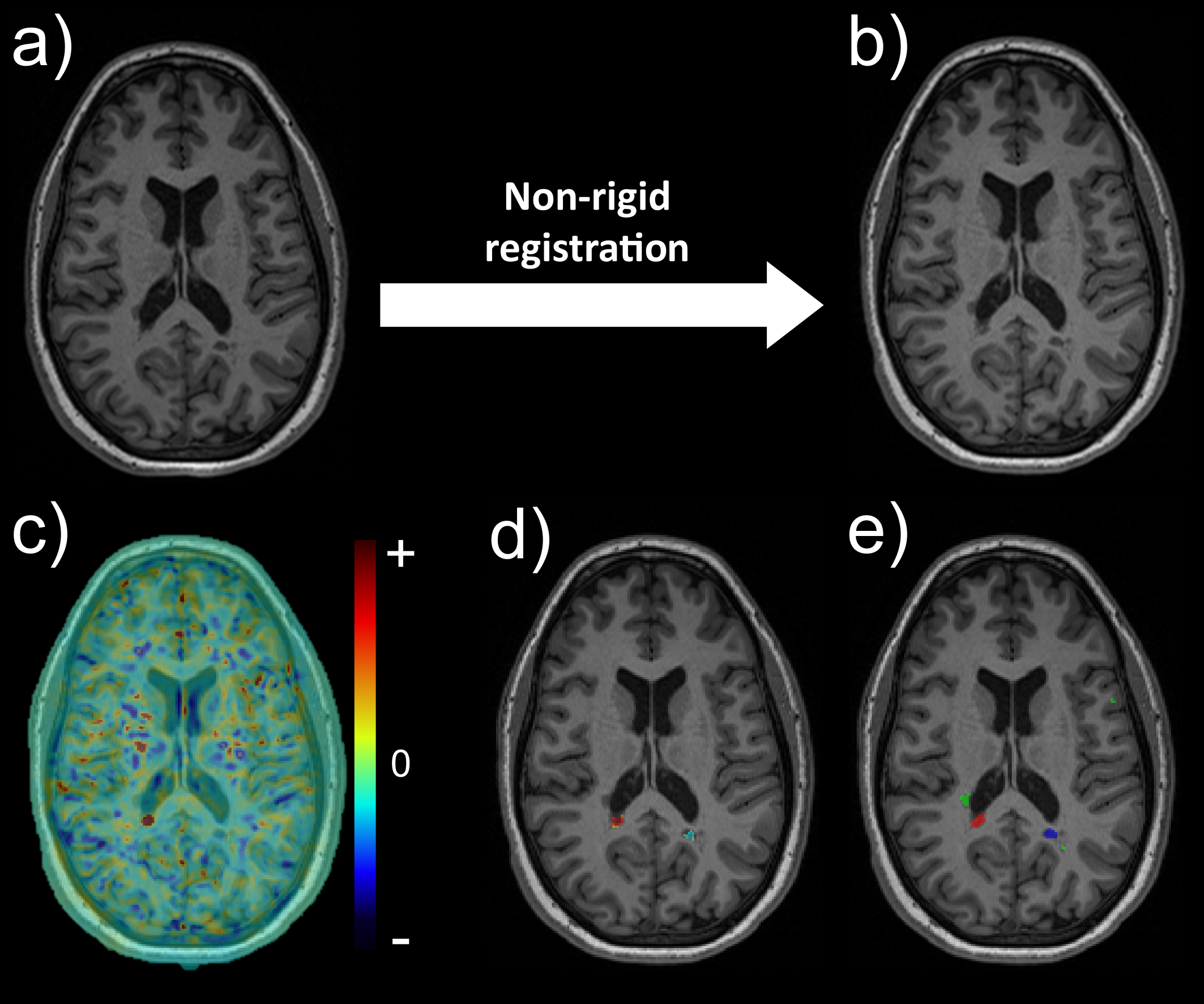
43 MS patients, with an MPRAGE at baseline and one-year follow-up, were included in this study. First, a deformation field was calculated by non-rigidly registering intra-subject baseline and follow-up MPRAGE images using ANTs.5 The Jacobian of this deformation field, normalized over a year, was used to compute local tissue expansion and shrinkage, as previously described.6 More specifically, expanding — resp. shrinking — lesional tissue was described as contiguous regions within lesions exhibiting positive — resp. negative — local volume change of at least 12%, extended by the surrounding area showing positive — resp. negative — volume change of at least 4%. Expanding lesions (EL) — resp. shrinking lesions (SL) — were defined as non-active chronic lesions displaying only or a majority (60%) of expansion — resp. shrinkage. Lesions showing neither expansion nor shrinkage were considered as stable, while ambiguous lesions showing both were discarded. The algorithm is represented in Figure 1. Overall, 1229 lesions (196 EL, 203 SL, 830 stable) were included in the analysis.Microstructural tissue integrity at baseline was investigated using quantitative MP2RAGE-derived tissue longitudinal relaxation time (T1), a known marker of myelination and axonal density,7 and multi-shell diffusion MRI (TR=4842 ms, TE=77 ms, Δ=35.7 ms, δ=22.9 ms, 64 gradients at b=1000, 32 at b=2000,3000,5000 s/mm2). Multi-shell diffusion data were preprocessed with denoising,8 motion and eddy-currents correction,9,10 skull-stripping using FSL BET,11 and then processed by two advanced diffusion models — Neurite Orientation Dispersion and Density Imaging (NODDI)12 and Microstructure Fingerprinting (MF)13 — to study NODDI’s extra-cellular volume fraction (ecvf) and MF’s weighted fiber volume fraction (wfvf).
Results
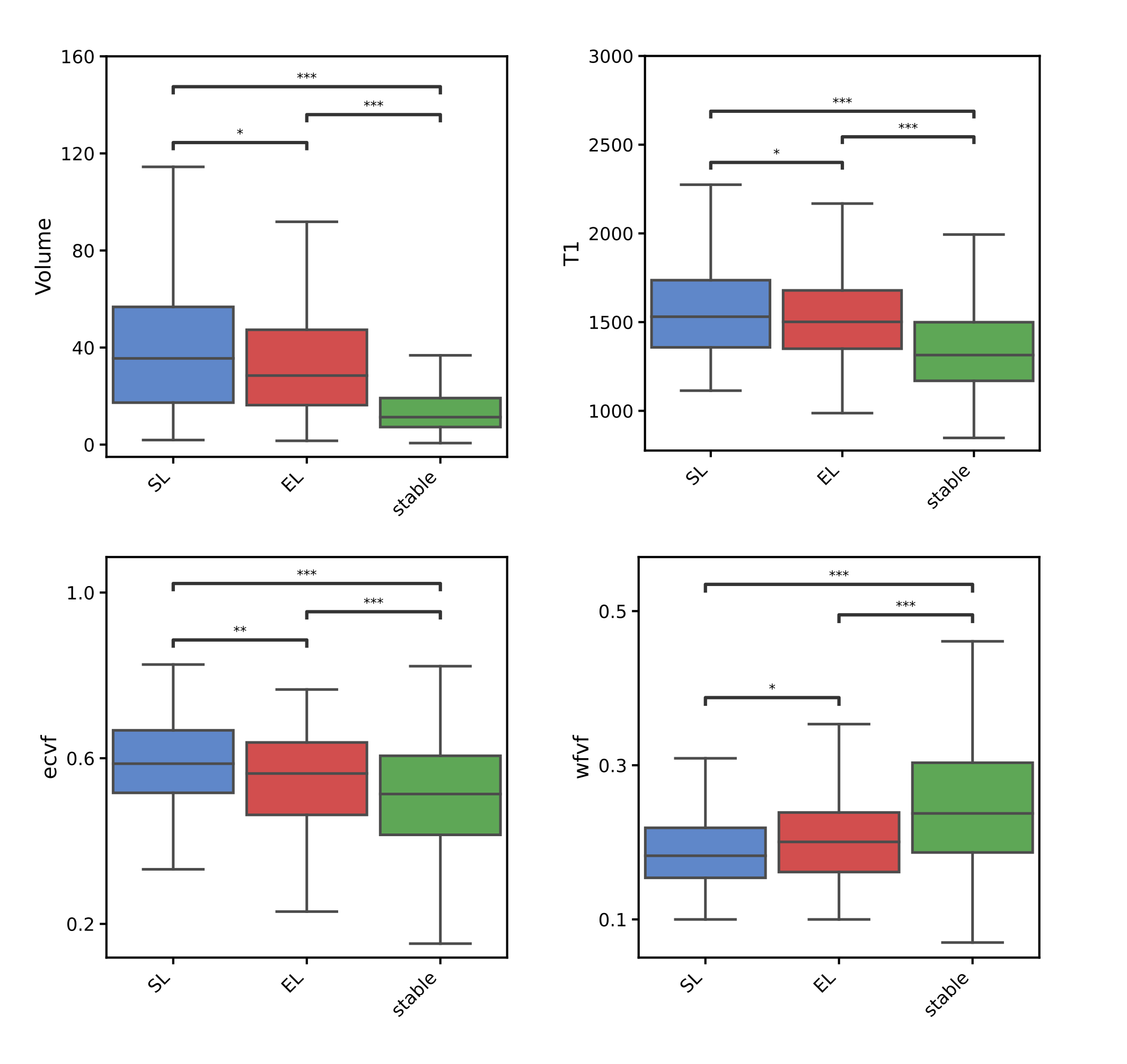
Results are displayed in Figure 2. At baseline, SL were bigger (p=0.013) than EL and showed longer T1 values (p=0.020). In addition, SL were characterized by more prominent extra-axonal cell infiltrate with higher NODDI’s ecvf (p=0.004) and by lower axonal integrity with lower MF’s wfvf (p=0.010). In comparison, stable lesions were considerably smaller than evolving lesions and characterized by a less destructive phenotype with shorter T1 values, lower ecvf, and higher wfvf (p<0.001).Discussion
Our results confirm that MRI-evolving lesions, whether shrinking or expanding, are considerably more destructive than stable lesions, underlying the dynamic nature of MS pathology. Interestingly, SL were characterized by a more destructive phenotype at baseline when compared to EL, with a more prominent accumulation of extra-axonal cells as the most significant difference, hypothesized to reflect a more important presence of astrogliosis in SL. These results are in line with previous findings on chronic shrinking lesions,1 but further investigations are needed to confirm that the most prevalent pathological process in chronic lesions is indeed tissue loss.1This preliminary study underlies the importance of considering all MRI-evolving lesions, whether shrinking or expanding, to gain a more comprehensive understanding of the dynamics of tissue damage within MS lesions. To build upon these initial findings, further investigations with a larger sample size are planned to investigate the association of MRI-evolving lesions, lesional age and other biomarkers of smoldering inflammation as well as with disability scores.
Acknowledgements
The authors thank the study participants; the neuroimmunology clinic of Cliniques universitaires Saint-Luc for recruiting and evaluating the patients and for coordinating the scans; Stefan Skare (Karolinska University Hospital), Thierry Duprez, Sébastien de Laever (Cliniques universitaires Saint-Luc), Laurence Dricot (Université catholique de Louvain) and Julie Poujol (GE Healthcare) for assistance with 3T MRI scan acquisition and analysis.References
1. Sethi, V. et al. Slowly eroding lesions in multiple sclerosis. Mult. Scler. J. 23, 464–472 (2017).
2. Reich, D. S., Lucchinetti, C. F. & Calabresi, P. A. Multiple Sclerosis. N. Engl. J. Med. 378, 169–180 (2018).
3. Calvi, A. et al. Association of Slowly Expanding Lesions on MRI With Disability in People With Secondary Progressive Multiple Sclerosis. Neurology 98, e1783–e1793 (2022).
4. Calvi, A. et al. Relationship between paramagnetic rim lesions and slowly expanding lesions in multiple sclerosis. Mult. Scler. Houndmills Basingstoke Engl. 29, 352–362 (2023).
5. Avants, B., Tustison, N. & Song, G. Advanced normalization tools (ANTS). Insight J 1–35, (2008).
6. Elliott, C. et al. Slowly expanding/evolving lesions as a magnetic resonance imaging marker of chronic active multiple sclerosis lesions. Mult. Scler. Houndmills Basingstoke Engl. 25, 1915–1925 (2019).
7. Kolb, H. et al. 7T MRI Differentiates Remyelinated from Demyelinated Multiple Sclerosis Lesions. Ann. Neurol. 90, 612–626 (2021).
8. Veraart, J. et al. Denoising of diffusion MRI using random matrix theory. NeuroImage 142, 394–406 (2016).
9. Andersson, J. L. R., Skare, S. & Ashburner, J. How to correct susceptibility distortions in spin-echo echo-planar images: application to diffusion tensor imaging. NeuroImage 20, 870–888 (2003).
10. Andersson, J. L. R. & Sotiropoulos, S. N. An integrated approach to correction for off-resonance effects and subject movement in diffusion MR imaging. NeuroImage 125, 1063–1078 (2016).
11. M, J. BET2 : MR-Based Estimation of Brain, Skull and Scalp Surfaces. Elev. Annu. Meet. Organ. Hum. Brain Mapp. 2005 (2005).
12. Zhang, H., Schneider, T., Wheeler-Kingshott, C. A. & Alexander, D. C. NODDI: practical in vivo neurite orientation dispersion and density imaging of the human brain. NeuroImage 61, 1000–1016 (2012).
13. Rensonnet, G. et al. Towards microstructure fingerprinting: Estimation of tissue properties from a dictionary of Monte Carlo diffusion MRI simulations. NeuroImage 184, 964–980 (2019).
Figures