2974
Assessment of Periventricular Gradient of Myelin Content in Multiple Sclerosis Using Synthetic MRI1Radiology, Juntendo University, Tokyo, Japan, 2Radiology, The University of Tokyo, Tokyo, Japan, 3Neurology, Juntendo University, Tokyo, Japan, 4Radiology, Toho University Omori Medical Center, Tokyo, Japan
Synopsis
Keywords: Multiple Sclerosis, Multiple Sclerosis
Motivation: Though periventricular gradient (PVG) mapping has been demonstrated to be useful, previous studies have employed 2D magnetization transfer ratios and T1 maps, which are not specific to myelin.
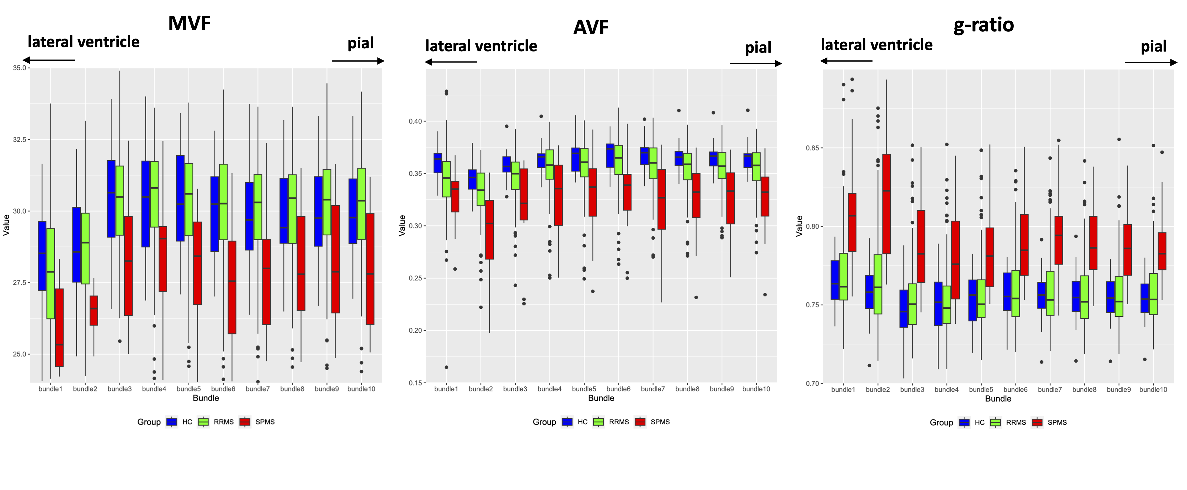
Goal(s): This study investigates the use of synthetic MRI myelin maps to assess the periventricular gradient (PVG) of myelin volume fraction (MVF) in multiple sclerosis (MS) patients.
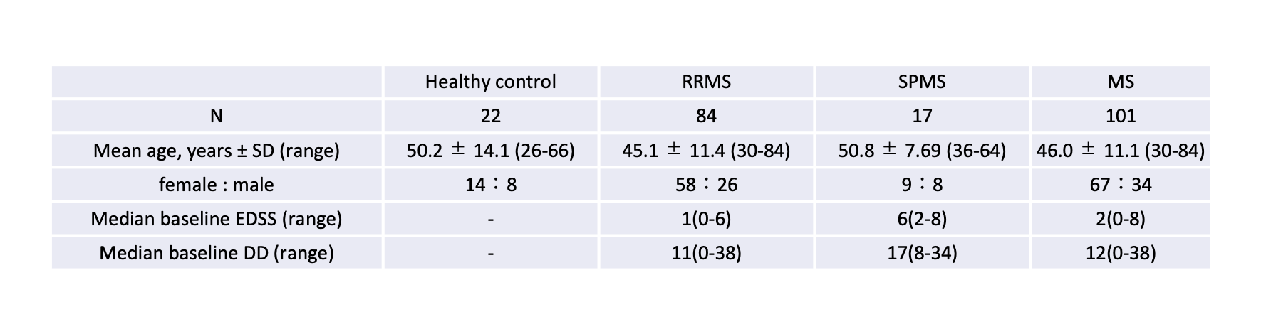
Approach: This study includes 22 healthy individuals, 84 patients with relapsing-remitting MS (RRMS), and 17 patients with secondary progressive MS (SPMS).
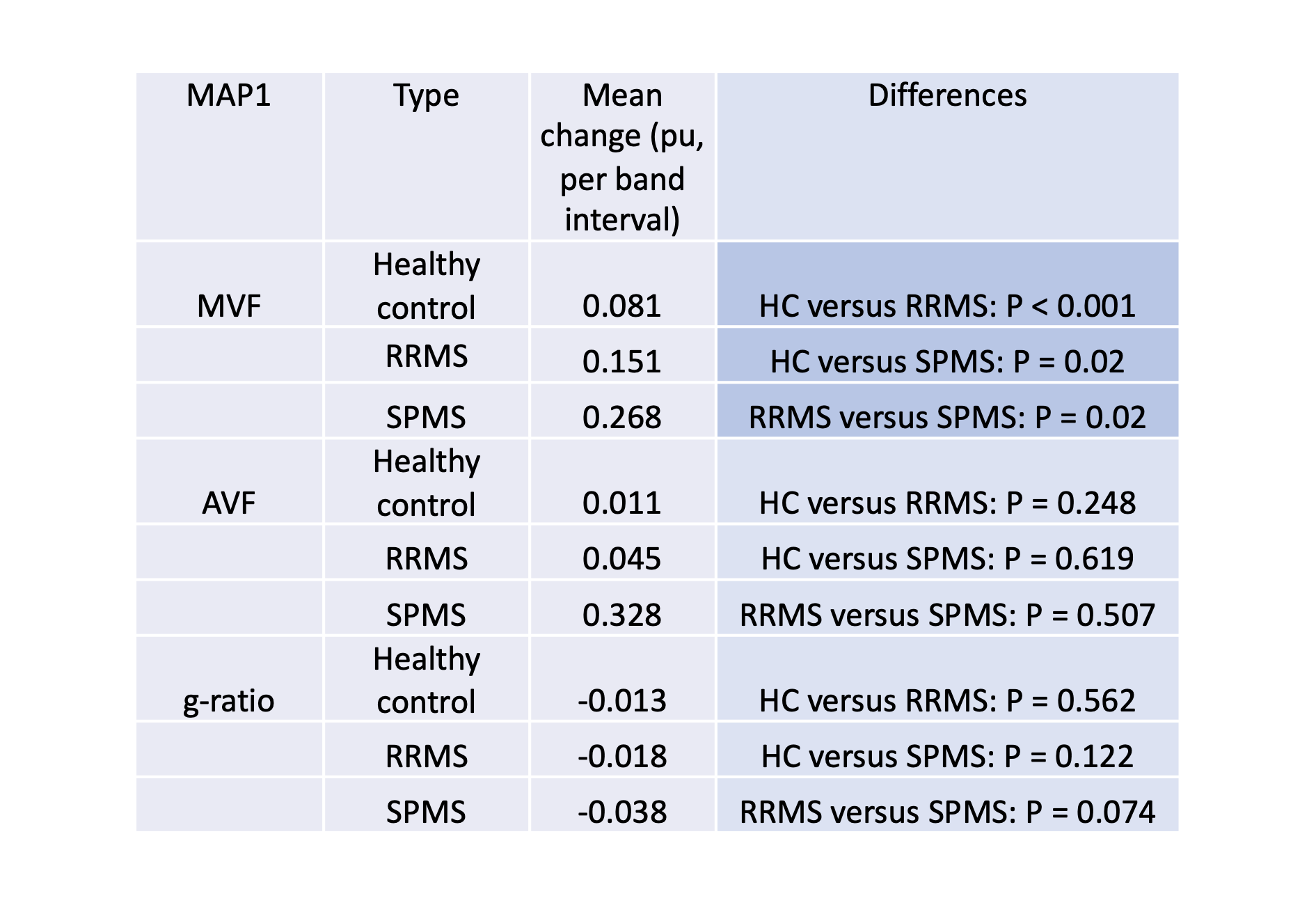
Results: PVGs of RRMS and SPMS were higher than that of healthy individuals, with SPMS showing higher PVG than RRMS.
Impact: In conclusion, the influence of demyelination in MS appears to be more prominent in the vicinity of the ventricles.
INTRODUCTION
Multiple sclerosis (MS) is characterized by an inflammatory process that originates from the brain's surface1. The damage to the lateral periventricular white matter exhibits a distinct ventricular-dominant gradient2. Though periventricular gradient (PVG) mapping has been demonstrated to be useful, previous studies have employed 2D magnetization transfer ratios and T1 maps3–6, which are not specific to myelin. Our study seeks to utilize the synthetic MRI myelin map; namely, the myelin volume fraction (MVF) to gauge the quantity of myelin fro evaluating PVG.METHODS
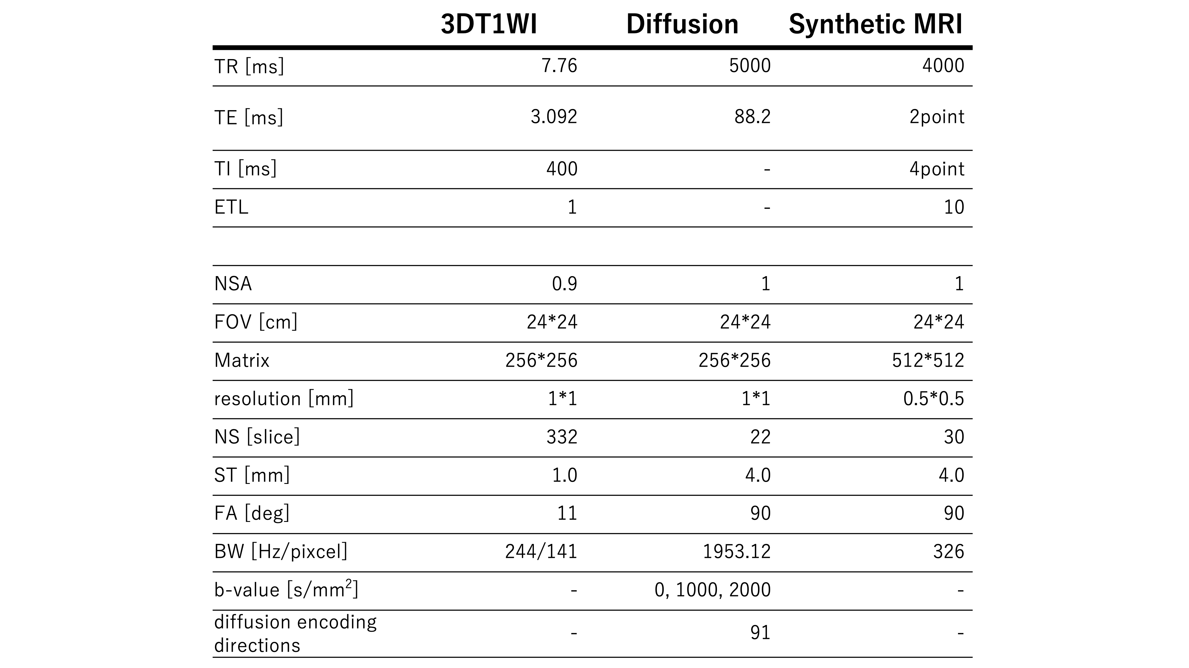
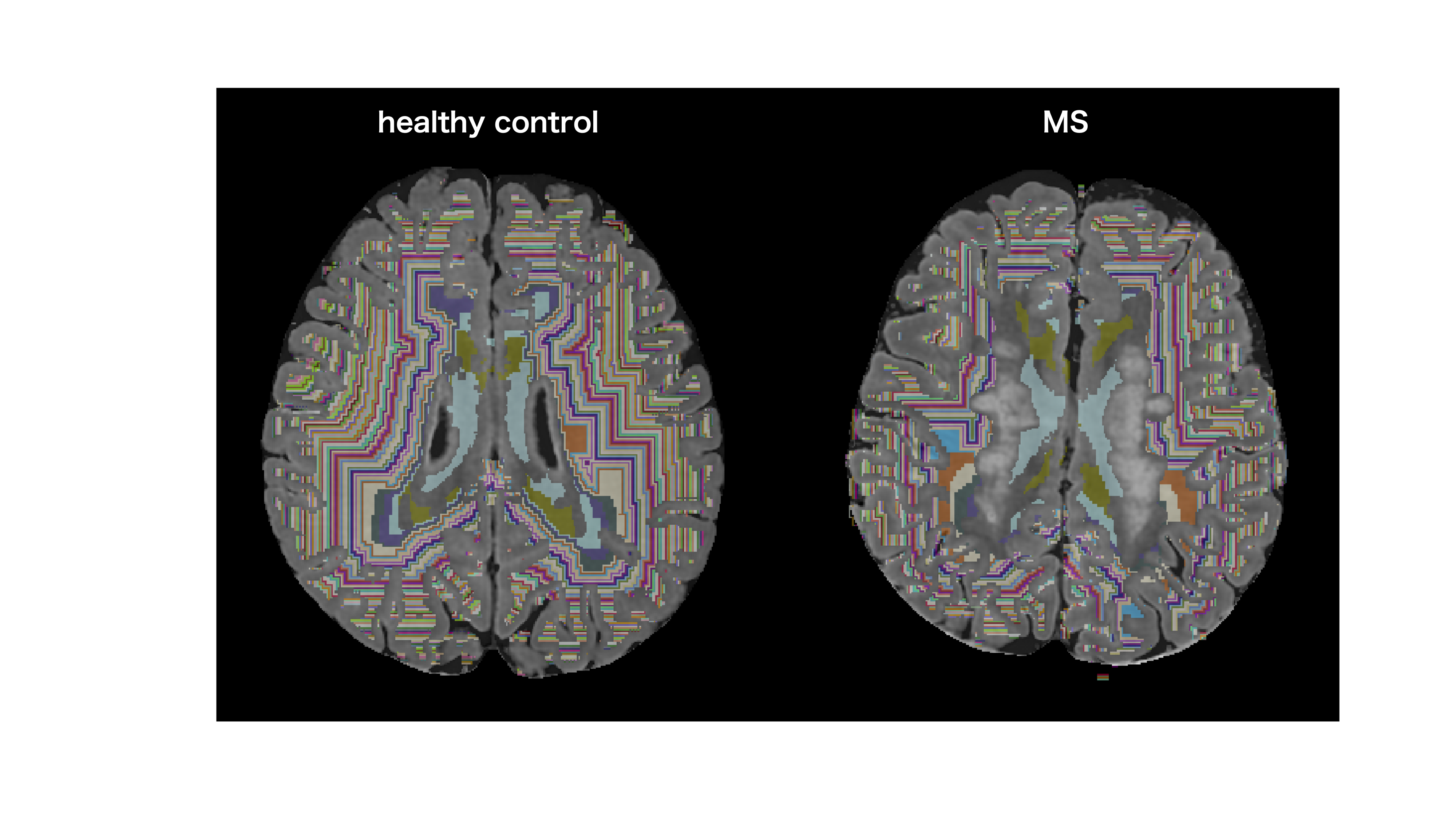
Participants and MRI: 22 healthy volunteers (8 male and 14 female, mean age 50.2 years, age range 26–66 years), 84 patients with relapsing-remitting multiple sclerosis (RRMS) (26 male and 58 female, mean age 45.1 years, age range 30–84 years), and 17 patients with secondary progressive multiple sclerosis (SPMS) (8 male and 9 female, mean age 50.8 years, age range 36–64 years) were included in this study. All patients underwent synthetic MRI relaxometry and diffusion-weighted on a 3-T MR scanner (Discovery MR750w; GE Healthcare, Milwaukee, Wisconsin). The FLAIR and myelin volume fraction (MVF) also synthesized with SyMRI software (version 11.0.7; SyntheticMR, Linköping, Swedens). Subsequently, the axonal volume fraction (AVF) and g-ratio were obtained as described by Stikov et al.7 using synthetized MVF and spatially co-registered neurite orientation dispersion and density imaging (NODDI) parameters obtained from the 2-shell diffusion data. In this study, for all subjects, hyperintense lesions were systematically segmented on synthetic FLAIR images employing the lesion-prediction algorithm implemented in the Lesion Segmentation Toolbox (LST), Version 2.0.13 (http://www.applied-statistics.de/lst.html)8, integrated within the Statistical Parametric Mapping (SPM12; Wellcome Trust Centre for Neuroimaging, London, UK; https://www.fil.ion.ucl.ac.uk/spm/software/spm12/) framework. Segmented lesions were used as the regions of interest (ROI). To create normal-appearing white matter (NAWM) mask, the ROI of lesion was subtracted from the manually corrected whole white matter mask defined based on ‘wm.seg.mgz’, a byproduct of the FreeSurfer pipeline (v.6.0.0, http://surfer.nmr.mgh.harvard.edu). Using FreeSurfer, the binarized lateral ventricule mask was also created and iteratively dilated by one-voxel until 100 utilizing the ‘fslmaths’ function with ‘-dilM’ option implemented in the FMRIB Software Library (http://www. fmrib.ox.ac.uk/fsl/). One-voxel thick NAWM bands were assigned with sequential integer labels by adding all dilated masks and applying a NAWM mask for each participant. Finally, for all subjects, the NAWM was segmented into concentric single-voxel bands, from ventricular boundary to the cortical surface. In this study, only 10 bands from the surface of lateral ventricles were used, after excluding one NAWM band closest to the lateral ventricles to eliminate the voxels affected by the partial volume effects. The analysis was confined to the tissues above the insula. PVG was defined as the gap of the mean parameter within a band between band 1 and 10. PVGs of MVF, AVF, and g-ratio were compared between three groups using Kruskal-Wallis test (post-hoc : Wilcoxon signed-rank test). We assessed the correlation between the PVG and expanded disabilitys status scale (EDSS), which reflects clinical disability, using Spearman’s correlation anlaysis. The false discovery rate-corrected p-value < 0.05 was considered as significant.RESULTS
PVGs of MVF for RRMS and SPMS were higher than that of healthy individuals (p < 0.001 and p = 0.02, respectively), with SPMS showing higher PVG than RRMS (p = 0.02). In the multiple sclerosis group combining RRMS and SPMS, the PVG of MVF correlated with EDSS (p = 0.005, Spearman's ρ = 0.28). For the PVG of axon volume fraction (AVF) and g-ratio, there was no significant difference and there was no significant correlation between PVG and EDSS.DISCUSSION
PVGs of RRMS and SPMS were higher than that of healthy individuals, with SPMS showing higher PVG than RRMS.This finding corroborates the previous pathological finding that NAWM in the vicinity of the lateral ventricles is preferentially affected by neuronal and glial degeneration in MS1. In addition, previous studies have used MTR, which may have affected both axons and myelin. In our study, the absence of significant findings as to AVF and g-ratio suggest that the myelin, rather than the axons, is the primary target of MS.CONCLUSION
The influence of neuronal and glial degeneration in MS appears to be more prominent in the vicinity of the ventricles, and the gradient of myelin damage seemed to be associated with clinical disability.Acknowledgements
No acknowledgement found.References
1. Wegner, C., Esiri, M. M., Chance, S. A., Palace, J. & Matthews, P. M. Neocortical neuronal, synaptic, and glial loss in multiple sclerosis. Neurology 67, 960–967 (2006) .
2. Magliozzi, R. et al. A Gradient of neuronal loss and meningeal inflammation in multiple sclerosis. Ann. Neurol. 68, 477–493 (2010).
3. Liu, Z. et al. Magnetization transfer ratio measures in normal-appearing white matter show periventricular gradient abnormalities in multiple sclerosis. Brain 138, 1239–1246 (2015).
4. Pardini, M. et al. Relationship of grey and white matter abnormalities with distance from the surface of the brain in multiple sclerosis. J. Neurol. Neurosurg. Psychiatry 87, 1212–1217 (2016).
5. Brown, J. W. L. et al. An abnormal periventricular magnetization transfer ratio gradient occurs early in multiple sclerosis. Brain 140, 387–398 (2017).
6. Poirion, E. et al. Structural and Clinical Correlates of a Periventricular Gradient of Neuroinflammation in Multiple Sclerosis. Neurology 96, e1865–e1875 (2021).
7. Stikov, N. et al. In vivo histology of the myelin g-ratio with magnetic resonance imaging. Neuroimage 118, 397–405 (2015).
8. Schmidt, P. et al. An automated tool for detection of FLAIR-hyperintense white-matter lesions in Multiple Sclerosis. Neuroimage 59, 3774–3783 (2012).
Figures