2972
Characterization of brain structure and perfusion in patients with systemic sclerosis by magnetic resonance imaging1School of Clinical Medicine, Tsinghua University, Beijing, China, 2Department of Rheumatology and Clinical Immunology, Chinese Academy of Medical Sciences & Peking Union Medical College, Beijing, China, 3Center for Biomedical Imaging Research, Tsinghua University, Beijing, China
Synopsis
Keywords: Multiple Sclerosis, Perfusion, systemic sclerosis
Motivation: Cerebral involvement in systemic sclerosis (SSc) was reported increasingly but lacks evidence of specific imaging biomarkers.
Goal(s): To characterize cerebral structure and perfusion in SSc patients using MR imaging.
Approach: Brain structure and perfusion were characterized using T1w and pCASL MR sequences. The regional volume and cerebral blood flow (CBF) and voxel-wise CBF differences were compared between healthy volunteers, diffuse cutaneous (dcSSc) and limited cutaneous of SSc (lcSSc) patients.
Results: The SSc patients showed significantly lower voxel-wize CBF than healthy volunteers, and the dcSSc patients showed significantly lower CBF in both region-wise and voxel-wise level than the lcSSc patients.
Impact: Our study demonstrates the regional-wise and voxel-wise cerebral blood flow decrease in SSc patients, which might provide the new insights into the central nervous system involvement and related clinical manifestations in SSc.
Introduction
Systemic sclerosis (SSc) is one of the systemic autoimmune diseases which frequently leads to fibrosis of skin and multiple organs including lung and heart [1]. Although neurological complications were thought to be rare in the past, some research has reported a high incidence (up to 40%) of neurological complications recently [2]. Central nervous system (CNS) damage in SSc can manifest as encephalopathy, vasculitis, headache, and cognitive impairment [3]. However, the specific imaging characteristics of CNS damage in SSc patients are still unclear. This study aimed to characterize cerebral impairment in SSc patients using MR imaging.Methods
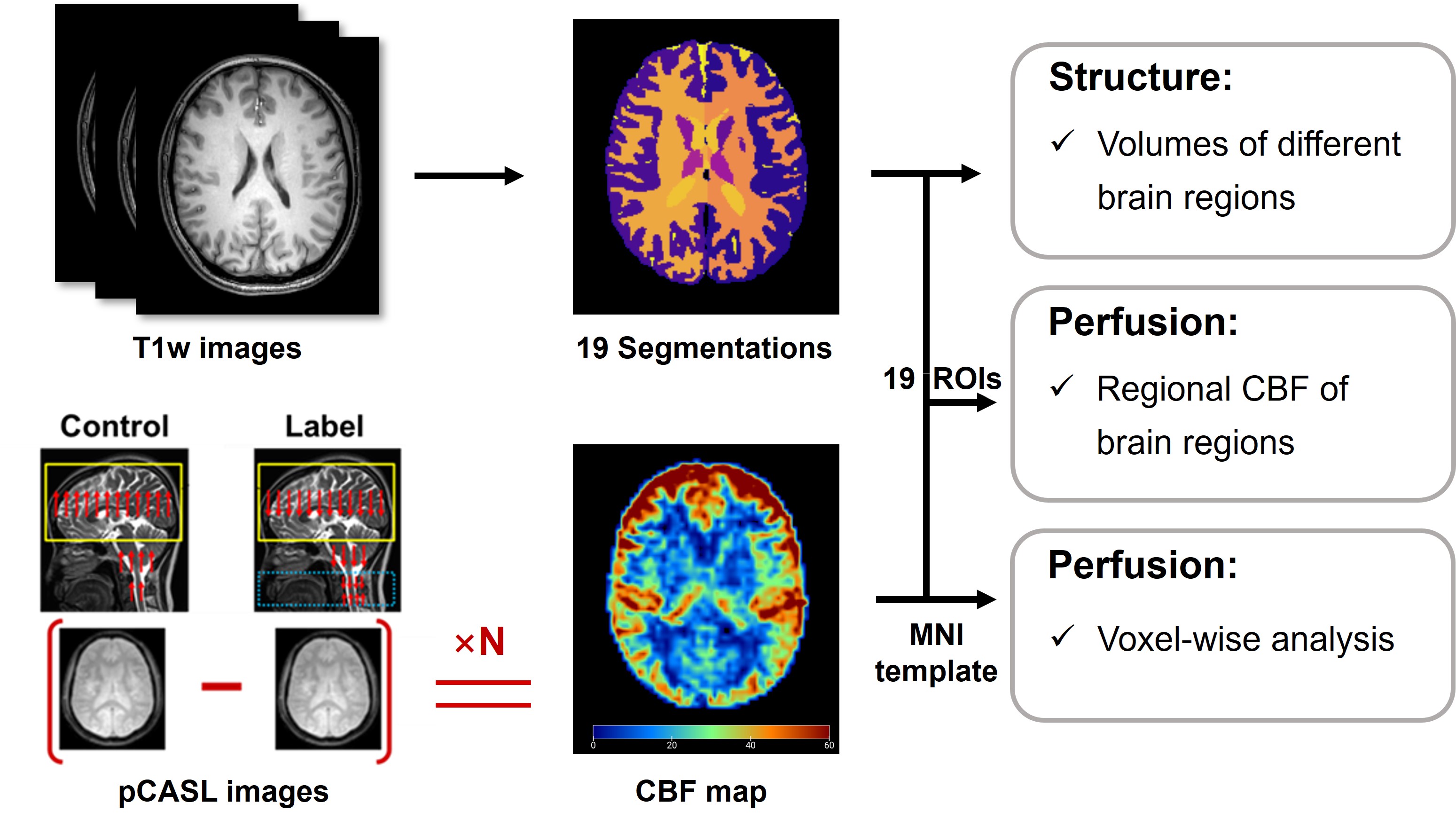
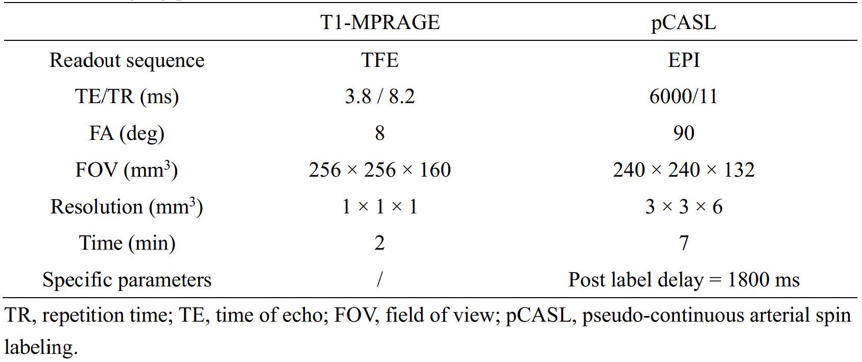
Study sample: Patients who were diagnosed with SSc and age- and sex-matched healthy volunteers were recruited and underwent brain MR imaging. Clinical characteristics of patients were collected. The study protocol was approved by institutional review board and written consent form was obtained from all patients and healthy volunteers. MR imaging protocol: Brain MR imaging was performed on a 3T MR scanner (Ingenia CX, Philips Healthcare, The Netherlands) with a 32-channel head coil. The MR brain imaging protocol includes T1-magnetization prepared rapid gradient echo imaging (T1-MPRAGE) and pseudo-continuous arterial spin labeling (pCASL) perfusion imaging. Detailed parameters of the imaging protocol are listed in Table 1. MR Image post-processing: For T1w images, an automatic processing tool of MRICloud (https://mricloud.org, Johns Hopkins University, Baltimore, MD, USA) was utilized to generate 19 segmentations of different brain regions. The percentage of normalized brain volume (pNBV) of these brain regions were measured and normalized by the whole brain volume. For pCASL images, cerebral blood flow (CBF) maps and the CBF values of different brain regions were calculated using MRICloud. The CBF maps registered to the Montreal Neuro-logical Institute (MNI) space were utilized to perform voxel-wise analysis by SPM12 toolbox (https://www.fil.ion.ucl.ac.uk/spm/software/spm12/). The illustration of whole post-processing analysis is shown in Figure 1. Statistical analysis: Above regional measurements were compared among diffuse cutaneous SSc (dcSSc) patients, limited cutaneous SSc (lcSSc) patients, and healthy volunteers using One-way ANOVA or Kruskal Wallis H test adjusted by LSD method. The statistical analyses were conducted using SPSS 27.0 (SPSS Inc. Chicago, IL, USA). Two sample t test was utilized to compare the voxel differences of CBF maps between SSc patients and healthy volunteers, and between dcSSc patients and lcSSc patients using SPM12.Results
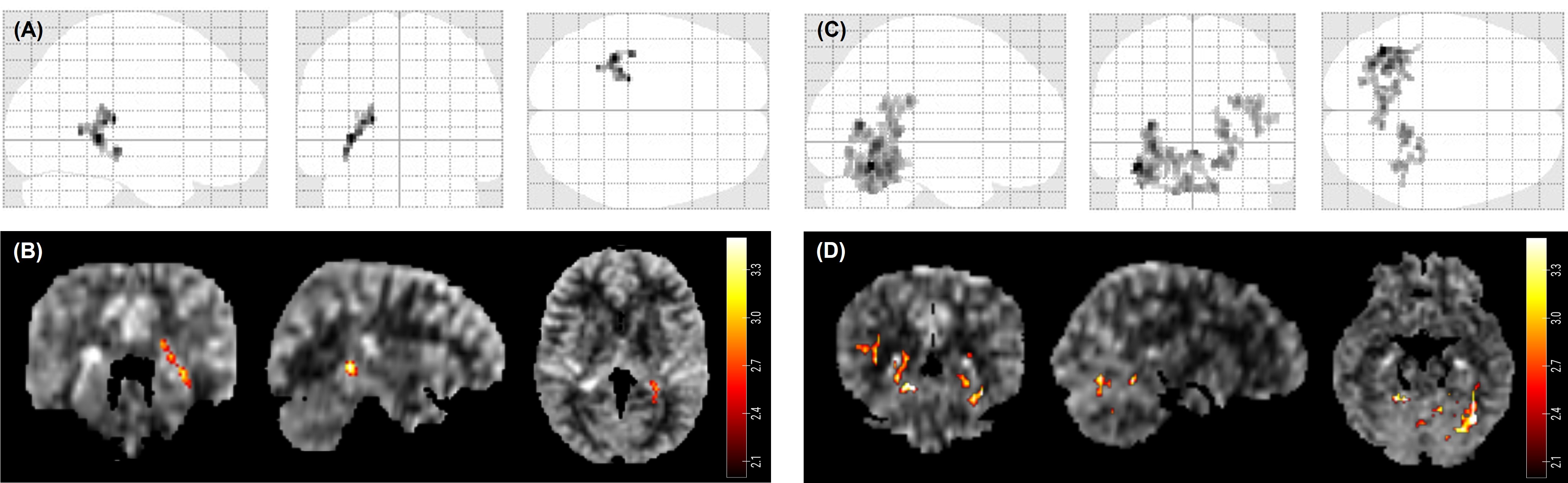
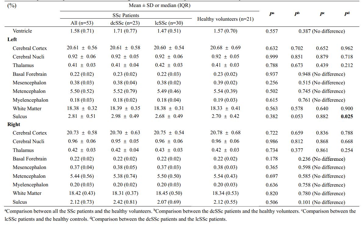
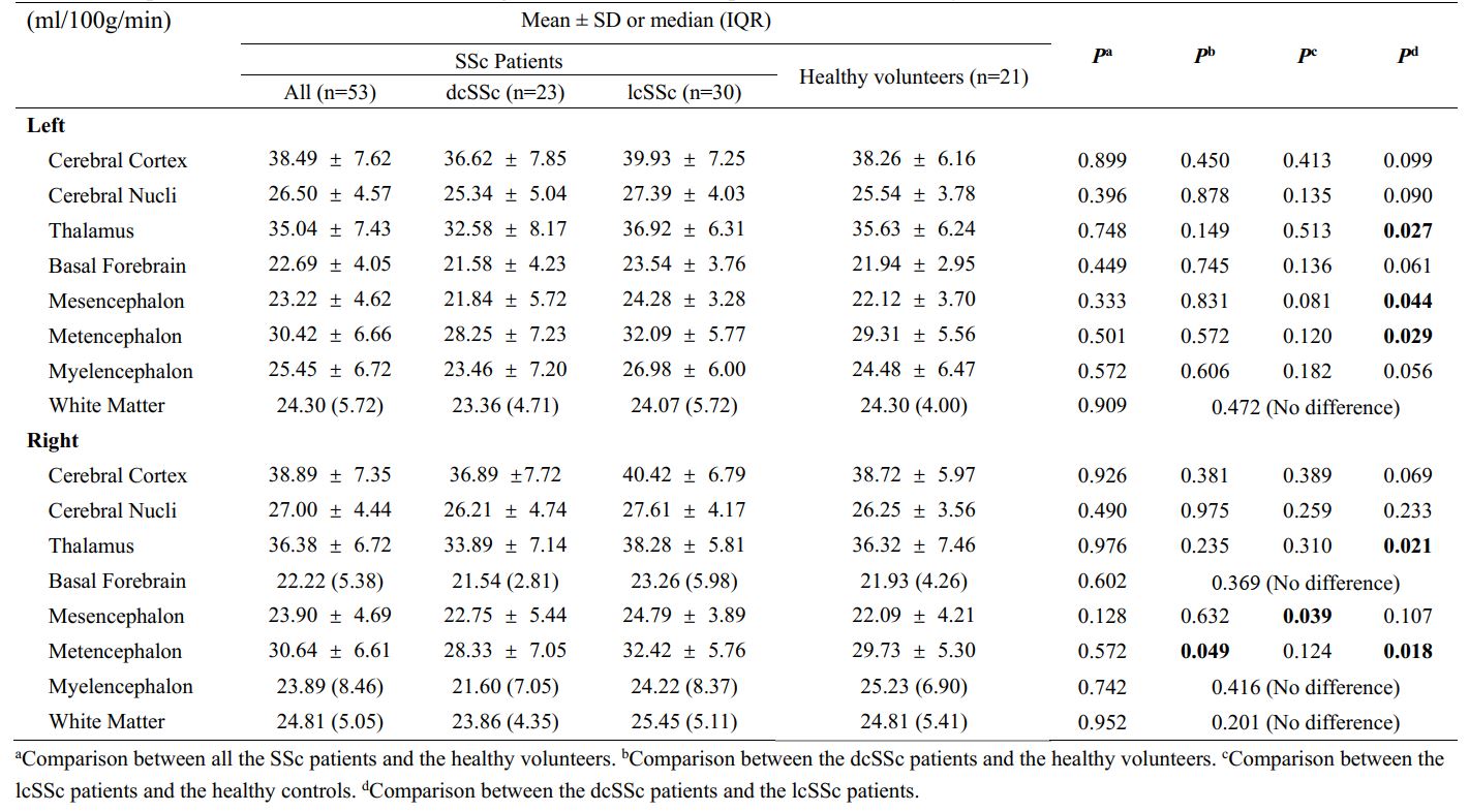
A total of 53 patients (mean age: 46.8 ± 12.9 years, 7 males) and 21 healthy volunteers (mean age: 43.9 ± 13.9 years, 3 males) were enrolled. Table 2 and Table 3 presented the comparison results of the pNBV and CBF values between the SSc patients and the healthy volunteers. Compared to the healthy volunteers, the dcSSc patients showed significantly lower CBF values in the right metencephalon (28.33 ± 7.05 ml/100g/min vs. 29.73 ± 5.30 ml/100g/min, P = 0.049), while the lcSSc patients showed significantly higher CBF values in the right mesencephalon (24.79 ± 3.89 ml/100g/min vs. 22.09 ± 4.21 ml/100g/min, P = 0.039). Compared to the lcSSc patients, the dcSSc patients showed significantly larger sulcus volume in the left brain (2.98 ± 0.49 % vs. 2.68 ± 0.49 %, P = 0.025), and lower CBF values in the thalamus (left: P = 0.027, Right: P = 0.021), metencephalon (left: P = 0.027, Right: P = 0.018), and left mesencephalon (P = 0.044). Figure 2 showed the voxel-wise differences in CBF. Compared to healthy volunteers, the SSc patients showed significantly lower CBF near the hippocampus. The dcSSc patients showed significantly lower CBF than lcSSc patients at the cerebellum and fusiform (non-corrected, P < 0.01).Discussion and Conclusion
We found that the SSc patients showed significantly lower voxel-wise CBF than healthy volunteers, and the dcSSc patients showed significantly lower CBF in both region-wise and voxel-wise level than the lcSSc patients. The widespread microvascular damage caused by SSc [4] might be the reason of CBF decrease, and the significant decrease near the hippocampus may be associated with the cognitive dysfunction reported previously [5]. According to the previous clinical cohort studies, dcSSc patients were reported to have globally severer disease and vascular involvement [6-7]. Our studies similarly indicated that the dcSSc patients had more severe cerebral involvement. Future studies are warranted to further investigate the brain function with larger sample size of SSc patients. In conclusion, the cerebral blood flow quantified by MR pCASL imaging might be effective imaging indicators for SSc patients.Acknowledgements
No acknowledgement found.References
1. Volkmann ER, Andréasson K, Smith V. Systemic sclerosis. Lancet. 2023; 401(10373):304-318.
2. Simeoni S, Puccetti A, Tinazzi E, et al. Systemic sclerosis and superficial siderosis of the central nervous system: casuality or causality? Rheumatol Int. 2008;28(8):815-818.
3. Olah C, Schwartz N, Denton C, et al. Cognitive dysfunction in autoimmune rheumatic diseases. Arthritis Res Ther. 2020;22:78.
4. McNair S, Hategan A, Bourgeois JA, Losier B. Neuropsychiatric symptoms in scleroderma. Psychosomatics. 2013;54(4):382-386.
5. Wuriliga, Xu D, He Y, et al. Mild cognitive impairment in patients with systemic sclerosis and features analysis, Rheumatology, 2022;61(6):2457-2463.
6. Frantz C, Huscher D, Avouac J, et al. Outcomes of limited cutaneous systemic sclerosis patients: Results on more than 12,000 patients from the EUSTAR database. Autoimmun Rev. 2020;19(2):102452.
7. Foeldvari I, Klotsche J, Kasapcopur O, et al. Differences sustained between diffuse and limited forms of juvenile systemic sclerosis in an expanded international cohort. Arthritis Care Res (Hoboken). 2022;74(10):1575-84.
Figures