2969
Computing myelin and axonal volume fractions in gray and white matters and g-ratio map using multiple-inversion MRF and NODDI1The University of Hong Kong, Hong Kong, Hong Kong
Synopsis
Keywords: Multiple Sclerosis, Diffusion Tensor Imaging, multiple sclerosis; mrf; g-ratio
Motivation: Multiple sclerosis effect both white matter and gray matter.
Goal(s): Using mIR-MRF with NODDI to generate myelin water fraction map, g-ratio map, and axonal volume fraction gray matter map.
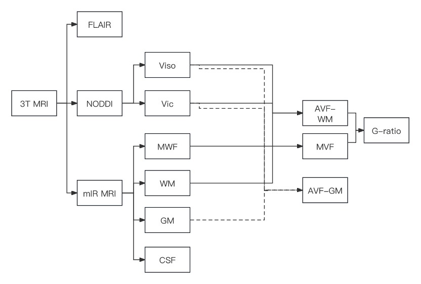
Approach: White matter, gray matter, cerebrospinal fluid (CSF), and myelin water fraction (MWF) map were generated by mIR MRF. NODDI was used to divide each voxel into isotropic (Viso) and anisotropically restricted volume fraction (Vic) map. These maps were used to generate the g-ratio and AVF-gm map.
Results: The WM lesions were in consistency within FLAIR and MWF but have a variance in g-ratio maps. AVF-gm map depicted the axonal fiber in GM well.
Impact: The mIR MRF with NODDI could provide insight of demyelination, axonal loss and the change in GM of MS.
Introduction
The g-ratio, which is the ratio of the axon diameter to the fiber diameter, plays a crucial role in determining the fiber conduction velocity [1, 2]. It can also be helpful in identifying de- and remyelination in individuals with multiple sclerosis (MS) [3]. However, measuring g-ratio in vivo is challenging because of the extremely small size of axonal fibers (~ μm). An area-weighted g-ratio using MRI, which determines axonal and fiber volume fraction in a voxel, has been proposed [4, 5]. The myelin volume fraction (MVF) and axonal volume fraction (AVF) are measured using the myelin or myelin-water detection and diffusion methods, respectively. One of the popular diffusion methods is NODDI. While white matter is the primary area affected by MS, gray matter may also suffer from atrophy, which could significantly impact clinical-cognitive functions [6, 7]. Recently, multiple inversion recovery (mIR) magnetic resonance fingerprinting (MRF) has been shown to separate brain water compartments, including myelin water, gray matter, white matter, and cerebrospinal fluid [8]. Therefore, this study aims to use mIR MRF and NODDI to obtain a myelin water fraction map, g-ratio, and axonal volume fraction in both gray and white matters.Method
All experiments were conducted using A 3T MRI (GE, SIGNA Premier) with a 48-channel brain coil. FLAIR was used as clinical reference for white matter lesion. A multiple-inversion-recovery fast imaging with steady-state precession (FISP) sequence was used to perform the mIR MRF scan [8]. The inversion was achieved by a slice selective Shinnar-Le Roux pulse. A non-negative least-square (NNLS) algorithm that included a reweighting iteration to update the joint distribution of T1/T2 across the slice/volume was used to multiple compartment analysis [9]. White matter (WM), gray matter (GM), cerebrospinal fluid (CSF), and myelin water fraction (MWF) map could be achieved at one time. For NODDI analysis, two different b-values were utilized: 1000 and 2000 s/mm2. The scan with b-value of 1000 s/mm2 involved 30 diffusion directions, while the one with b-value of 2000 s/mm2 used 64 diffusion directions. To calculate isotropic (Viso) and anisotropically restricted volume fraction (Vic) of each voxel, the NODDI Matlab toolbox from UCL microstructure imaging group was utilized. The following equations were used to obtain AVF, MVF, and g-ratio:1) MVF = (MWF * 0.86) / (MWF * (0.86-0.36) + 0.36)
2) AVF-wm = (WM fraction) * (1 - Viso) * Vic
3) AVF-gm = (GM fraction) * (1 - Viso) * Vic
4). g-ratio = sqrt (1/(1 + MVF/AVF-wm))
Details of the data processing pipeline is shown in Figure 1.
Result
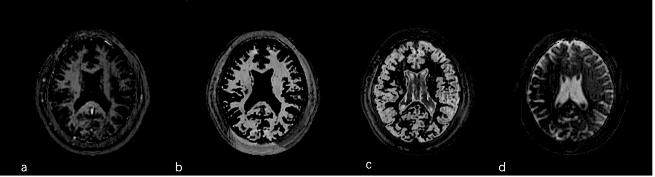
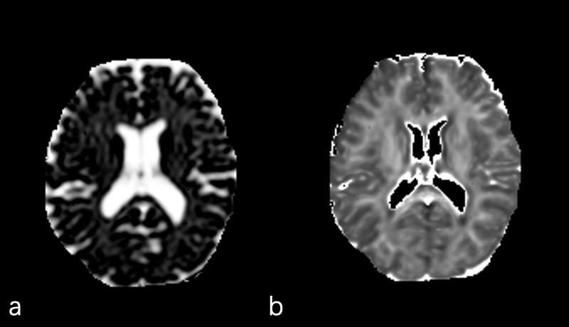
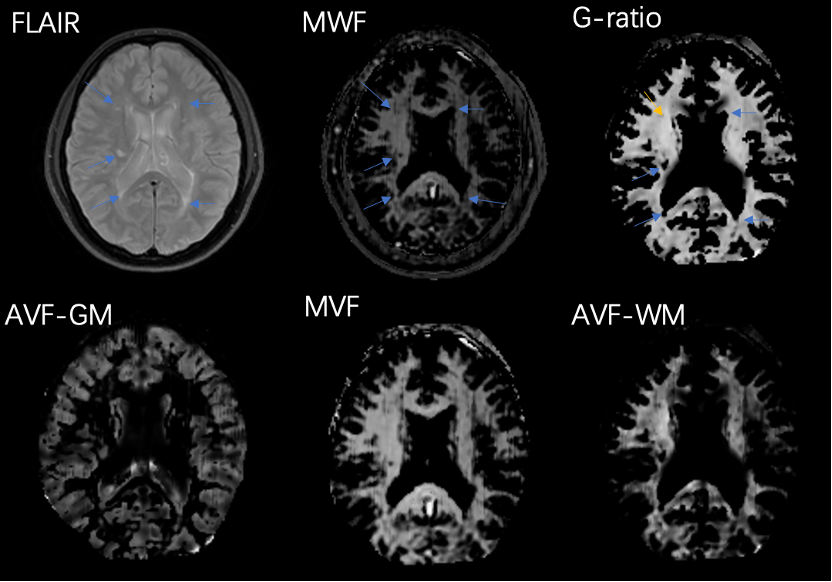
Results shown in Figure 2 are the maps generated by mIR MRF for WMF GM, WM, and CSF. Figure 3 displays the isotropic (Viso) and anisotropically restricted volume fraction (Vic). Additionally, Figure 4 provides the results for FLAIR, MWF, AVF, AVF-gm, and g-ratio maps. While the white matter (WM) lesions were consistent in FLAIR and MWF, they varied in the g-ratio maps.Discussion
In this study, we have demonstrated a method that combines mIR MRF with NODDI to produce MWF maps, g-ratio maps, AVF-wm and AVF-gm maps. We found that the MWF map was comparable to FLAIR in identifying WM lesions. However, the g-ratio map did not show an obvious decrease in WM lesions, which suggests that it has the potential to differentiate between WM lesions dominated by demyelination from those with concordant axonal and myelin loss. Additionally, since MS can lead to GM damage, the AVF-gm could provide insight into gray matter changes.Conclusion
In conclusion, our method not only provides insight into demyelination and axonal loss in white matter, but also provides information on the intracellular axonal volume of gray matter, which has the potential to evaluate the atrophy of gray matter.Acknowledgements
This work is supported by Hong Kong HMRF grant number 09201346.References
Reference:
1. Goldman, L. and J.S. Albus, Computation of Impulse Conduction in Myelinated Fibers; Theoretical Basis of the Velocity-Diameter Relation. Biophysical journal, 1968. 8(5): p. 596-607.
2. Chomiak, T. and B. Hu, What Is the Optimal Value of the g-Ratio for Myelinated Fibers in the Rat CNS? A Theoretical Approach. PloS one, 2009. 4(11): p. e7754-e7754.
3. Jung, W., et al., Whole brain g-ratio mapping using myelin water imaging (MWI) and neurite orientation dispersion and density imaging (NODDI). NeuroImage (Orlando, Fla.), 2018. 182: p. 379-388.
4. Berman, S., et al., Evaluating g-ratio weighted changes in the corpus callosum as a function of age and sex. NeuroImage (Orlando, Fla.), 2018. 182: p. 304-313.
5. Dean, D.C., et al., Mapping an index of the myelin g-ratio in infants using magnetic resonance imaging. NeuroImage (Orlando, Fla.), 2016. 132: p. 225-237.
6. Laule, C., et al., Water content and myelin water fraction in multiple sclerosis - A T-2 relaxation study. Journal of neurology, 2004. 251(3): p. 284-293.
7. Fiore, A., et al., Correspondence among gray matter atrophy and atlas-based neurotransmitter maps is clinically relevant in multiple sclerosis. Molecular psychiatry, 2023. 28(4): p. 1770-1782.
8. Cui, D., E.S. Hui, and P. Cao, A multi-inversion-recovery magnetic resonance fingerprinting for multi-compartment water mapping.Magnetic resonance imaging, 2021. 81: p. 82-87.
9. Nagtegaal, M.A., P. Koken, T. Amthor, and M. Doneva, Fast multi-component analysis using a joint sparsity constraint for MR fingerprinting. Magnetic resonance in medicine, 2020. 83(2): p. 521-534.
Figures