2960
Current Stimulation and Brain Connectivity in Prodromal Multiple Sclerosis: A Simultaneous tDCS-MRI Study1Bernard and Irene Schwartz Center for Biomedical Imaging, Department of Radiology, New York University Grossman School of Medicine, New York City, NY, United States, 2Center for Advanced Imaging Innovation and Research (CAI2R), Department of Radiology, New York University Grossman School of Medicine, New York City, NY, United States, 3Neurology, New York University Grossman School of Medicine, New York City, NY, United States, 4Research and Development, Soterix Medical Inc, Woodbridge Township, NJ, United States, 5Biomedical Engineering, City College of New York, New York City, NY, United States
Synopsis
Keywords: Multiple Sclerosis, Neurodegeneration
Motivation: The extent of tDCS’s impact on brain functional connectivity(FC) in individuals with prodromal multiple sclerosis(MS) remains largely unknown.
Goal(s): To investigate the acute tDCS effects on brain network and FC using resting state functional MRI(rs-fMRI) in MS prodromal patients.
Approach: The study involved a concurrent tDCS-MRI session, in which rs-fMRI data were acquired prior to and during tDCS (2mA, DLPFC left anodal).
Results: During tDCS, we noted a significant increase in FC between hippocampus and frontal pole as well as lateral parietal cortex in the left hemisphere. Similar increases were observed between frontal left regions and cortical and subcortical areas.
Impact: The observed effects of tDCS on brain network dynamics and resting state functional connectivity in prodromal MS could potentially influence its future clinical applications as a treatment option in such early stages of the disease.
Introduction
Transcranial direct current stimulation (tDCS) is an emerging therapeutic approach that modulates cortical excitability via safe and well-tolerated weak electrical currents [1-3]. Recently, it has gained attention for its potential benefits in several neurological conditions [4-6], such as reducing fatigue[7], improving motor function[8] and cognitive performance[9] in multiple sclerosis (MS) patients. However, little is known about its effects on brain functional connectivity in patients with prodromal MS, characterized by MS-related signs or symptoms predating its clinical definitive diagnosis [10,11]. Recent advancements in neuroimaging allow for real-time measurement of neuronal activity during tDCS using MRI (or simultaneous tDCS-MRI). However, previous studies have been focused on global neuronal or cerebrovascular responses resulting from tDCS[12-15], while a few studies have investigated changes of functional connectivity in healthy individuals both during rest and cognitive tasks[16-18]. In this pilot study, we used simultaneous tDCS-MRI to examine the acute effects of tDCS on frontal and whole brain networks in a cohort of prodromal MS patients.Methods
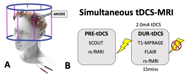
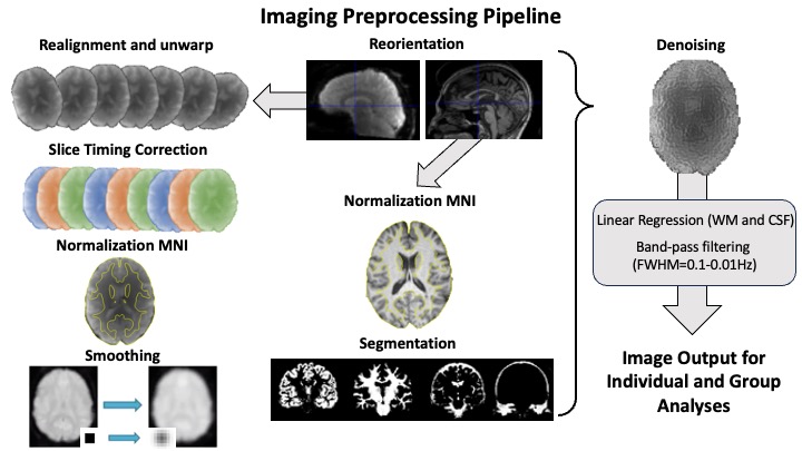
10 Patients with an either radiologically or clinically isolated prodromal MS diagnosis or recent (<5 years) MS diagnosis (age: 35±8 years, 5 males), were recruited for a simultaneous tDCS-MRI scan. Images were acquired in a 3T scanner and a 64 channels head coil. Stimulation was administered using an MR-compatible tDCS device (Soterix Medical) with a 2.0mA current applied to the left anodal dorsolateral prefrontal cortex (DLPC; F3; Fig.1A) for 15 minutes. The MRI protocol includes resting state functional MRI (rs-fMRI) images acquired before and during tDCS for functional connectivity analyses as well as a 3D T1 MPRAGE for structural imaging (Fig.1B). Resting-state functional data was analyzed using CONN toolbox 21.a [19] using SPM12 (http://www.fil.ion.ucl.ac.uk/spm/) and running in Matlab 2022.a (The Mathworks Inc, USA). A default imaging preprocessing pipeline was used, and blood-oxygen-level-dependent (BOLD) signal was band-pass filtered (0.01-0.1 Hz) to mitigate noise effects (Fig 2). Subsequently, we conducted seed-to-voxel and ROI-to-ROI analyses using predefined seeds and regions of interest (ROIs) from CONN toolbox, with each functional connectivity (FC) corrected for multiple comparison using false discovery rate (FDR) and significance threshold set at 0.05.Results
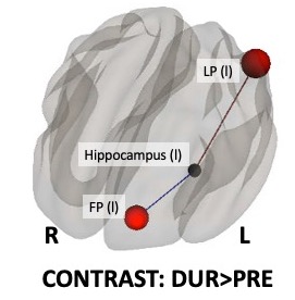
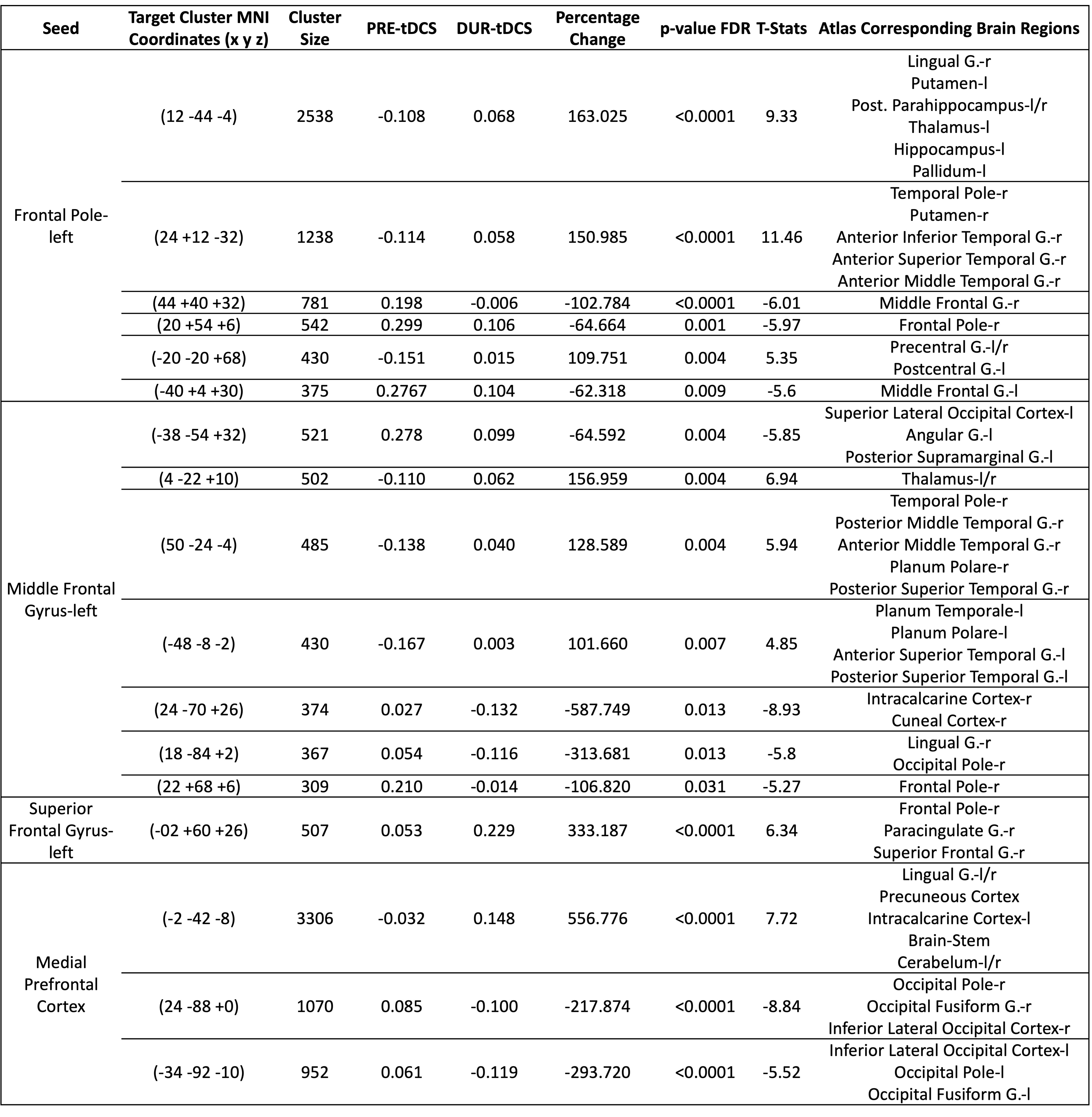
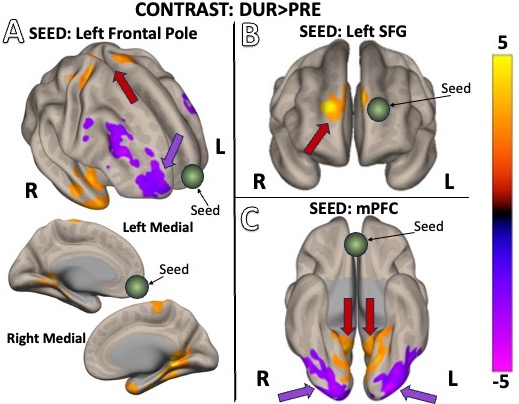
We observed a significant increase (beta=0.25; t-stat=6.34; p-FDR=0.0053) between left lateral parietal cortex and left hippocampus from pre- (-0.225±0.156 a.u.) to during-tDCS (0.228±0.124 a.u; Fig.3). In addition, functional connectivity analysis between frontal regions, where tDCS is applied, and the rest of brain regions showed a significant increase of FC strength (beta=0.23; t-stat=4.87; p-FDR=0.0173) between the left frontal pole and the left hippocampus from pre- (-0.169±0.150 a.u.) to during-tDCS (0.064±0.144 a.u; Fig.3). Seed-to-voxel analysis showed many significant changes in FC as response to tDCS. The most relevant are showed in figure 4. Additionally, we found increased FC between the left frontal pole and bilateral precentral gyrus (t-stat=6.66, p-FDR<0.0001; Fig.5A); increased connectivity between left superior frontal gyrus and voxels mainly in the contralateral region (right superior frontal gyrus; SFG: t-stat=6.34, p-FDR<0.0001;Fig.5B); increased medial prefrontal cortex FC with voxels mainly part of the precuneal cortex (t-stat=7.72, p-FDR<0.0001) and decreased functional connectivity with part of the right occipital cortex (t-stat=-8.84, p-FDR<0.0001) and left occipital cortex (t-stat-5.52,p-FDR<0.0001), represented in figure 5C.Discussion
In this study we showed that tDCS can be used to modulate functional connectivity in the brain of patients with early-stage MS. The acute increased fictional connectivity between the left hippocampus and left lateral parietal cortex as well as left frontal pole is in line with our hypothesis that tDCS effects would be mainly localized in the same hemisphere of the stimulation, left frontal anodal montage in this case. Moreover, whilst the hippocampus is notably associated with verbal and spatial memory[20], the frontal pole and left lateral parietal cortex have been linked to goals management (or multitasking)[21] and attention[22-23], respectively. Therefore, our results suggest that the tDCS-induced increase in functional connectivity between these specific areas might have significant clinical and cognitive consequences for prodromal MS subjects and tDCS has already been reported to improve memory function[24]. Moreover, whole brain seed-to-voxel analyses showed interesting increases in connectivity between the left frontal pole and bilateral precentral gyri. Due to the importance of the precentral gyri in motor task execution[25], this increase in connectivity with areas responsible for action selection (e.g. frontal pole) [26] might imply important clinical effects of tDCS on MS patients.Conclusion
In conclusion, our study provides useful and important insights into the potentially beneficial effects that tDCS has on the whole brain functional connectivity of MS patients in the early stages of the disease. We showed increased connectivity between major cortical and subcortical brain regions involved in cognitive and motor functions.Acknowledgements
This study was funded by National Multiple Sclerosis Society (RFA-2104-37483) and was performed under the rubric of the Center for Advanced Imaging Innovation and Research (CAI2R, www.cai2r.net), an NIBIB National Center for Biomedical Imaging and Bioengineering (NIH P41 EB017183).References
1. Dedoncker J, Baeken C, De Raedt R, Vanderhasselt MA. Combined transcranial direct current stimulation and psychological interventions: State of the art and promising perspectives for clinical psychology. Biol Psychol. 2021;158:107991.
2. Nitsche MA, Cohen LG, Wassermann EM, Priori A, Lang N, Antal A, et al. Transcranial direct current stimulation: State of the art 2008. Brain Stimul. 2008;1(3):206-23.
3. Bikson M, Grossman P, Thomas C, Zannou AL, Jiang J, Adnan T, et al. Safety of Transcranial Direct Current Stimulation: Evidence Based Update 2016. Brain Stimul. 2016;9(5):641-61.
4. Fregni, F., El-Hagrassy, M.M., Pacheco-Barrios, K., Carvalho, S., Leite, J., Simis, M., Brunelin, J., Nakamura-Palacios, E.M., Marangolo, P., Venkatasubramanian, G. and San-Juan, D., 2021. Evidence-based guidelines and secondary meta-analysis for the use of transcranial direct current stimulation in neurological and psychiatric disorders. International Journal of Neuropsychopharmacology, 24(4), pp.256-313.
5. Khedr, E.M., Shawky, O.A., El-Hammady, D.H., Rothwell, J.C., Darwish, E.S., Mostafa, O.M. and Tohamy, A.M., 2013. Effect of anodal versus cathodal transcranial direct current stimulation on stroke rehabilitation: a pilot randomized controlled trial. Neurorehabilitation and neural repair, 27(7), pp.592-601.
6. Breitling, C., Zaehle, T., Dannhauer, M., Bonath, B., Tegelbeckers, J., Flechtner, H.H. and Krauel, K., 2016. Improving interference control in ADHD patients with transcranial direct current stimulation (tDCS). Frontiers in Cellular Neuroscience, 10, p.72.
7. Charvet, L.E., Dobbs, B., Shaw, M.T., Bikson, M., Datta, A. and Krupp, L.B., 2018. Remotely supervised transcranial direct current stimulation for the treatment of fatigue in multiple sclerosis: results from a randomized, sham-controlled trial. Multiple Sclerosis Journal, 24(13), pp.1760-1769.
8. Pilloni, G., Choi, C., Shaw, M.T., Coghe, G., Krupp, L., Moffat, M., Cocco, E., Pau, M. and Charvet, L., 2020. Walking in multiple sclerosis improves with tDCS: a randomized, double‐blind, sham‐controlled study. Annals of Clinical and Translational Neurology, 7(11), pp.2310-2319.
9. Simani, L., Roozbeh, M., Shojaei, M., Ramezani, M., Roozbeh, M., Gharehgozli, K. and Rostami, M., 2022. The effectiveness of anodal tDCS and cognitive training on cognitive functions in multiple sclerosis; a randomized, double-blind, parallel-group study. Multiple Sclerosis and Related Disorders, 68, p.104392.
10. Matthews, W.B. and MacAlpine, D., 1985. McAlpine's multiple sclerosis: a reappraisal. Livingstone.
11. Tremlett, H., Munger, K.L. and Makhani, N., 2022. The multiple sclerosis prodrome: evidence to action. Frontiers in Neurology, 12, p.761408.
12. Merzagora AC, Foffani G, Panyavin I, Mordillo-Mateos L, Aguilar J, Onaral B, et al. Prefrontal hemodynamic changes produced by anodal direct current stimulation. Neuroimage. 2010;49(3):2304-10.
13. Jamil A, Batsikadze G, Kuo HI, Meesen RLJ, Dechent P, Paulus W, et al. Current intensity- and polarity-specific online and aftereffects of transcranial direct current stimulation: An fMRI study. Hum Brain Mapp. 2020;41(6):1644-66.
14. Zheng X, Alsop DC, Schlaug G. Effects of transcranial direct current stimulation (tDCS) on human regional cerebral blood flow. Neuroimage. 2011;58(1):26-33.
15. Muccio, M., Masters, L.W., Pilloni, G., He, P., Krupp, L., Datta, A., Bikson, M., Charvet, L. and Ge, Y., 2022. Cerebral metabolic rate of oxygen (CMRO2) changes measured with simultaneous tDCS-MRI in healthy adults. Brain Research, 1796, p.148097.
16. Vecchio, F., Miraglia, F., Rodella, C., Alù, F., Miniussi, C., Rossini, P.M. and Pellicciari, M.C., 2021. tDCS effects on brain network properties during physiological aging. Pflügers Archiv-European Journal of Physiology, 473, pp.785-792.
17. Li, L.M., Violante, I.R., Leech, R., Ross, E., Hampshire, A., Opitz, A., Rothwell, J.C., Carmichael, D.W. and Sharp, D.J., 2019. Brain state and polarity dependent modulation of brain networks by transcranial direct current stimulation. Human brain mapping, 40(3), pp.904-915.
18. Park, C.H., Chang, W.H., Park, J.Y., Shin, Y.I., Kim, S.T. and Kim, Y.H., 2013. Transcranial direct current stimulation increases resting state interhemispheric connectivity. Neuroscience letters, 539, pp.7-10.
19. Whitfield-Gabrieli, S. and Nieto-Castanon, A., 2012. Conn: a functional connectivity toolbox for correlated and anticorrelated brain networks. Brain connectivity, 2(3), pp.125-141.
20. Ezzati, A., Katz, M.J., Zammit, A.R., Lipton, M.L., Zimmerman, M.E., Sliwinski, M.J. and Lipton, R.B., 2016. Differential association of left and right hippocampal volumes with verbal episodic and spatial memory in older adults. Neuropsychologia, 93, pp.380-385.
21. Dreher, J.C., Koechlin, E., Tierney, M. and Grafman, J., 2008. Damage to the fronto-polar cortex is associated with impaired multitasking. PLoS One, 3(9), p.e3227.
22. Humphreys, G.F. and Lambon Ralph, M.A., 2015. Fusion and fission of cognitive functions in the human parietal cortex. Cerebral Cortex, 25(10), pp.3547-3560.
23. Tumati, S., Martens, S., de Jong, B.M. and Aleman, A., 2019. Lateral parietal cortex in the generation of behavior: Implications for apathy. Progress in neurobiology, 175, pp.20-34.
24. Fregni, F., Boggio, P.S., Nitsche, M., Bermpohl, F., Antal, A., Feredoes, E., Marcolin, M.A., Rigonatti, S.P., Silva, M.T., Paulus, W. and Pascual-Leone, A., 2005. Anodal transcranial direct current stimulation of prefrontal cortex enhances working memory. Experimental brain research, 166, pp.23-30.
25. Banker L, Tadi P. Neuroanatomy, Precentral Gyrus. 2023 Jul 24. In: StatPearls [Internet]. Treasure Island (FL): StatPearls Publishing; 2023 Jan–. PMID: 31334938.
26. Kovach, C.K., Daw, N.D., Rudrauf, D., Tranel, D., O'Doherty, J.P. and Adolphs, R., 2012. Anterior prefrontal cortex contributes to action selection through tracking of recent reward trends. Journal of Neuroscience, 32(25), pp.8434-8442.
Figures