2950
Assessment of region-specific oxidative stress in multiple sclerosis using short-TE STEAM at 7T1Montreal Neurological Institute-Hospital, McGill University, Montreal, QC, Canada, 2Medical Biophysics, Sunnybrook Research Institute, Toronto, ON, Canada, 3Centre de recherche de l'Institut universitaire de gériatrie de Montréal, Université de Montréal, Montreal, QC, Canada
Synopsis
Keywords: Multiple Sclerosis, Multiple Sclerosis, MR spectroscopy, Oxidative Stress, Glutathione
Motivation: Brain oxidative stress has an important role in the pathophysiology of multiple sclerosis (MS).
Goal(s): This work assessed oxidative stress in relapsing remitting MS (RRMS) by measuring the major brain antioxidant, glutathione (GSH).
Approach: GSH concentrations were measured using 7T MRS in the posterior cingulate cortex (PCC) and centrum semiovale white matter (CSWM) of 10 RRMS patients and 13 matched healthy controls. The relationship between GSH concentrations and functional measures was also investigated.
Results: This preliminary investigation showed no significant difference in GSH concentration between RRMS and healthy participants, and no significant relationship between GSH concentration and functional measures.
Impact: Oxidative stress was not detected in our RRMS participants, concordant with one previous study. Prior work has shown low GSH in progressive MS. Further work with larger sample sizes will investigate oxidative stress in RRMS participants at risk of progression.
Introduction
MS is a chronic inflammatory disease associated with multiple lesions disseminated in the central nervous system at different times. Inflammation and mitochondrial dysfunction in MS result in increased production of reactive oxygen species (ROS). When ROS production exceeds the brain’s antioxidant capacity, a state of oxidative stress occurs, which can drive further pathology. GSH, the main antioxidant in the brain, is vital for protecting against ROS-induced damage. Given that brain cells are particularly sensitive to oxidative damage due to the brain’s substantial metabolic requirements, reduced GSH availability can result in oxidative damage, particularly to oligodendrocytes and neurons.The application of MRS at ultra-high field (B0≥7T) offers substantial advantages for reliable quantification of low-concentration metabolites such as GSH, due to the enhanced signal-to-noise ratio and spectral dispersion at higher field strengths1. This work aimed to assess region-specific GSH alterations using short-TE STEAM MRS in two brain regions relevant to MS pathology, the PCC and CSWM, in RRMS patients compared to age-matched healthy controls. In addition, the associations between GSH levels and MS functional composite measures, including the 9-Hole Peg Test (9HPT), Symbol Digit Modalities Test (SDMT), and Timed 25-foot walk (T25-FW), were investigated.
Methodology
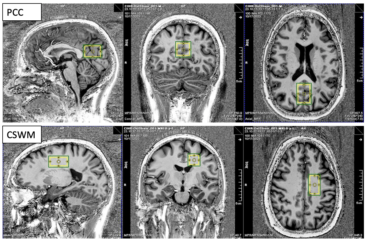
We recruited 10 RRMS patients (3 male, 7 female; age=51±6.8) followed at the Montreal Neurological Institute-Hospital, and 13 age-matched healthy controls (6 male, 7 female, age=49.2±10.7) with no history of brain disease.Data acquisition was performed on a 7T Siemens Terra scanner using a 1Tx/32Rx head coil (Nova Medical). A T1-weighted MP2RAGE anatomical scan2 was acquired (TR/TE=5000/2.33ms, TI1/TI2=900/2750ms, 0.7mm3 isotropic, flip angle=4°) and used to guide placement of MRS voxels in the PCC and CSWM regions (Figure 1). STEAM spectra were acquired using the following parameters: TR/TM/TE=5000/40/8ms, spectral width=10kHz, 64 averages, vector-size=4096, delta frequency=-1.7ppm and VAPOR water suppression. Water-unsuppressed reference data were also acquired (8 averages, delta frequency = 0ppm). First and second order B0 shimming was performed using FAST(EST) MAP3.
MRS data were preprocessed using the FID-A toolkit4. Analysis of the preprocessed data was performed using LCmodel v6.35 in the range [0.5-4.2ppm] with the water-scaling and eddy current correction options enabled. The STEAM basis set included the spectra of 19 brain metabolites and 9 simulated macromolecule (MM) resonances6.
Statistical analysis was performed using R. Linear regression was carried out with absolute GSH concentrations as the response variable and age as the predictor variable. To determine if there were significant differences in GSH concentrations between MS patients and healthy controls, an independent samples one tailed t-test was conducted (significance level set at ɑ=0.05). To assess relationships between GSH levels and MS functional composite scores, separate linear regression analyses were performed in PCC and CSWM for the combined group, with age as a covariate.
Results
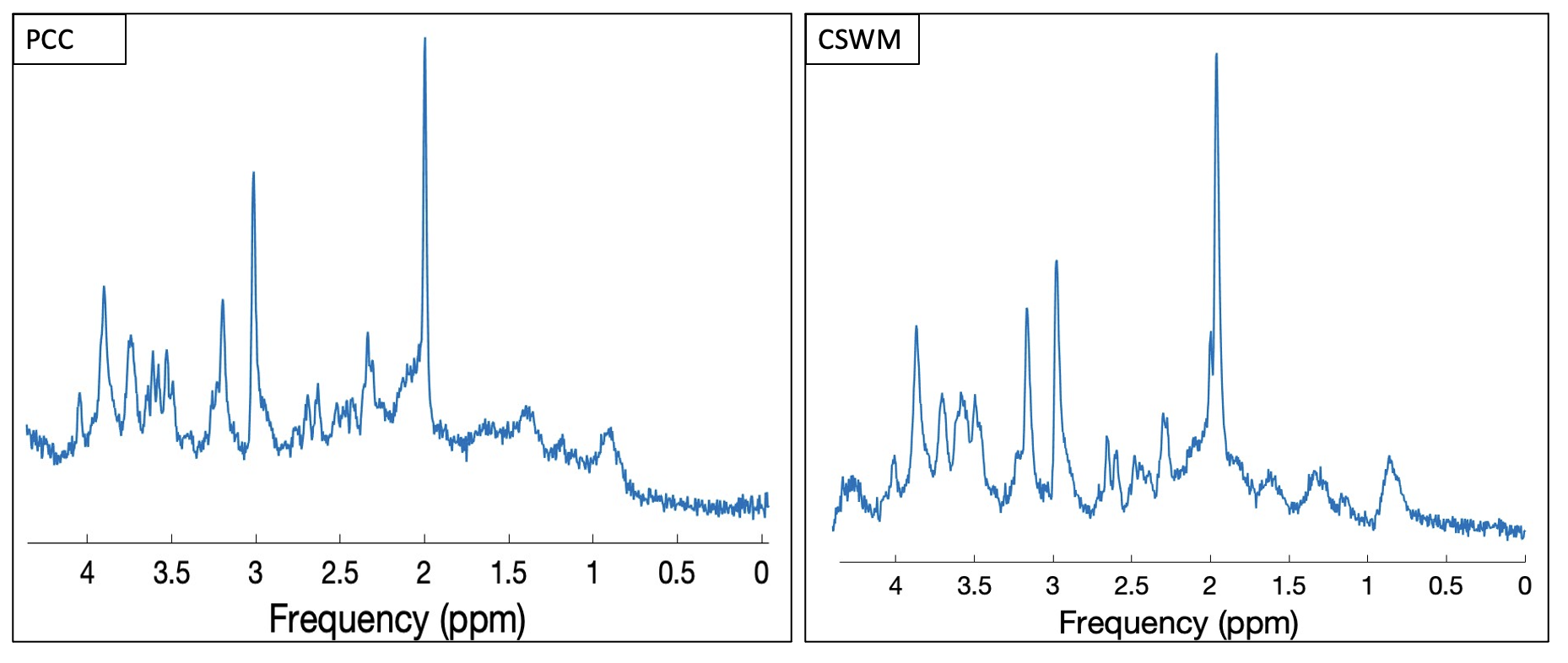
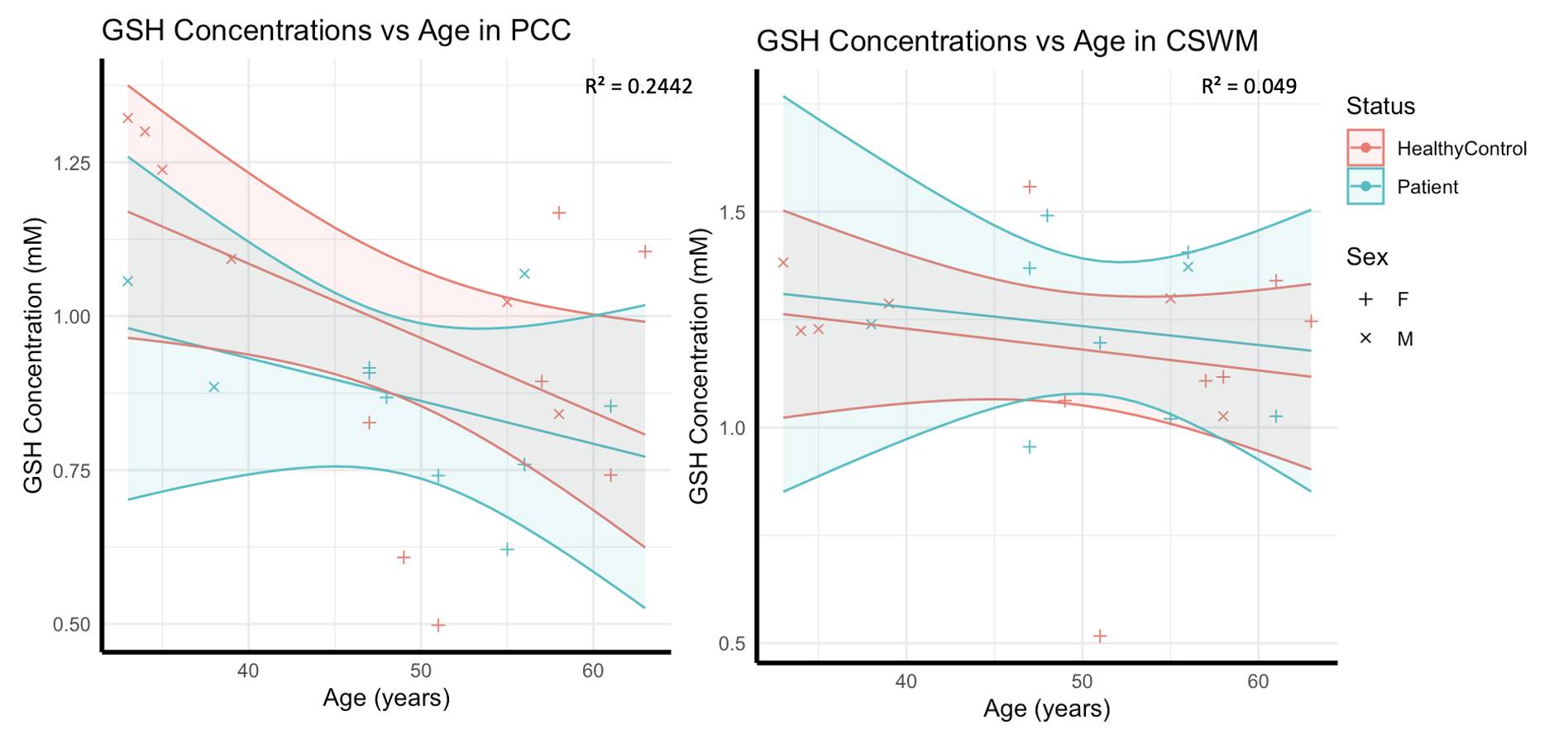
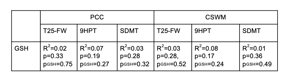
The representative STEAM spectra obtained for each cortical region is shown in Figure 2. In the PCC, mean GSH concentrations in the healthy control and RRMS sub-groups were 0.97±0.26mM and 0.87±0.14mM (p>0.05), respectively. In the CSWM, mean GSH concentrations were 1.18±0.25mM for healthy controls and 1.23±0.19mM for RRMS patients (p>0.05). Figure 3 shows the plot of GSH concentrations versus age for both subgroups with confidence intervals. No significant difference in GSH levels was observed between the RRMS and controls groups. Figure 4 summarises the results of the analyses of relationships between GSH and functional scores, showing no significant relationship between GSH concentration and functional measures.Discussion
To our knowledge, this is the first study investigating oxidative stress in MS that takes into account the macromolecular background when quantifying GSH. The accurate estimation of MM is an important prerequisite for reliable quantification of metabolites. MM resonances overlapping with GSH have previously been reported to be altered in neurological conditions6, which could potentially introduce inaccuracies in GSH quantifications.We found age-related GSH decreases in both RRMS and controls in PCC. However, GSH levels were not significantly lower in RRMS patients in either region, concordant with a previous study using a multiple quantum filtering technique at 3T7. While oxidative stress within lesions has been observed even in early MS8, evidence of oxidative stress in normal-appearing tissues has been associated with progressive MS7.
Future directions will include expanding the cohort and looking at differences between different subtypes of MS, and whether oxidative stress in subgroups of RRMS predicts progression.
Acknowledgements
This work was supported by CIHR (SN), QBIN (AB), PRSS-CRIUGM (AB), Fondation Courtois (AB), FRQS (SS and FD) and Brain Canada (Platform Support Grant to the McConnell Brain Imaging Centre, MNI).References
1. Deelchand DK, Marjańska M, Hodges JS et al. Sensitivity and specificity of human brain glutathione concentrations measured using short‐TE 1H MRS at 7 T. NMR Biomed. 2016;29(5):600-6.
2. Marques JP, Kober T, Krueger G, van der Zwaag W, Van de Moortele PF, Gruetter R. MP2RAGE, a self bias-field corrected sequence for improved segmentation and T1-mapping at high field. Neuroimage. 2010;49(2):1271-1281. doi:10.1016/j.neuroimage.2009.10.002
3. Gruetter R. Automatic, localized in vivo adjustment of all first- and second-order shim coils. Magn Reson Med. 1993;29(6):804-811. doi:10.1002/mrm.1910290613
4. Simpson R, Devenyi GA, Jezzard P, Hennessy TJ, Near J. Advanced processing and simulation of MRS data using the FID appliance (FID-A)-An open source, MATLAB-based toolkit. Magn Reson Med. 2017;77(1):23-33. doi:10.1002/mrm.26091
5. Provencher SW. Automatic quantitation of localized in vivo 1H spectra with LCModel. NMR Biomed. 2001;14(4):260-264. doi:10.1002/nbm.698
6. Cudalbu C, Behar KL, Bhattacharyya PK, et al. Contribution of macromolecules to brain 1 H MR spectra: Experts' consensus recommendations. NMR Biomed. 2021;34(5):e4393. doi:10.1002/nbm.4393
7. Choi IY, Lee P, Adany P, et al. In vivo evidence of oxidative stress in brains of patients with progressive multiple sclerosis. Mult Scler. 2018;24(8):1029-1038. doi:10.1177/1352458517711568
8. Carvalho AN, Lim JL, Nijland PG, Witte ME, Van Horssen J. Glutathione in multiple sclerosis: more than just an antioxidant? Mult Scler. 2014;20(11):1425-31.
Figures