2947
GABA and Glx levels in relapsing-remitting multiple sclerosis correlate with clinical disability1Departments of Magnetic Resonance, The First Affiliated Hospital of Harbin Medical University, Harbin, China, 2Philips Healthcare, Beijing, China
Synopsis
Keywords: Multiple Sclerosis, Multiple Sclerosis, Relapsing-remitting multiple sclerosis; Gamma- aminobutyric acid; Glutamine–glutamate complex; Magnetic resonance spectroscopy
Motivation: Multiple sclerosis (MS) is a leading cause for clinical disability in youth and middle-aged people.
Goal(s): Multiple researches have implicated glutamine–glutamate complex (Glx) and gamma- aminobutyric acid (GABA) as key roles in neuronal signalling and other central functions. In MS patients, dysfunctional Glx excitation and/or GABA inhibition may contribute to neurological symptoms and disease progression.
Approach: To identify the relationship between metabolic abnormalities and clinical disability, the metabolism of brain tissue was investigated by using MEscher-GArwood Point RESolved Spectroscopy.
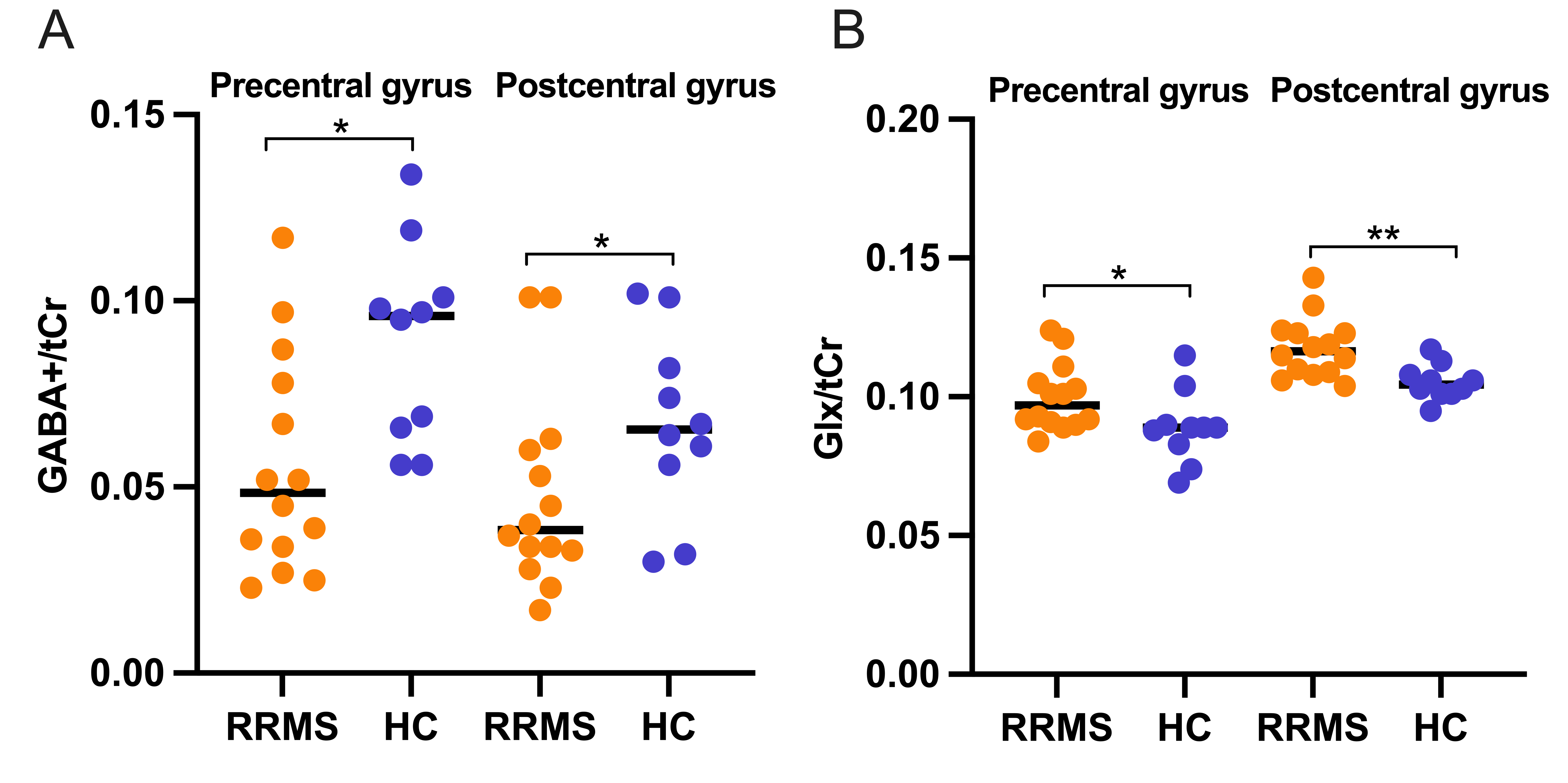
Results: Decreasing levels of GABA+/tCr and increasing levels of Glx/tCr were found in the precentral gyrus and postcentral gyrus VOIs of MS patients.
Impact: GABA and Glx detected by MEGA-PRESS MRS were utilized for investigating correlations between metabolic abnormalities in brain tissue and clinical disability.
Introduction
Multiple sclerosis (MS) is an inflammatory demyelinating disease of the central nervous system (CNS) leading to physical disability and cognitive dysfunction[1]. The pathological processes contributing to clinical disability in MS are intricate, including neuronal and glial changes with associated structural and metabolic abnormalities[2]. However, the high degree of radiological and symptom heterogeneity in MS presents challenges for doctors when evaluating the effectiveness of treatments for MS patients [3,4]. Concentrations of metabolites within various brain regions can be non-invasively measured in humans using magnetic resonance spectroscopy (MRS) [5]. Previous studies have emphasized the potential role of gamma- aminobutyric acid (GABA) and glutamine–glutamate complex (Glx) in contributing to physical disability in patients with progressive MS and relapsing-remitting multiple sclerosis (RRMS) [2,6]. In this study, we aim to measure the GABA and Glx level in precentral gyrus and postcentral gyrus in patients with RRMS by MEscher-GArwood Point RESolved Spectroscopy (MEGA-PRESS), in order to investigate the potential role of GABA and Glx in physical disability.Methods
14 patients diagnosed RRMS were recruited according to McDonalds’ criteria, who had no history of relapse and steroids treatment within the preceding 3 months and then underwent MRI from March to September 2023. The Expanded Disability Status Scale (EDSS) was evaluated before MR scanning for measuring clinical disability. Exclusion criteria were: (1) a history of head trauma or neurological or psychiatric disorder; (2) previous or current substance abuse; and (3) intake of GABAergic agents before enrolment. Age- and gender-matched healthy controls (HCs) were enrolled. MRI were performed on a 3.0 T MR scanner (Ingenia Elition; Philips Healthcare, Best, the Netherlands) with a 32-channel phased-array head coil. T1-weighted 3D TFE, T2-weighted 3D FLAIR was acquired for MRS voxel localization, subsequent tissue segmentation and volume of brain lesions measurement (scan parameters shown in Table1). MRS data for GABA and Glx were acquired from two lesion free anatomical locations of precentral gyrus and postcentral gyrus, as shown in Figure 1 and Table1. Levels of GABA, Glx and total creatine (tCr) were acquired with the MEGA-PRESS sequence. Spectral analysis was performed according to previous study [7]. Statistical analysis were carried out using SPSS 26.0. Normally distributed continuous variables were quoted as mean±SD values and compared by Student’s t-test. Non-normally distributed continuous variables were quoted as median (interquartile range) and verified by Mann-Whitney U test. Categorical variables were described as numbers (percentages) and compared via Fisher's exact analysis. p< 0.05 was considered statistically significant.Result
Demographic, clinical, and whole brain structural MRI characteristics of participants are displayed in Table 2. RRMS (n=14) and HC (n=10) groups did not differ significantly in age, sex, or normalized brain volume (P>0.05). Lower grey matter (GM) ratio, white matter (WM) ratio, brain parenchymal fraction, cortical thickness were calculated in RRMS group. Table 3 displays the corrected metabolite concentrations as well as structural information on the precentral gyrus and postcentral gyrus volume of interest (VOIs). The fitting errors of metabolites, the full-widths at half-maximum (FWHM), GM volume, WM volume, CSF volume and GM/(GM + WM) in both VOIs did not differ between patients and HCs (P>0.05). A decrease in GABA+/tCr levels was found in the precentral gyrus and postcentral gyrus VOIs of patients compared with HCs (Figure2A, P=0.044 and 0.043, respectively). Conversely, Glx/tCr levels in precentral gyrus and postcentral gyrus VOIs increased in patients compared with HCs (Figure2B, P=0.047 and 0.005, respectively).Discussion
By assessing GABA and Glx levels in a clinical population with highly heterogeneous in structural damage and symptoms, this study provides support that levels of these neurometabolites in the human brain may have regionally specific relationships to clinical disability. Prior research has indicated that GABA ratios in the thalamus and GABA level in hippocampus, posterior cingulated cortex, or sensorimotor were significantly lower in RRMS patients than in HCs, which raises the possibility that GABA may be a marker of neurodegeneration in the brain [2,4,6]. Conversely, GABA level in the sensorimotor region was observed an increase in the MS group compared to HCs, possibly contribute to poorer motor performance [9]. Compared to HC, Glx levels in MS patients were significantly decreased in sensorimotor and hippocampus [7,10]. MS-induced demyelination would result in significant decreases in synaptic density and the levels of neuronal proteins required for glutamatergic and GABAergic neurotransmission.Conclusions
Decreased GABA+/tCr levels and increased Glx/tCr levin the precentral gyrus and postcentral gyrus region were identified in RRMS patients, which indicate that GABA+/tCr and Glx/tCr levels might be a potential metabolic feature of clinical disability in patients with RRMS.Acknowledgements
No acknowledgement found.References
1. Chiaravalloti ND, DeLuca J. Cognitive impairment in multiple sclerosis. Lancet Neurol. 2008 Dec;7 (12):1139–1151.2. Cawley Niamh,Solanky Bhavana S,Muhlert Nils et al. Reduced gamma-aminobutyric acid concentration is associated with physical disability in progressive multiple sclerosis.[J] .Brain, 2015, 138: 2584-95.3. Filippi Massimo,Rocca Maria A,MR imaging of multiple sclerosis.[J] .Radiology, 2011, 259: 659-81.4. Prinsen Hetty,de Graaf Robin A,Mason Graeme F et al. Reproducibility measurement of glutathione, GABA, and glutamate: Towards in vivo neurochemical profiling of multiple sclerosis with MR spectroscopy at 7T.[J] .J Magn Reson Imaging, 2017, 45: 187-198.5. Duarte João M N,Lei Hongxia,Mlynárik Vladimír et al. The neurochemical profile quantified by in vivo 1H NMR spectroscopy.[J] .Neuroimage, 2012, 61: 342-62.6. Cao Guanmei,Edden Richard A E,Gao Fei et al. Reduced GABA levels correlate with cognitive impairment in patients with relapsing-remitting multiple sclerosis.[J] .Eur Radiol, 2018, 28: 1140-1148.7. Arm Jameen,Oeltzschner Georg,Al-Iedani Oun et al. Altered in vivo brain GABA and glutamate levels are associated with multiple sclerosis central fatigue.[J] .Eur J Radiol, 2021, 137: 109610.8. Kantorová E,Hnilicová P,Bogner W et al. Neurocognitive performance in relapsing-remitting multiple sclerosis patients is associated with metabolic abnormalities of the thalamus but not the hippocampus- GABA-edited 1H MRS study. Neurol Res, 2022, 44: 57-64.9. Nantes Julia C,Proulx Sébastien,Zhong Jidan et al. GABA and glutamate levels correlate with MTR and clinical disability: Insights from multiple sclerosis.[J] .Neuroimage, 2017, 157: 705-715.10. Gao Fei,Yin Xuntao,Edden Richard A E et al. Altered hippocampal GABA and glutamate levels and uncoupling from functional connectivity in multiple sclerosis.[J] .Hippocampus, 2018, 28: 813-823.Figures