2943
Cross-Sectional and Longitudinal Evaluation of White Matter Microstructure Damages by Automated Fiber Quantification in Multiple Sclerosis1Department of Radiology,, the First Affiliated Hospital of Chognqing Medical University, Chongqing, China
Synopsis
Keywords: Multiple Sclerosis, Multiple Sclerosis
Motivation: Multiple sclerosis (MS) is characterized by a series of pathological processes mainly caused by white matter (WM) lesions. Thus, the correct and comprehensive understanding of WM in RRMS patients is essential for clinical practice.
Goal(s): To characterize the WM fiber tracts by automated fiber quantification (AFQ) cross-sectionally and longitudinally, and explore the correlation between the cognitive performance.
Approach: The DTI metrics ectracted by AFQ were investigated cross-sectionally and longitudinal in entire and pointwise manners. The partial correlation analyses were performed between the abnormal metrics and the cognitive performance.
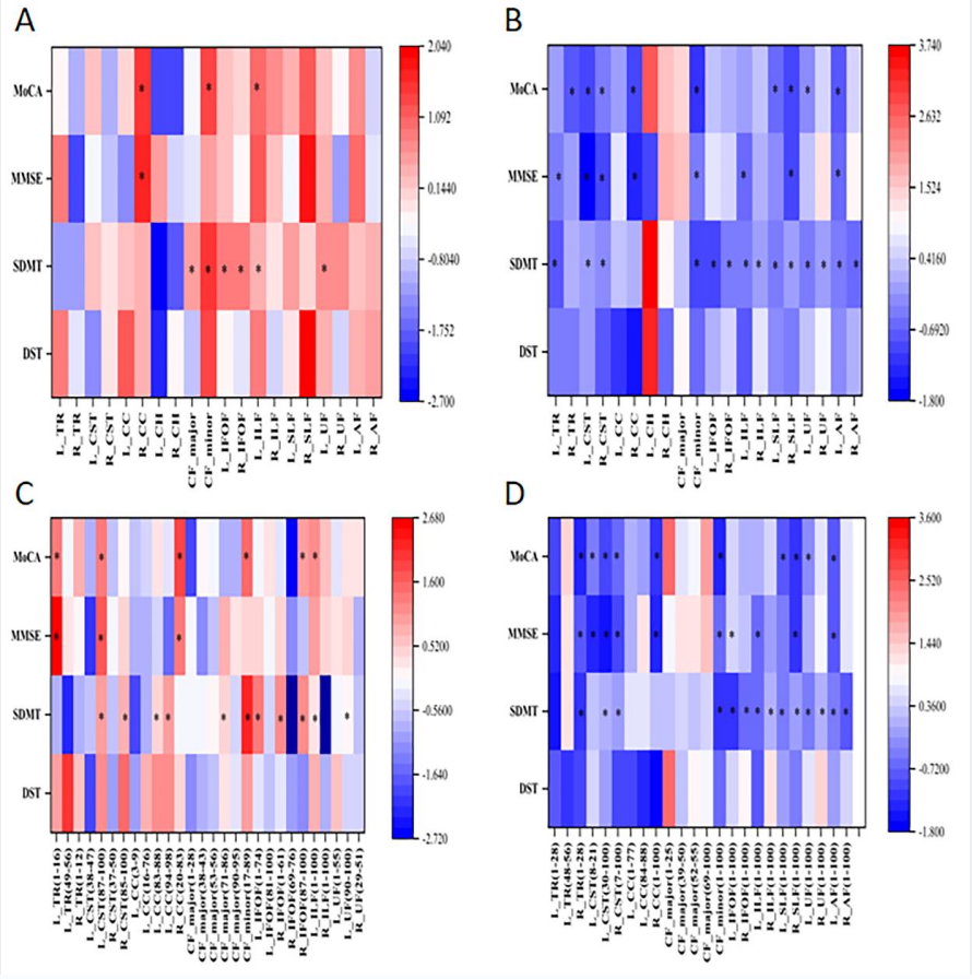
Results: MS patients showed a widespread WM microstructure alteration, and widely correlated with cognitive performance.
Impact: RRMS patients showed a widespread WM microstructure alteration, and the altered metrics were widely correlated with cognitive performance, which will enhance our understanding of WM microstructure damages in RRMS patients.
Introduction
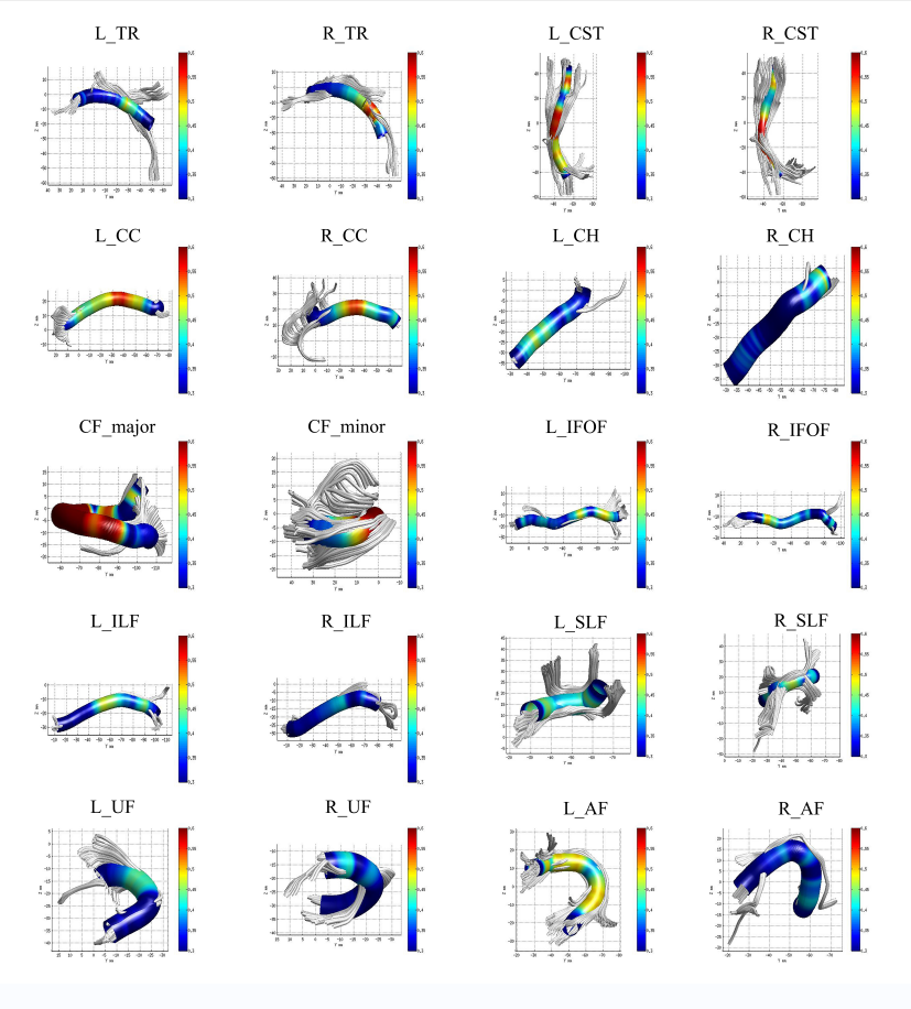
Multiple sclerosis (MS) is the most common inflammatory demyelinating disease of the central nervous system, characterized by a series of pathological processes mainly caused by focal lesions in gray and white matter (WM) in the entire brain, including inflammation, demyelination, axonal loss, and remyelination.Although there are some discrepancies between the findings of previous studies, it has been suggested that diffusion tensor imaging (DTI) can be used to detect alterations in DTI scalars as a biomarker of WM microstructural damages in patients with RRMS. Among these techniques for DTI analyses which have merits and demerits respectively, AFQ is a novel algorithm that can automated perform the reconstruction and segmentation of WM fiber tracts at anatomically multiple equivalent locations along their trajectories.
To our knowledge, there are few applications of AFQ for patients with RRMS, which results in a more detailed cross-sectional and longitudinal characterization of whole-brain WM fiber tracts and its correlation with cognitive performance still requires further investigation.
Methods
This retrospective study was approved by the Institutional Review Board of the First Affiliated Hospital of Chongqing Medical University, Chongqing, China, and written informed consent was obtained from each participant before MRI scans.All MR scans were performed on a 3.0 T MR scanner (Magnetom Skyra, Siemens Healthcare GmbH, Erlangen, Germany) using a 32-channel head coil. The MRI protocol included a 3-dimensional (3D) T1-weighted magnetization prepared rapid gradient echo (MPRAGE) sequence, a 3D fluid-attenuated inversion recovery (FLAIR) sequence and a diffusion kurtosis imaging (DKI) sequence.
For the DTI data, it was preprocessed by using FMRIB Software Library (FSL) software (https://fsl.fmrib.ox.ac.uk/fsl/fslwiki/). The pre-processed DTI and T1MPRAGE images were fed into the open-source MATLAB version of AFQ software (https://github.com/jyeatman/AFQ) to identify and quantify 100 equally spaced nodes of 20 specific WM fiber tracts for each participant.
All statistical analyses were performed by using the SPSS software (version 25.0; SPSS, Chicago, IL, United States).
Results
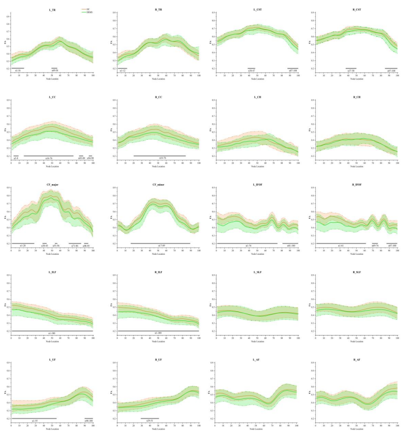
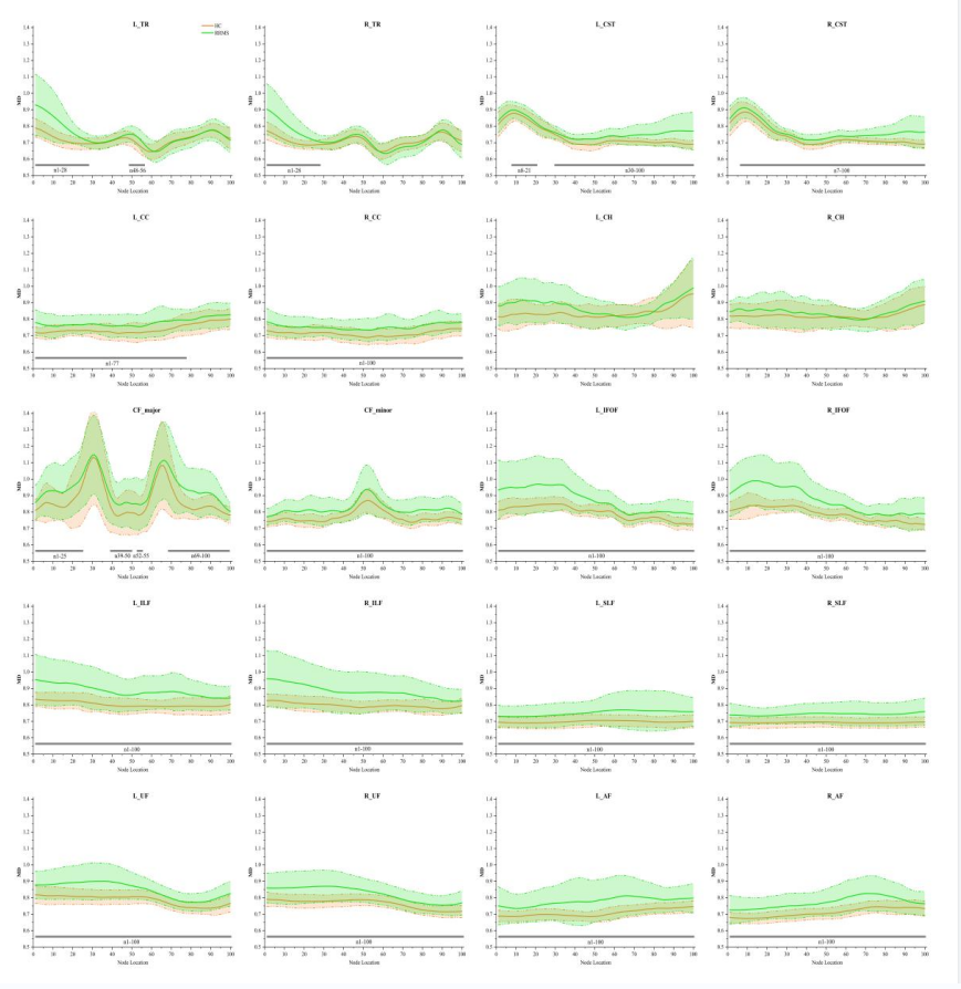
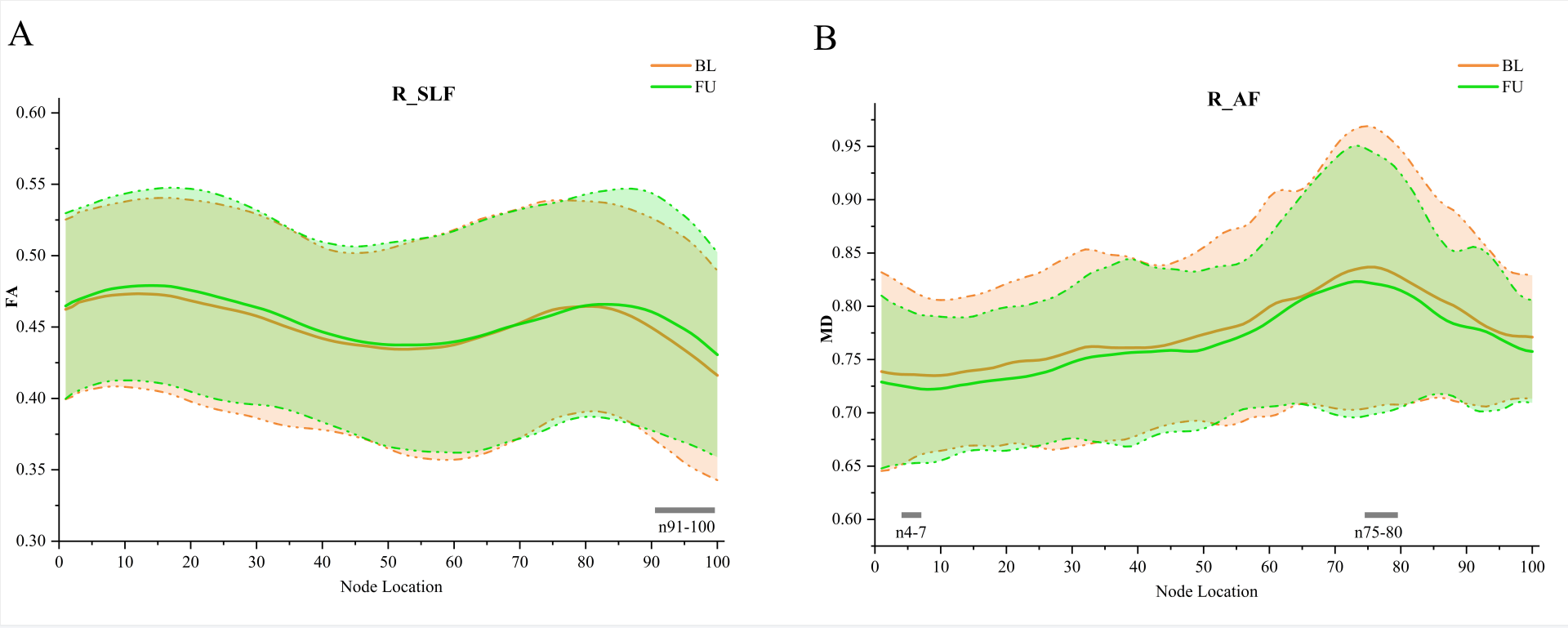
At baseline, compared with HC, RRMS showed a widespread FA decrease and MD increase among tracts. While in the pointwise comparison, within the more detailed abnormalities localized to specific positions. At follow up, compared with baseline, although there was no statistical difference on FA and MD values in entire manner, FA reduced in the posterior portion of the right superior longitudinal fasciculus (R_SLF) and MD elevated in the anterior and posterior portion of the right arcuate fasciculus (R_AF) in the pointwise analysis. Furthermore, the altered metrics were widely correlated with cognitive performance in RRMS.Discussion
In line with previous studies, at baseline comparison of entire WM fiber tracts, our finding revealed significant DTI metrics alterations (FA in 11/20 and MD in 18/20) in WM fiber tracts, which may indicate a widespread microstructural damage in RRMS. The MD metrics were seemed to be more sensitive to WM damage than FA, consistent with the previous studies. As to the tracts (bilateral CST, CC, IFOF, ILF; L_UF and CF) altered significantly both in FA and MD, it may result from the loss of axonal integrity and demyelination or edema in fiber tracts.At follow-up, although there were no significant mean DTI metrics changes in patients with RRMS, there was a FA reduction in the posterior portion of R_SLF and a MD elevation in the anterior and posterior portion of R_AF in the pointwise manner. This also further illustrates the high sensitivity of the AFQ algorithm.
To further understand the underlying mechanisms, the correlation analyses were performed between WM microstructure damages and cognitive performance with age, gender, and education level as covariates. After FDR correction, our results showed that the altered DTI metrics were widely correlated with cognitive performance in RRMS. It was consistent with our hypothesis above about the potential contribution on cognitive impairment by altered DTI metrics in the WM fiber tracts. Moreover, a wider range of results were shown compared to previous studies, which demonstrated the advantages of the AFQ algorithm and revealed more pathophysiological mechanisms associated with cognitive impairment.
Conclusion
In conclusion, the application of AFQ showed that RRMS patients had a widespread WM microstructure alteration at baseline and some certain regions alterations at a short-term follow-up, which will enhance our understanding of WM microstructural abnormalities in RRMS patients. And the correlation between altered DTI metrics and cognitive performance in RRMS can be used as imaging markers for early identification of RRMS cognitive impairment.Acknowledgements
We thank all the subjects who participated in this study.References
Amin, M., & Ontaneda, D. (2020). Thalamic Injury and Cognition in Multiple Sclerosis. Front Neurol, 11, 623914. https://doi.org/10.3389/fneur.2020.623914 Banfi, C., Koschutnig, K., Moll, K., Schulte-Körne, G., Fink, A., & Landerl, K. (2019). White matter alterations and tract lateralization in children with dyslexia and isolated spelling deficits. Hum Brain Mapp, 40(3), 765-776. https://doi.org/10.1002/hbm.24410 Browne, P., Chandraratna, D., Angood, C., Tremlett, H., Baker, C., Taylor, B. V., & Thompson, A. J. (2014). Atlas of Multiple Sclerosis 2013: A growing global problem with widespread inequity. Neurology, 83(11), 1022-1024. https://doi.org/10.1212/wnl.0000000000000768 Bubb, E. J., Metzler-Baddeley, C., & Aggleton, J. P. (2018). The cingulum bundle: Anatomy, function, and dysfunction. Neurosci Biobehav Rev, 92, 104-127. https://doi.org/10.1016/j.neubiorev.2018.05.008 Cao, Y., Diao, W., Tian, F., Zhang, F., He, L., Long, X., . . . Jia, Z. (2021). Gray Matter Atrophy in the Cortico-Striatal-Thalamic Network and Sensorimotor Network in Relapsing-Remitting and Primary Progressive Multiple Sclerosis. Neuropsychol Rev, 31(4), 703-720. https://doi.org/10.1007/s11065-021-09479-3 Chen, H. F., Huang, L. L., Li, H. Y., Qian, Y., Yang, D., Qing, Z., . . . Xu, Y. (2020). Microstructural disruption of the right inferior fronto-occipital and inferior longitudinal fasciculus contributes to WMH-related cognitive impairment. CNS Neurosci Ther, 26(5), 576-588. https://doi.org/10.1111/cns.13283 Chen, X., Roberts, N., Zheng, Q., Peng, Y., Han, Y., Luo, Q., . . . Li, Y. (2021). Progressive brain microstructural damage in patients with multiple sclerosis but not in patients with neuromyelitis optica spectrum disorder: A cross-sectional and follow-up tract-based spatial statistics study. Mult Scler Relat Disord, 55, 103178. https://doi.org/10.1016/j.msard.2021.103178 Chylińska, M., Karaszewski, B., Komendziński, J., Wyszomirski, A., Hałas, M., Szurowska, E., & Sabisz, A. (2023). The association between white matter tract structural connectivity and information processing speed in relapsing-remitting multiple sclerosis. Neurol Sci. https://doi.org/10.1007/s10072-023-06817-6 Confavreux, C., Vukusic, S., Moreau, T., & Adeleine, P. (2000). Relapses and progression of disability in multiple sclerosis. N Engl J Med, 343(20), 1430-1438. https://doi.org/10.1056/nejm200011163432001 Deloire, M. S., Bonnet, M. C., Salort, E., Arimone, Y., Boudineau, M., Petry, K. G., & Brochet, B. (2006). How to detect cognitive dysfunction at early stages of multiple sclerosis? Mult Scler, 12(4), 445-452. https://doi.org/10.1191/1352458506ms1289oa Dobson, R., & Giovannoni, G. (2019). Multiple sclerosis - a review. Eur J Neurol, 26(1), 27-40. https://doi.org/10.1111/ene.13819 Dou, X., Yao, H., Feng, F., Wang, P., Zhou, B., Jin, D., . . . Liu, Y. (2020). Characterizing white matter connectivity in Alzheimer's disease and mild cognitive impairment: An automated fiber quantification analysis with two independent datasets. Cortex, 129, 390-405. https://doi.org/10.1016/j.cortex.2020.03.032 Gazzaniga, M. S. (2000). Cerebral specialization and interhemispheric communication: does the corpus callosum enable the human condition? Brain, 123 ( Pt 7), 1293-1326. https://doi.org/10.1093/brain/123.7.1293 Ge, Y., Law, M., & Grossman, R. I. (2005). Applications of diffusion tensor MR imaging in multiple sclerosis. Ann N Y Acad Sci, 1064, 202-219. https://doi.org/10.1196/annals.1340.039 Gois, L. C. P., Pimentel-Silva, L. R., Damasceno, B. P., & Damasceno, A. (2021). Associations between cognitive and clinical disability across MS subtypes: The role of the underlying brain damage. Mult Scler Relat Disord, 48, 102701. https://doi.org/10.1016/j.msard.2020.102701 Grothe, M., Jochem, K., Strauss, S., Langner, S., Kirsch, M., Hoffeld, K., . . . Lotze, M. (2022). Performance in information processing speed is associated with parietal white matter tract integrity in multiple sclerosis. Front Neurol, 13, 982964. https://doi.org/10.3389/fneur.2022.982964 Harel, A., Sperling, D., Petracca, M., Ntranos, A., Katz-Sand, I., Krieger, S., . . . Inglese, M. (2018). Brain microstructural injury occurs in patients with RRMS despite 'no evidence of disease activity'. J Neurol Neurosurg Psychiatry, 89(9), 977-982. https://doi.org/10.1136/jnnp-2017-317606 Hori, M., Maekawa, T., Kamiya, K., Hagiwara, A., Goto, M., Takemura, M. Y., . . . Aoki, S. (2022). Advanced Diffusion MR Imaging for Multiple Sclerosis in the Brain and Spinal Cord. Magn Reson Med Sci, 21(1), 58-70. https://doi.org/10.2463/mrms.rev.2021-0091 Jin, Y., Huang, C., Daianu, M., Zhan, L., Dennis, E. L., Reid, R. I., . . . Thompson, P. M. (2017). 3D tract-specific local and global analysis of white matter integrity in Alzheimer's disease. Hum Brain Mapp, 38(3), 1191-1207. https://doi.org/10.1002/hbm.23448 Kim, S. H., Kwak, K., Hyun, J. W., Joung, A., Lee, S. H., Choi, Y. H., . . . Kim, H. J. (2017). Diffusion tensor imaging of normal-appearing white matter in patients with neuromyelitis optica spectrum disorder and multiple sclerosis. Eur J Neurol, 24(7), 966-973. https://doi.org/10.1111/ene.13321 Liu, Y., Duan, Y., Huang, J., Ren, Z., Ye, J., Dong, H., . . . Li, K. (2015). Multimodal Quantitative MR Imaging of the Thalamus in Multiple Sclerosis and Neuromyelitis Optica. Radiology, 277(3), 784-792. https://doi.org/10.1148/radiol.2015142786 Meca-Lallana, V., Gascón-Giménez, F., Ginestal-López, R. C., Higueras, Y., Téllez-Lara, N., Carreres-Polo, J., . . . Pérez-Miralles, F. (2021). Cognitive impairment in multiple sclerosis: diagnosis and monitoring. Neurol Sci, 42(12), 5183-5193. https://doi.org/10.1007/s10072-021-05165-7 Meoded, A., & Huisman, T. (2019). Diffusion Tensor Imaging of Brain Malformations: Exploring the Internal Architecture. Neuroimaging Clin N Am, 29(3), 423-434. https://doi.org/10.1016/j.nic.2019.03.004 Mistri, D., Cacciaguerra, L., Valsasina, P., Pagani, E., Filippi, M., & Rocca, M. A. (2023). Cognitive function in primary and secondary progressive multiple sclerosis: A multiparametric magnetic resonance imaging study. Eur J Neurol. https://doi.org/10.1111/ene.15900 Ontaneda, D., Raza, P. C., Mahajan, K. R., Arnold, D. L., Dwyer, M. G., Gauthier, S. A., . . . Azevedo, C. J. (2021). Deep grey matter injury in multiple sclerosis: a NAIMS consensus statement. Brain, 144(7), 1974-1984. https://doi.org/10.1093/brain/awab132 Paul, L. K., Brown, W. S., Adolphs, R., Tyszka, J. M., Richards, L. J., Mukherjee, P., & Sherr, E. H. (2007). Agenesis of the corpus callosum: genetic, developmental and functional aspects of connectivity. Nat Rev Neurosci, 8(4), 287-299. https://doi.org/10.1038/nrn2107 Peterson, D. S., & Fling, B. W. (2018). How changes in brain activity and connectivity are associated with motor performance in people with MS. Neuroimage Clin, 17, 153-162. https://doi.org/10.1016/j.nicl.2017.09.019 Schmidt, P., Gaser, C., Arsic, M., Buck, D., Förschler, A., Berthele, A., . . . Mühlau, M. (2012). An automated tool for detection of FLAIR-hyperintense white-matter lesions in Multiple Sclerosis. Neuroimage, 59(4), 3774-3783. https://doi.org/10.1016/j.neuroimage.2011.11.032 Schmierer, K., Wheeler-Kingshott, C. A., Boulby, P. A., Scaravilli, F., Altmann, D. R., Barker, G. J., . . . Miller, D. H. (2007). Diffusion tensor imaging of post mortem multiple sclerosis brain. Neuroimage, 35(2), 467-477. https://doi.org/10.1016/j.neuroimage.2006.12.010 Shu, M., Yu, C., Shi, Q., Li, Y., Niu, K., Zhang, S., & Wang, X. (2021). Alterations in white matter integrity and asymmetry in patients with benign childhood epilepsy with centrotemporal spikes and childhood absence epilepsy: An automated fiber quantification tractography study. Epilepsy Behav, 123, 108235. https://doi.org/10.1016/j.yebeh.2021.108235 Šoda, J., Pavelin, S., Vujović, I., & Rogić Vidaković, M. (2023). Assessment of Motor Evoked Potentials in Multiple Sclerosis. Sensors (Basel), 23(1). https://doi.org/10.3390/s23010497 Sun, H., Lui, S., Yao, L., Deng, W., Xiao, Y., Zhang, W., . . . Gong, Q. (2015). Two Patterns of White Matter Abnormalities in Medication-Naive Patients With First-Episode Schizophrenia Revealed by Diffusion Tensor Imaging and Cluster Analysis. JAMA Psychiatry, 72(7), 678-686. https://doi.org/10.1001/jamapsychiatry.2015.0505 Thompson, A. J., Banwell, B. L., Barkhof, F., Carroll, W. M., Coetzee, T., Comi, G., . . . Cohen, J. A. (2018). Diagnosis of multiple sclerosis: 2017 revisions of the McDonald criteria. Lancet Neurol, 17(2), 162-173. https://doi.org/10.1016/s1474-4422(17)30470-2 Tu, T. W., Williams, R. A., Lescher, J. D., Jikaria, N., Turtzo, L. C., & Frank, J. A. (2016). Radiological-pathological correlation of diffusion tensor and magnetization transfer imaging in a closed head traumatic brain injury model. Ann Neurol, 79(6), 907-920. https://doi.org/10.1002/ana.24641 Yamout, B. I., & Alroughani, R. (2018). Multiple Sclerosis. Semin Neurol, 38(2), 212-225. https://doi.org/10.1055/s-0038-1649502 Yan, Z., Wang, X., Zhu, Q., Shi, Z., Chen, X., Han, Y., . . . Li, Y. (2022). Alterations in White Matter Fiber Tracts Characterized by Automated Fiber-Tract Quantification and Their Correlations With Cognitive Impairment in Neuromyelitis Optica Spectrum Disorder Patients. Front Neurosci, 16, 904309. https://doi.org/10.3389/fnins.2022.904309 Yeatman, J. D., Dougherty, R. F., Myall, N. J., Wandell, B. A., & Feldman, H. M. (2012). Tract profiles of white matter properties: automating fiber-tract quantification. PLoS One, 7(11), e49790. https://doi.org/10.1371/journal.pone.0049790 Zhang, H., Li, H., Yin, L., Chen, Z., Wu, B., Huang, X., . . . Gong, Q. (2022). Aberrant White Matter Microstructure in Depressed Patients with Suicidality. J Magn Reson Imaging, 55(4), 1141-1150. https://doi.org/10.1002/jmri.27927 Zhang, X., Sun, Y., Li, W., Liu, B., Wu, W., Zhao, H., . . . Zhang, B. (2019). Characterization of white matter changes along fibers by automated fiber quantification in the early stages of Alzheimer's disease. Neuroimage Clin, 22, 101723. https://doi.org/10.1016/j.nicl.2019.101723Figures