2930
The effect of shift working on workers brain morphometric changes from a voxel-wise comparison1Department of Biomedical Engineering, Yonsei University, Wonju, Korea, Republic of, 2Department of Preventive Medicine, College of Medicine, Chung-Ang University, Seoul, Korea, Republic of
Synopsis
Keywords: Gray Matter, Neuroscience
Motivation: Compared to non-shift workers, shift workers are more likely to be at risk for accidents due to decreased performance, attention to work, and concentration.
Goal(s): We aim to examine the health effects of shift work from a neuroscientific perspective.
Approach: T1w-MPRAGEs were utilized to generate VBM maps to investigate regional volumetric changes between shift and non-shift workers. GLM was created to compare VBM between two groups.
Results: The cerebellum region was found to be significantly larger in non-shift work compared to shift work while shift workers had larger volume of the inferior parietal region compared to non-shift workers.
Impact: In this study, our aim was to examine the neuroscientific evidence explaining various health issues among shift workers. To the best of our knowledge, this is the first study to investigate structural differences between shift and non-shift workers.
Introduction
It is widely known that shift work has a negative impact on the health of workers. In comparison to non-shift workers, shift workers are more likely to suffer from sleep disorders, depression, and suicide, and are more at risk for accidents due to decreased performace and attention to work and concentration [1]. While there are many observational studies linking these various health problems to shift work, there has been a lack of evidence on the basic science. In this study, we aim to examine the health effects of shift work from a neuroscience perspective.Materials and Methods
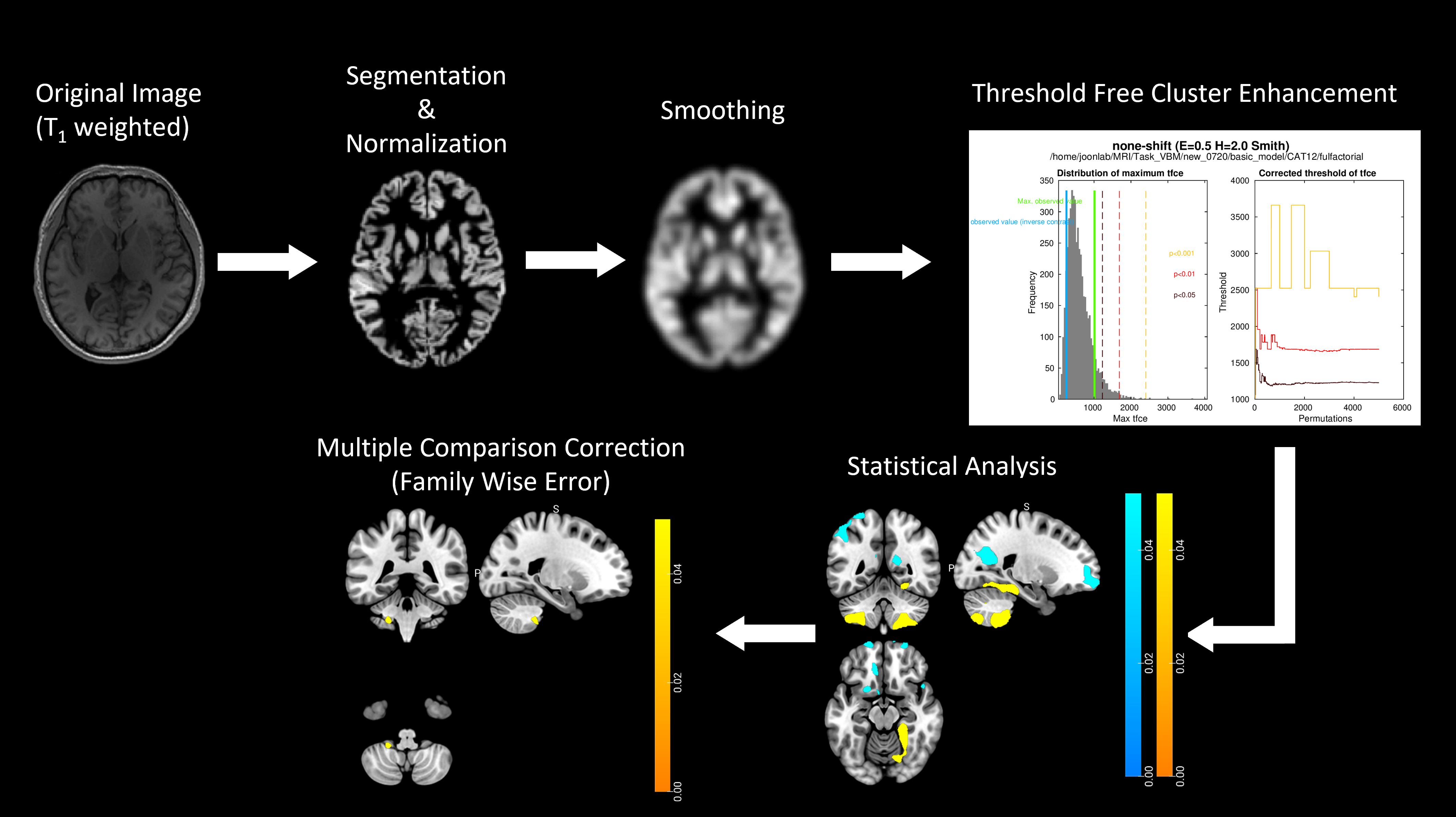
Data acquisition and processing: T1w-MPRAGE was acquired from 32 shift workers and 79 non-shift workers in a medical facility with the following parameters: TR=1970 ms, TE=2.84 ms, TI=991 ms, FOV=256x256, FA=9, in-plane resolution=0.5x0.5x1 mm3, the number of slices=192, and the scan time=4 min 34 sec. The acquired T1w-MPRAGE images were utilized to generate voxel-based morphometry (VBM) maps to investigate regional volumetric changes in the brain [2]. As shown in Fig.1, we performed segmentation, normalization, and smoothing for the preprocessing of VBM using the computation anatomy toolbox (CAT12) in statistical parametric mapping (SPM). In this study, VBM analysis was conducted within the gray matter (GM). After brain segmentation, the segmented gray matter images were spatially normalized to a T1w-MNI 152 template image using non-linear registration. Finally, Gaussian smoothing with an 8 mm full width at half maximum (FWHM) was applied to reduce noise, improve registration, and enhance statistical validity [2].Statistical analysis: To investigate structural changes associated with shift and non-shift work, a general linear model (GLM) based on two sample t-tests was created to compare VBM between two groups using CAT12. In the GLM, total intracranial volume (TIV), age, and gender were used as covariates. Considering the inherent individual differences in brain volume, we included TIV as one of the covariates. Whole-brain results from GLM were evaluated using threshold free cluster enhancement (TFCE) inference for multiple comparison correction (Smith and Nichols, NI, 2009). Both uncorrected and family-wise error (FWE) corrected thresholds with p < 0.05 were explored. The approach of combining GLM with TFCE has been recommended as the optimal configuration for advanced VBM analysis.
Results
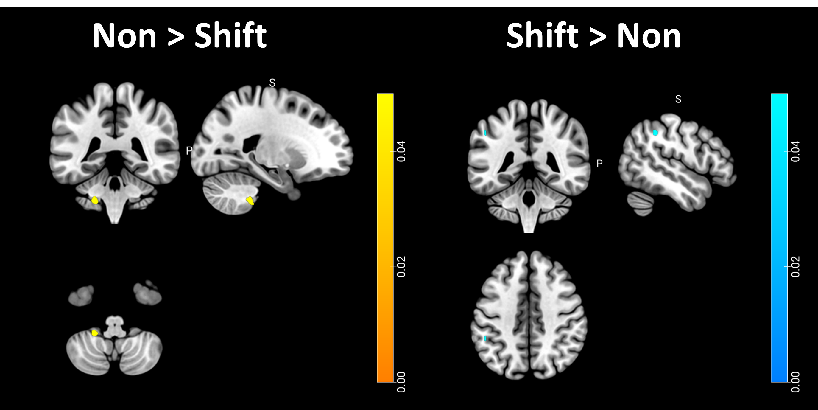
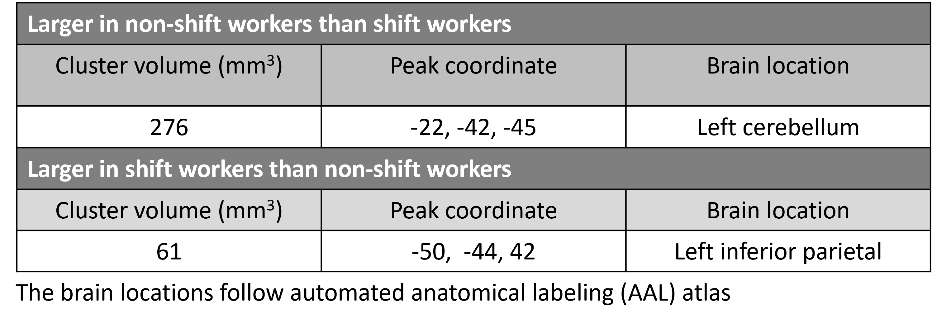
When comparing the volume of non-shift work with that of shift work, the cerebellum region was found to be significantly larger in non-shift work compared to shift work (Fig.2 left)(p < 0.05, FWE corrected) while shift workers had larger volume of the inferior parietal region compared to non-shift workers (Fig.2 right)(p < 0.05, FWE corrected). Table 1 summarizes the significant clusters and their brain locations.Discussion and Conclusion
This study examined the changes in brain MRI structure between shift workers and non-shift workers. As the study aimed to examine changes in normal structure, not clinical abnormalities, there were many difficulties in clearly identifying the differences between the two groups, but we were able to identify meaningful differences in some areas. None-shift workers showed significantly increased clusters in the left cerebellum than shift workers. The cerebrum participates in higher levels of thinking and action [3]. The cerebellum has several functions relating to movement and coordination, including maintaining balance, coordination movement, vision, motor learning and others (some role in thinking, including processing language and mood) [4]. As a result of the close relationship between the cerebellum and movement, the most common signs of cerebellar disorder involve a disturbance in muscle control such as lack of muscle control and coordination, difficulties with walking and mobility, slurred speech or difficulty speaking, abnormal eye movements, headaches. This provides a neurological explanation for the decreased performance, headaches and increased risk of accidents observed in shift workers. Shift workers showed significantly increased clusters in the left inferior parietal lobe than none-shift workers. The inferior parietal lobe is a key neural substrate underlying diverse mental processes, from basic attention to language and social cognition, that define human interactions [5]. We expected shift work to have only negative neurological effects, but in fact the brain areas associated with the main work performance (patient care and related tasks) were enhanced. This suggests that the stimulation from the acquired occupation may have contributed to strengthening some of the brain structures. In this study, we aimed to observe the evidence of neuroscientific explanation for several unhealthy conditions in shift workers. Further research on the level of connectivity and activation of synapses, as well as simple brain areas, will provide scientific evidence for the potential health effects of shift work.Acknowledgements
This work was supported by the Ministry of Education of the Republic of Korea and the National Research Foundation of Korea (NRF-2021R1C1C1008871) and by the National Research Foundation of Korea(NRF) grant funded by the Korea government(MSIT)(RS-2023-00251484). The funder had no role in either the direction or methodology of the study.
References
[1] G Costa, The impact of shift and night work on health, Applied Ergonomics, Volume 27, 9-16, 1996
[2] Ashburner J, Friston KJ, Voxel-based morphometry-the methods, Neuroimage, 805- 821, 2000
[3] Hehui Li, Junjie Wu, Rebecca A Marks, et al. Functional mapping and cooperation between the cerebellum and cerebrum during word reading, cerebral Cortex, Volume 32, 5175-5190, 2022
[4] Randy L Buckner, The cerebellum and Cognitive Function:25 Years of insight from Anatomy and Neuroimaging, Neuron, 2013
[5] Bzdok D, Hartwigsen G, Reid A, et al. Left inferior parietal lobe engagement in social cognition and language, Neuroscience & Biobehavioral Reviews, Volume 68, 319-334, 2016
Figures