2926
Brain gray matter alterations in individuals with convergence insufficiency1Department of Radiology and Huaxi MR Research Center (HMRRC), Functional and Molecular Imaging Key Laboratory of Sichuan Province, West China Hospital, Sichuan University, Chengdu 610041, Sichuan Province, China, 2Department of Radiology, Suining Central Hospital, Suining, Sichuan 629099, China, 3Department of Ophthalmology, Laboratory of Optometry and Vision Sciences, West China Hospital, Sichuan University, Chengdu 610041, Sichuan Province, China, 4MR Research Collaboration, Siemens Healthineers, Beijing, China
Synopsis
Keywords: Gray Matter, Nervous system, Convergence insufficiency;Brain;Structure
Motivation: Whether abnormal visual experiences in individuals with convergence insufficiency (CI) would be associated with brain structural alterations.
Goal(s): To explore altered cortical structural characteristics in individuals with CI and its relationship with visual measures and clinical symptoms.
Approach: All participants underwent high-resolution T1-weighted brain magnetic resonance imaging scanning and cortical thickness, surface area, and volume were calculated and compared between two groups.
Results: There are altered cortical gray matter volume and surface area in the brain regions related to the function of eye movement in CI than healthy controls, one of which was correlated with near point of convergence.
Impact: Our results shed light on the impact of binocular vision dysfunctions on human brain structure, explicate the neural mechanism of CI, and reveal the potential utility of brain MRI in assessing the neural mechanism of ocular diseases.
Introduction
The eyes of individuals with convergence insufficiency (CI) have a strong tendency to drift outward with difficulty turning the eyes inward during near work 1. The visual system uses specific neural circuits to mediate eye movement 2. Previous studies have demonstrated that abnormal visual experiences can induce alterations in the brain structure and function of the visual system 3, and acquired brain injury can also lead to visual dysfunctions, such as vergence abnormalities. Thus, the present aim is to explore if abnormal visual experiences in CI would be associated with brain structural alterations.Methods
Thirty-four participants with CI and 35 health controls (HCs) were recruited and underwent brain high-resolution T1-weighted 3.0T MRI scans. CI dysfunction was examined and diagnosed by an optometrist. Using FreeSurfer, we calculated cortical thickness, surface area, and volume and completed the whole-brain between-group analyses with corrections for multiple comparisons utilizing permutation simulation. Spearman partial correlation analyses were performed to explore the relationships between structural features and clinical symptoms of CI in all participants, with age and sex as covariates.Results
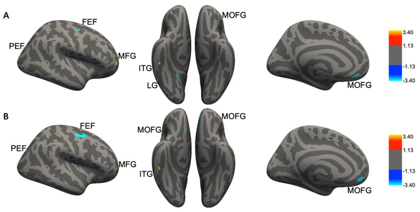
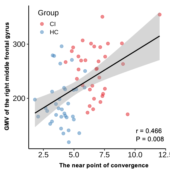
Compared to HCs, participants with CI had reduced cortical gray matter volume in the right frontal eye field, parietal eye field, lingual gyrus, and left medial orbitofrontal cortex, while increased gray matter volume in the right middle frontal gyrus and inferior temporal gyrus. Similarly, gray matter surface area in right frontal eye fields, parietal eye field, and bilateral medial orbitofrontal gyrus of individuals with CI was decreased compared to HCs, while the surface area in the right middle frontal gyrus was increased (Figure 1). Further, the gray matter volume of right middle frontal gyrus showed a positive correlation with near point of convergence (r = 0.466, p-corrected = 0.008) (Figure 2).Discussion
The visual system comprises a three-level hierarchy for controlling saccadic eye movement in which the frontal eye field and parietal eye field play a significant role in triggering eye movement as the highest level 4. The lingual gyrus works on conveying visual information as the second visual cortex (V2). Previous studies found that individuals with CI exhibited reduced activation in the frontal eye field, parietal eye field, and V2 during convergence tasks 5-7. Together with these functional findings, our results with reduced surface area and cortical volume in the frontal eye field, parietal eye field, and V2 may indicate that CI might not only affect the brain function but also be associated with brain structural alterations in brain regions related to eye movements in individuals with CI.People with CI often experience varying degrees of visual fatigue symptoms8. The human brain stimulator system maintains performance by enhancing motor input from the primary motor cortex to muscles during periods of physical fatigue8. In chronic fatigue patients, metabolic, functional and structural damage of key brain regions of the promoting system, such as orbitofrontal gyrus and prefrontal cortex, may underlie their dysfunction9. Thus, the reduced surface area and volume of the orbitofrontal gyrus we found in CI may reflect a neurobiological mechanism of aggregation dysfunction that further exacerbates visual fatigue.The prefrontal gyrus belongs to the prefrontal cortex, which plays a supervisory role in controlling eye movement 10, 11. Previous study demonstrated a positive correlation between changes of deoxygenated hemoglobin concentration, total hemoglobin concentration in the frontal lobe during convergence task and near point of convergence, which means that the larger near point of convergence, the worse convergence function, and the more metabolic activity increased in the frontal lobe 10. In line with this, our results revealed increased cortical surface area and gray matter volume in the anterior portion of the right medial frontal gyrus in CI and a positive correlation between gray matter volume and near point of convergence. These results may be conducive to the supervised control of eye movement, helping patients perceive the eye movement cycle and complete the entire eye movement process.The inferior temporal gyrus selectively responds to three-dimensional objects under binocular viewing conditions, which will provide information to direct convergence eye movement 12, 13. Thus, the increase of cortical surface area and volume of the inferior temporal gyrus may enhance the parallax information processing of three-dimensional objects in CI, facilitating the initiation of convergence eye movements.Conclusion
Our results shed light on the impact of binocular vision dysfunctions on human brain structure, explicate the neural mechanism of CI, and reveal the potential utility of brain MRI in assessing the neural mechanism of ocular diseases.Acknowledgements
No acknowledgement found.References
1. Hashemi, H.; Nabovati, P.; Khabazkhoob, M.; Ostadimoghaddam, H.; Doostdar, A.; Shiralivand, E.; Yekta, A., The prevalence of convergence insufficiency in Iran: a population-based study. Clin Exp Optom 2017, 100 (6), 704-709.2. Seung, S.; Yuste, R., Principles of neural science (5th ed.). McGraw-Hill Companies 2013.3. Chan, S. T.; Tang, K. W.; Lam, K. C.; Chan, L. K.; Mendola, J. D.; Kwong, K. K., Neuroanatomy of adult strabismus: a voxel-based morphometric analysis of magnetic resonance structural scans. Neuroimage 2004, 22 (2), 986-94.4. Munoz, D. P.; Coe, B. C., Saccade, search and orient--the neural control of saccadic eye movements. Eur J Neurosci 2011, 33 (11), 1945-7.5. Jaswal, R.; Gohel, S.; Biswal, B. B.; Alvarez, T. L., Task-modulated coactivation of vergence neural substrates. Brain Connect 2014, 4 (8), 595-607.6. Alvarez, T. L.; Jaswal, R.; Gohel, S.; Biswal, B. B., Functional activity within the frontal eye fields, posterior parietal cortex, and cerebellar vermis significantly correlates to symmetrical vergence peak velocity: an ROI-based, fMRI study of vergence training. Front Integr Neurosci 2014, 8, 50.7. Alvarez, T. L.; Vicci, V. R.; Alkan, Y.; Kim, E. H.; Gohel, S.; Barrett, A. M.; Chiaravalloti, N.; Biswal, B. B., Vision therapy in adults with convergence insufficiency: clinical and functional magnetic resonance imaging measures. Optom Vis Sci 2010, 87 (12), E985-1002.8. Ovenseri-Ogbomo, G. O.; Eguegu, O. P., Vergence findings and horizontal vergence dysfunction among first year university students in Benin City, Nigeria. J Optom 2016, 9 (4), 258-63.9. Tanaka, M.; Ishii, A.; Watanabe, Y., Regulatory mechanism of performance in chronic cognitive fatigue. Med Hypotheses 2014, 82 (5), 567-71.10. Richter, H. O.; Forsman, M.; Elcadi, G. H.; Brautaset, R.; Marsh, J. E.; Zetterberg, C., Prefrontal Cortex Oxygenation Evoked by Convergence Load Under Conflicting Stimulus-to-Accommodation and Stimulus-to-Vergence Eye-Movements Measured by NIRS. Front Hum Neurosci 2018, 12, 298.11. Gu, C.; Corneil, B. D., Transcranial magnetic stimulation of the prefrontal cortex in awake nonhuman primates evokes a polysynaptic neck muscle response that reflects oculomotor activity at the time of stimulation. J Neurosci 2014, 34 (44), 14803-15.12. Tanaka, K.; Saito, H.; Fukada, Y.; Moriya, M., Coding visual images of objects in the inferotemporal cortex of the macaque monkey. J Neurophysiol 1991, 66 (1), 170-89.13. Searle, A.; Rowe, F. J., Vergence Neural Pathways: A Systematic Narrative Literature Review. Neuroophthalmology 2016, 40 (5), 209-218.Figures