2922
Altered cortical morphology and morphological similarity network patterns in classical trigeminal neuralgia patients1Department of Magnetic Resonance, Lanzhou University Second Hospital, Lanzhou, China, 2Second Clinical School, Lanzhou University, Lanzhou, China, 3Gansu Province Clinical Research Center for Functional and Molecular Imaging, Lanzhou, China, 4Philips Healthcare, Xi'an, China
Synopsis
Keywords: Gray Matter, Neuroscience, trigeminal neuralgia, grey-matter morphology, structural covariance network
Motivation: Morphological covariance in classical trigeminal neuralgia is not well understood.
Goal(s): To characterize the brain morphometry, and further construct individual-level morphological similarity networks.
Approach: We performed volume and surface-based morphometry analyses respectively. Using cortical indicators combined with Kullback-Leibler divergence, we further investigated the topological properties of structural covariance network.
Results: Patients presented decreased cortical indicators in salience and default mode network, along with increased volume and cortical complexity. Topological analysis revealed impaired information integration of the fractal dimension and sulcus depth networks, and the opposite trend in cortical thickness network. Gray matter covariation provides connectome evidence for central plasticity in chronic pain.
Impact: The present study, for the first time, revealed the impairments of individual-level morphological covariance networks in CTN chronic pain patients, highlighting the combined effects of pain and mood disorders. Additionally, volume and surface integration analyses help to provide complementary information.
Introduction
Aberrant neuroplasticity in the gray matter (GM) is an important feature of classical trigeminal neuralgia (CTN)1. However, evidence of fractal dimension (FD) remains limited. Additionally, exploring the synergistic patterns in brain morphology is valuable. Based on our previous findings2,3, the present study aims to further investigate the topological properties in morphological covariance network.Methods
A total of 43 CTN patients and 45 age, sex, and education-matched healthy controls were recruited. All subjects underwent 3.0T MR scanning (Ingenia CX, Philips healthcare, the Netherlands) with a 16-channel head coil for 3D-T1WI data acquisition. Images were preprocessed using the computational anatomy toolbox with default pipeline. In VBM analysis, GM maps were smoothed using an 8mm Gaussian kernel. In SBM analysis, FD, gyrification index (GI), sulcal depth (SD), and cortical thickness (CT) were obtained. The smooth nuclei were set to 15mm for CT and 25mm for other maps. In morphological networks, we defined nodes based on the DK40 atlas4 (68 regions). Symmetric similarity metrics based on KL divergence5 was computed as edge strength. The topological properties, including global (global and local efficiency (Eg and Eloc), small worldness (σ, γ, λ)) and local properties (nodal efficiency (Ne), degree centrality, betweenness centrality), were calculated using GRETNA, with sparsity range of 0.063 to 0.4 and interval of 0.01. We also explored ROI-wised SBM. The between-group comparisons of ROI-wised SBM and topological properties were performed using nonparametric permutation test (10,000 permutations), wherein ROI and nodal properties underwent FDR correction. Spearman's partial correlation analyses were also performed. Age, gender, education and TIV (only for VBM) were considered as covariates. Moreover, we conducted validation analyses: (1) using Jensen-Shannon divergence based similarity6 as edges; (2) defining nodes based on the Destrieux atlas7 (148 areas).Results
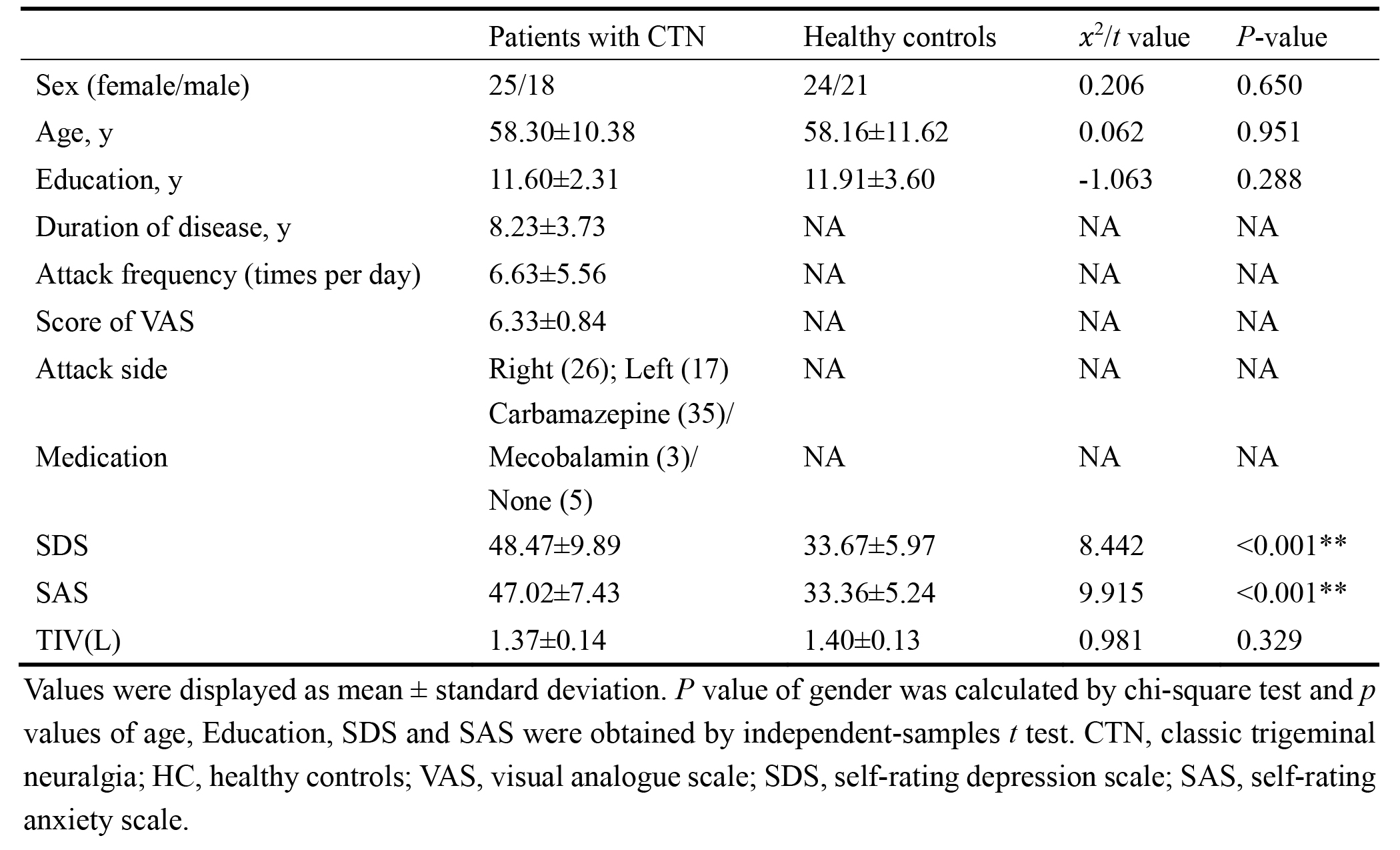
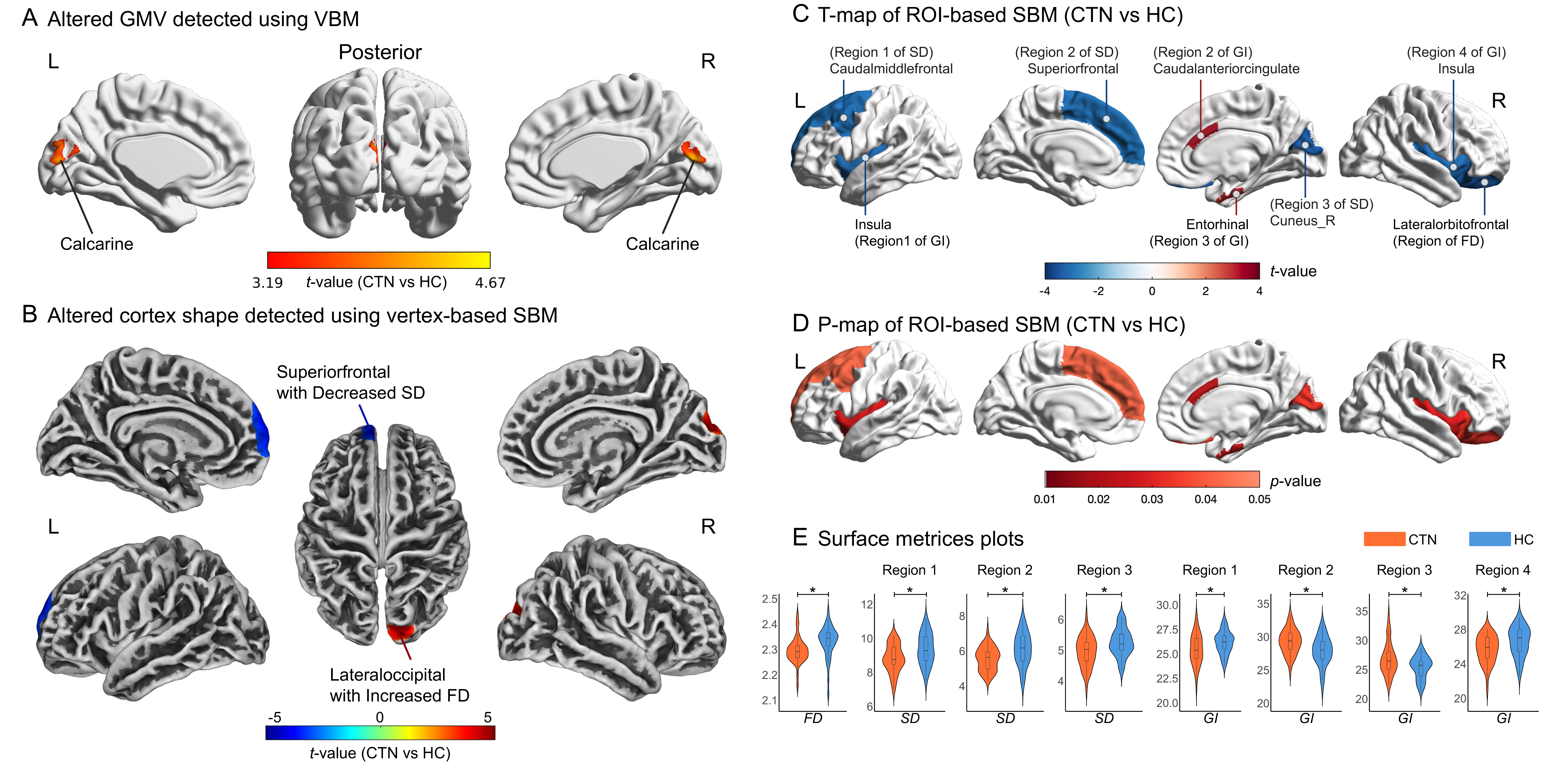
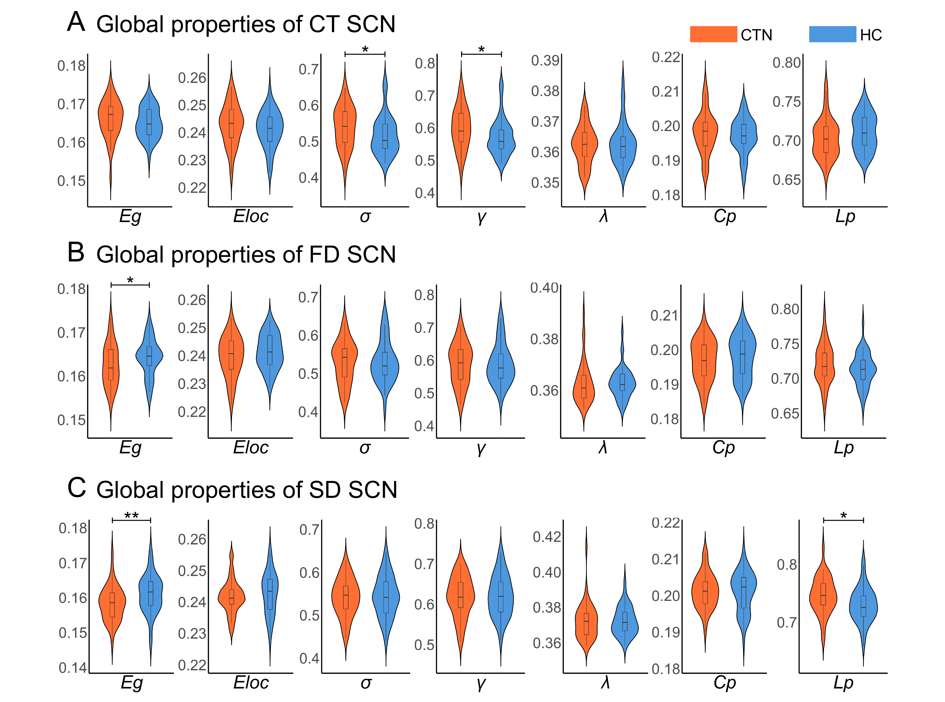
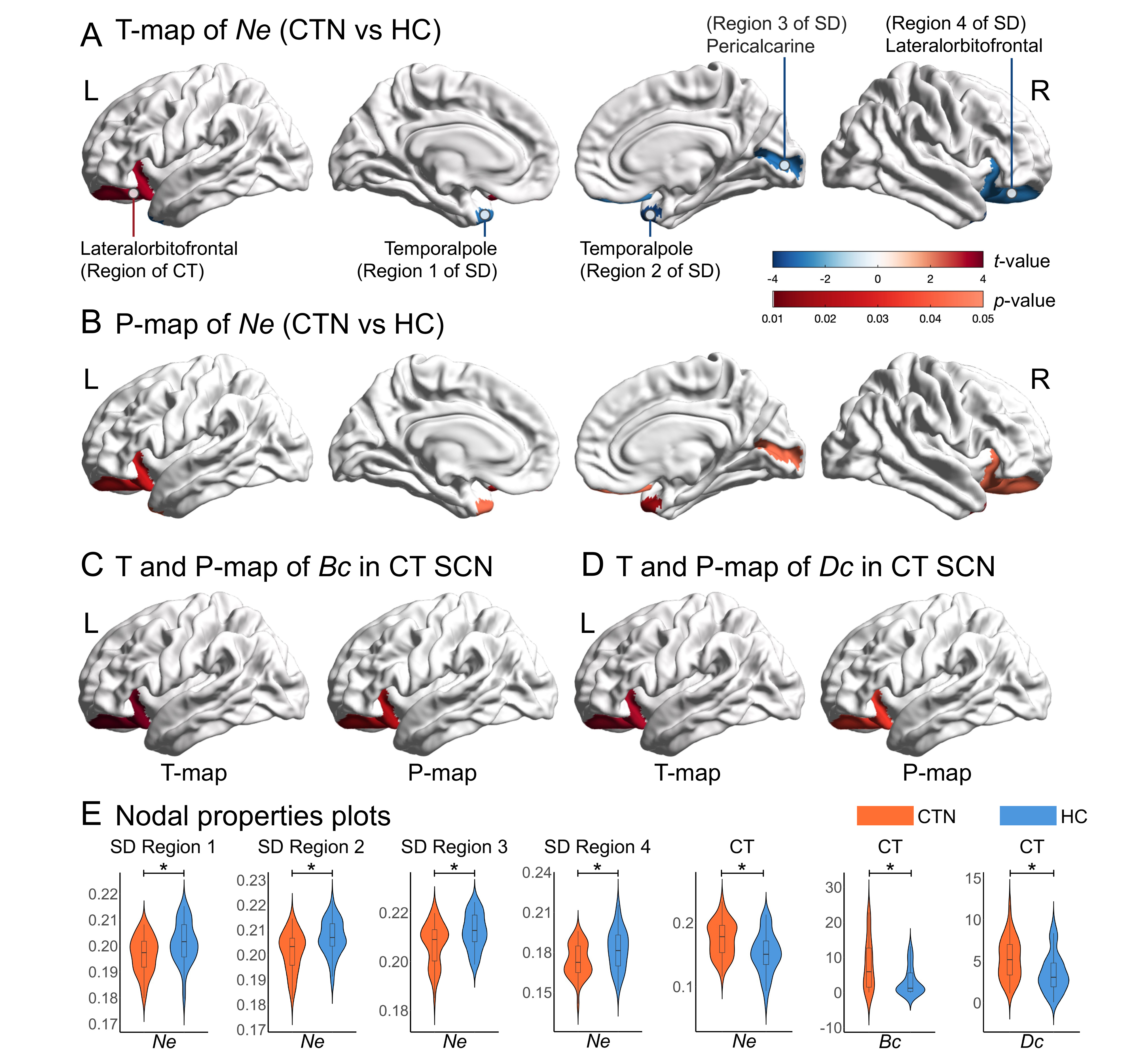
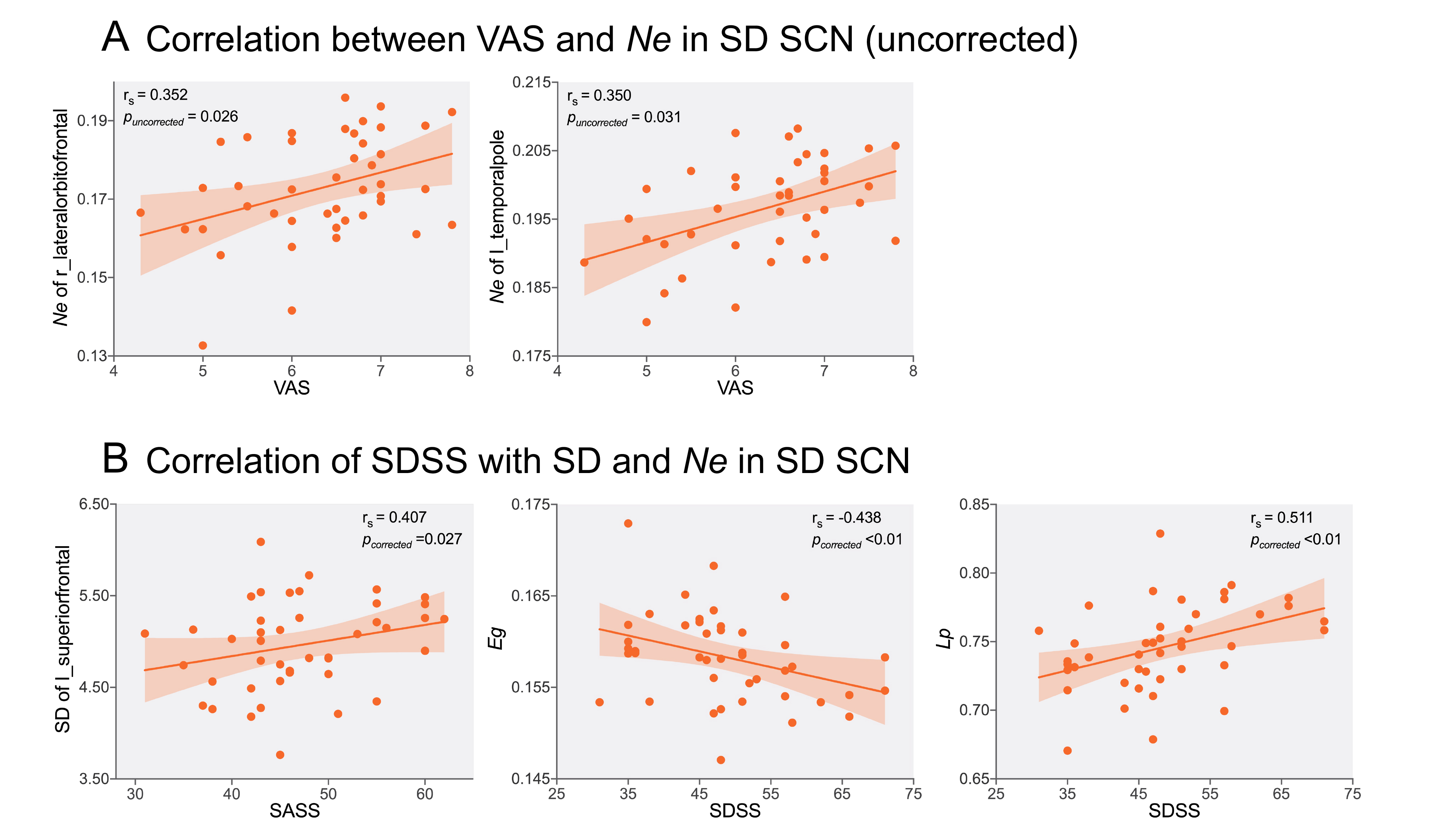
Table 1 showed the demographic and clinical characteristics. In VBM analyses, CTN patients presented increased GMV in the bilateral calcarine and the left cuneus lobe. Vertex-wised SBM analysis revealed reduced SD in the left superior frontal gyrus (SFG) and increased FD in the right lateral occipital cortex, superior parietal lobe, and cuneus lobe. In additional ROI-wised SBM analyses, patients showed reduced SD in the left SFG, caudal middle frontal gyrus, and right cuneus, with reduced GI in the bilateral insula (INS), and reduced FD in the right lateral orbitofrontal (lOFC). However, in the right caudal anterior cingulate cortex (ACC) and entorhinal cortex, patients had increased GI (Figure 1). Moreover, patients showed greater σ and γ in the CT network, also greater γ in the FD network. In contrast, significantly reduced Eg was found in the FD network, as well as higher Lp of the SD network (Figure 2). Patients showed higher nodal properties in the left lOFC of CT network. In the SD network, bilateral temporal poles, right pericalcarine and lOFC showed reduced Ne, which were further positively correlated with pain intensity (without correction). The mean SD of the left SFG was positively correlated with the anxiety score, while Eg and Lp within the SD network showed negative and positive correlations with the depression score, respectively (Figure 3-4). The results of the validation analyses were generally consistent.Discussion
The higher GMV and FD observed in the occipital lobe may reflect adaptive process in patient's brain to maintain the ability of sensory integration8, or it may be a compensation. Notably, the opposite alteration of GMV and SD in cuneus lobe may be partly due to that VBM is a comprehensive representation of gray matter morphology.The INS plays a key role in coding pain intensity9. The ACC is involved in pain related adaptation, whereas the OFC and the entorhinal cortex participate in emotional responses to pain10. In addition, the ACC and INS are both parts of salience network, which is responsible for maintaining a dynamic balance between brain activities and external stimuli11. While the SFG is involved in the default mode network, responsible for attention-driven descending analgesic mechanisms12. Additionally, multiple indicators combination can provide complementary information.
The FD and SD networks exhibited reduced information integration efficiency, which may further map to deep white matter damage. Eg and Lp correlation results may suggest the potential network basis for psychiatric disorders. The reduced Ne within the SD network may further exacerbate the formation of painful memories13. Correlation of Ne may indicate that with disease progression, the network will reorganize in multidimension to accommodate the processing of pain information.
Acknowledgements
This work was supported by the Natural Science Foundation of China (No. 81960309), Gansu Province Clinical Research Center for Functional and Molecular Imaging (No. 21JR7RA438) and Gansu Provincial Science and Technology Program Projects (Key Research and Development Program) (No. 23YFFA0041).References
1. Henssen D, Dijk J, Knepfle R et al (2019) Alterations in grey matter density and functional connectivity in trigeminal neuropathic pain and trigeminal neuralgia: A systematic review and meta-analysis. Neuroimage Clin 24:102039. doi:10.1016/j.nicl.2019.102039
2. Zhang Q, Zhang W, Zhang P et al (2022) Altered dynamic functional connectivity in rectal cancer patients with and without chemotherapy: a resting-state fMRI study. Int J Neurosci:1-11. doi:10.1080/00207454.2022.2130295
3. Zhang P, Wan X, Ai K et al (2022) Rich-club reorganization and related network disruptions are associated with the symptoms and severity in classic trigeminal neuralgia patients. Neuroimage Clin 36:103160. doi:10.1016/j.nicl.2022.103160
4. Desikan R S, Ségonne F, Fischl B et al (2006) An automated labeling system for subdividing the human cerebral cortex on MRI scans into gyral based regions of interest. Neuroimage 31:968-980. doi:10.1016/j.neuroimage.2006.01.021
5. Lai H, Kong X, Zhao Y et al (2022) Patterns of a structural covariance network associated with dispositional optimism during late adolescence. Neuroimage 251:119009. doi:10.1016/j.neuroimage.2022.119009
6. Li Y, Wang N, Wang H et al (2021) Surface-based single-subject morphological brain networks: Effects of morphological index, brain parcellation and similarity measure, sample size-varying stability and test-retest reliability. Neuroimage 235:118018. doi:10.1016/j.neuroimage.2021.118018
7. Destrieux C, Fischl B, Dale A et al (2010) Automatic parcellation of human cortical gyri and sulci using standard anatomical nomenclature. Neuroimage 53:1-15. doi:10.1016/j.neuroimage.2010.06.010
8. Parise M, Kubo T T, Doring T M et al (2014) Cuneus and fusiform cortices thickness is reduced in trigeminal neuralgia. J Headache Pain 15:17. doi:10.1186/1129-2377-15-17
9. Schreckenberger M, Siessmeier T, Viertmann A et al (2005) The unpleasantness of tonic pain is encoded by the insular cortex. Neurology 64:1175-1183. doi:10.1212/01.WNL.0000156353.17305.52
10. Gil-Gouveia R, Martins I P (2019) Cognition and Cognitive Impairment in Migraine. Curr Pain Headache Rep 23:84. doi:10.1007/s11916-019-0824-7
11. Isenburg K, Mawla I, Loggia M L et al (2021) Increased salience network connectivity following manual therapy is associated with reduced pain in chronic low back pain patients. The Journal of Pain 22:545-555. doi:10.1016/j.jpain.2020.11.007
12. Kucyi A, Davis K D (2015) The dynamic pain connectome. Trends Neurosci 38:86-95. doi:10.1016/j.tins.2014.11.006
13. Bauch E M, Rausch V H, Bunzeck N (2014) Pain anticipation recruits the mesolimbic system and differentially modulates subsequent recognition memory. Hum Brain Mapp 35:4594-4606. doi:10.1002/hbm.22497
Figures