2919
MRS Investigations of the Metabolomic Pathophysiology of Acute Respiratory Distress Syndrome (ARDS)1Radiology and Pathology, Massachusetts General Hospital, Harvard Medical School, Boston, MA, United States, 2Environmental Health, Harvard T.H. Chan School of Public Health, Boston, MA, United States
Synopsis
Keywords: Lung, Spectroscopy, Metabolomic imaging, acute respiratory distress syndrome, nuclear magnetic resonance, metabolomics, metabolites
Motivation: ARDS currently has unacceptable high mortality and long-term complications, which underscores the urgency of improving our understanding and management of ARDS.
Goal(s): To establish pathology-guided serum metabolomic profiles for ARDS patients by comparing them with profiles from patients without ARDS.
Approach: We measured ARDS serum using high-resolution magic angle spinning (HRMAS) magnetic resonance spectroscopy (MRS) to establish serum metabolomic profiles for ICU-patients with and without ARDS following various clinical outcomes.
Results: Serum specimens measured by HRMAS MRS enables the predictions of ARDS patient outcome in ICU.
Impact: Our initial results highlight the feasibility of HRMAS MRS in investigating ARDS metabolomic mechanisms, which may lay the basis for future diagnostic and therapeutic research, offering promising prospects for innovation in ARDS management.
Introduction
Acute Respiratory Distress Syndrome (ARDS) is a life-threatening clinical syndrome characterized by several clinical symptoms like rapid onset of respiratory symptoms, chest x-ray abnormalities, interstitial edema and severe oxygen deficiency, often requiring mechanical ventilation at an Intensive Care Unit (ICU).1,2ARDS can result from various causes, such as pneumonia, sepsis, or trauma, leading to uncontrolled systemic inflammation and general epithelial damage. Unacceptable high mortality and long-term complications underscore the urgency of improving our understanding and management of ARDS.3 However, to the best of our knowledge, only few studies have investigated the systemic metabolic changes in ARDS patients using nuclear magnetic resonance (NMR) spectroscopy.
We aim to measure ARDS serum using high-resolution magic angle spinning (HRMAS) MRS to establish serum metabolomic profiles for ICU-patients with and without ARDS following various clinical outcomes. Discovery and understanding of metabolomic changes in ARDS may facilitate the early diagnosis and contribute to the development of targeted interventions that exploit metabolomics mechanisms and thus enhance therapeutic outcomes.
Methods
The current institutional review boards–approved study included 174 serum samples from Intensive care unit (ICU) admitted patients with ARDS (n=74) and without ARDS (n=100) diagnosis obtained from the Molecular Epidemiology of ARDS Study (MEARDS) repository. All serum samples were stored at -80°C until analysis, defrosted for one hour in a water bath at 4°C and vigorously vortexed to ensure that all serum components were homogenous in solution.We conducted HRMAS proton MRS measurements on 20 microliters of serum specimens,
at 4°C with a spin rate of 5000 Hz, using a Bruker Avance 600 MHz spectrometer with 4mm rotors. Spectra were analyzed with Bruker Topspin 4.3.0, resulting in 37 spectral regions of interest (ROIs). Principal component analysis (PCA) was performed using JMP Pro (SAS). A p-value of ≤ 0.05 was considered statistically significant.
Results
Study PopulationAll patients were treated at an ICU with 74 of them including an ARDS diagnosis. Their 28-days-survival follow up is stratified in four groups with A (Discharged home with unassisted breathing), B (Death prior to home with unassisted breathing), C (Other/Still in hospital or discharged to another hospital or rehabilitation), D (ICU patients without ARDS diagnosis).
The measured samples included 74 ICU patients with diagnosed ARDS
[F=20, M=54, mean age (range) = 58 (19-88), 24 current, 22 former and 19 never smoker] and 100 ICU patients without ARDS [F=32, M=68, mean age (range) = 59.63 (93-18), 20 current, 26 former and 45 never smoker].
Metabolic regions significant for human ARDS
Figure 1 compares one pair of representative spectra between ARDS patients who survived (A) or died (B) during the 28-days follow up. Examining all 174 spectra, 37 regions of interest (ROI) were determined. An example of a significant ROI, from 2.14 - 2.12 ppm, was found to be capable of patient differentiation between groups (A) and (B).
Considering potential metabolomic differences between groups (A) and (B), we used them as a training cohort for PCA, and then applied PCA formulars and parameters thus obtained onto groups (C) and (D), as the testing cohort, with results shown in Figure 2. Figure 2 demonstrates a significant Analysis of Variance (ANOVA) result over all groups with p=0.0010 and a significant difference among groups. Of great importance, we observed that the metabolomic profiles of the testing cohort, groups (C) and (D), lie between that groups (A) and (B), as expected.
We further examined our metabolomic profiles in relationship with the APACHE III score, which is comprised of several symptoms, parameters and information on the medical history of intensive care patients, classifies them according to severity, and allows a prognosis of the individual probability of outcome in the further course.4 Figure 3 presents six such examples for ROIs that display significant linear relationships between the APACHE III score and the ROI intensities.
Discussion & Conclusion
The current study of human serum metabolomics for ICU patients with and without ARDS aims to evaluate the underlying biological mechanisms. Of note, our study focuses on discovering differences in ROIs rather than naming metabolic alterations characteristic for ARDS since a single metabolite may appear in various spectral regions. Our findings, whilst preliminary, seem promising in the discovery of metabolomics profiles which are based on ARDS. Based on our observed significant ROIs, either from univariate or multivariate statistical analyses, as shown above, investigations into their represented metabolites, as well as their involved and inferenced metabolic pathways, is currently underway in our laboratory, and will be presented at the meeting.Acknowledgements
NIH Grants: R01 AG070257 and R01 CA273010. MGH Martinos Center for Biomedical Imaging.References
- RANIERI, V. I. T. O., et al. Acute respiratory distress syndrome: the Berlin Definition. Jama, 2012, 307. Jg., Nr. 23, S. 2526-2533
- HUPPERT, Laura A.; MATTHAY, Michael A.; WARE, Lorraine B. Pathogenesis of acute respiratory distress syndrome. In: Seminars in respiratory and critical care medicine. Thieme Medical Publishers, 2019. S. 031-039.
- BELLANI, Giacomo, et al. Epidemiology, patterns of care, and mortality for patients with acute respiratory distress syndrome in intensive care units in 50 countries. Jama, 2016, 315. Jg., Nr. 8, S. 788-800.
- KNAUS, William A., et al. The APACHE III prognostic system: risk prediction of hospital mortality for critically III hospitalized adults. Chest, 1991, 100. Jg., Nr. 6, S. 1619-1636.
Figures
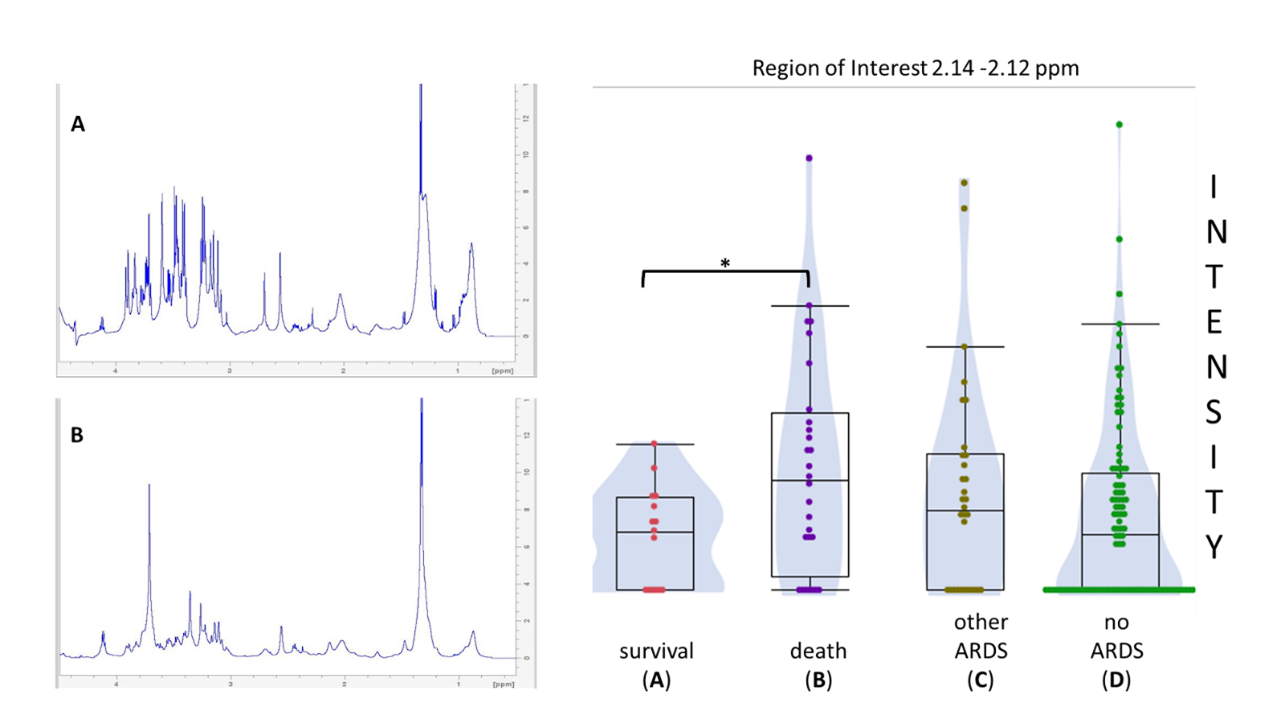
Figure 1. Spectra and ROI differentiation of patient groups. Comparison between representative spectra from ARDS patients who survived (A) or died (B) under 28-days follow up. Group plot for a specific ROI with significant
(p-value* <0.05) difference between (A) and (B).
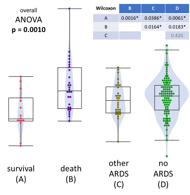
Figure 2. Differentiation of significance between respective patient groups. The table shows the Wilcoxon significant p-values for the compared groups.
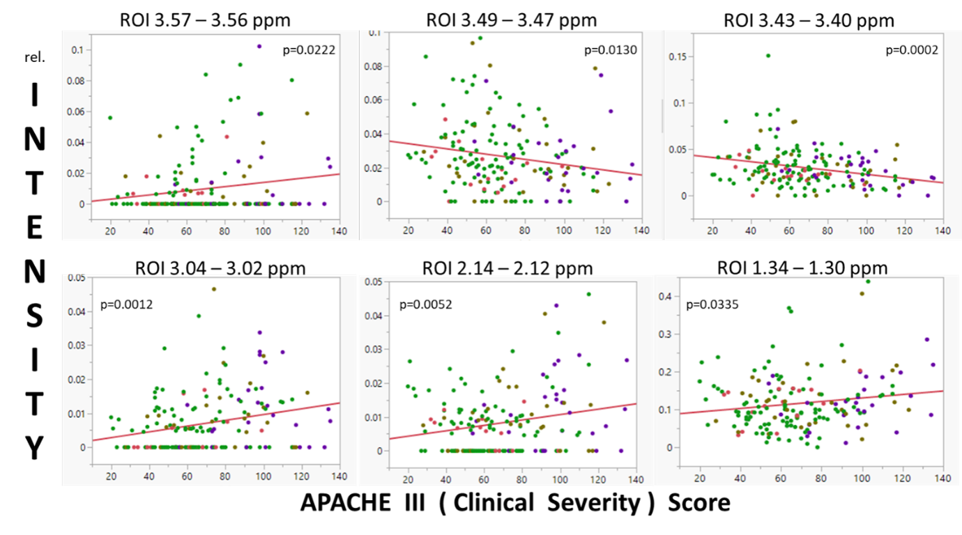
Figure 3. Linear regression of APACHE III scores. Relative intensity plotted against APACHE III (Clinical Severity) Score over representative ROIs with linear regression p-values. (APACHE III, a score including different symptoms providing risk stratification of critical illnesses, such as ARDS, is used in ICU.)