2911
Investigating SMS in dynamic MR for Free-Breathing Functional Lung Imaging1Computer Assisted Clinical Medicine, Medical Faculty Mannheim, Heidelberg University, Mannheim, Germany, 2Mannheim Institute for Intelligent Systems in Medicine, Medical Faculty Mannheim, Heidelberg University, Mannheim, Germany, 3MR Research Collaborations, Siemens Healthcare Limited, Melbourne, Australia, 4Department of Radiology, The University of Melbourne, Melbourne, Australia, 5Cardiovascular Predevelopment, Siemens Healthcare GmbH, Erlangen, Germany, 6EMEA Scientific Partnerships, Siemens Healthcare GmbH, Erlangen, Germany
Synopsis
Keywords: Lung, Lung, sms, functional, pulmonary
Motivation: Pulmonary functional imaging is critical for diagnosing lung diseases. However, sequential acquisition of multiple slices hinder the investigation of concurrent breathing dynamics while prolonging the overall the acquisition time.
Goal(s): Our goal is to investigate the use of simultaneous multi-slice (SMS) imaging as an alternative approach for accelerating dynamic acquisitions for functional lung imaging.
Approach: We obtained dynamic images using bSSFP and GRE acquisitions at 1.5T, from two healthy volunteers. Afterwards, registered images were analyzed using dynamic mode decomposition to generate pulmonary ventilation and perfusion maps.
Results: Functional maps were obtained using both pulse sequences with SMS in both sagittal and coronal views.
Impact: Dynamic lung imaging often requires multiple slices for volumetric coverage, which can be time-consuming. Simultaneous multi-slice (SMS) technique enables the acquisition of multiple slices at the same time, thus enabling the observation of concurrent breathing dynamics in an efficient manner.
Introduction
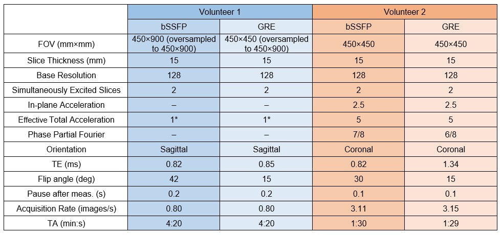
Pulmonary imaging plays a critical role for the diagnosis and follow-up of lung diseases, with non-contrast-enhanced MRI techniques offering safe and effective means to measure lung functions1. By exploiting the periodic signal changes related to respiration and cardiac pulsation, these methods can identify ventilation and perfusion related information2. While more advanced acquisition methods have been previously suggested for functional lung imaging3,4, current methods rely on parallel imaging-accelerated single-slice acquisitions and require sequential measurements for volumetric coverage.Here, we investigate the use of simultaneous multi-slice (SMS) imaging5,6 as an alternative approach for functional lung imaging. We present in vivo results from healthy volunteers obtained at 1.5T field strength using bSSFP and GRE pulse sequences while considering coronal and sagittal views.
Methods
To demonstrate the SMS technique in pulmonary functional imaging, measurements were performed with a research application sequence on a 1.5T scanner (Magnetom Aera, Siemens Healthineers, Germany). In vivo bSSFP and GRE acquisitions were obtained from two volunteers in supine position during free-breathing. Details of the pulse sequences can be found in Table 1. SMS imaging was implemented using RF phase-cycling based controlled aliasing in parallel imaging results in higher acceleration (CAIPIRINHA) encoding5,6. The bSSFP sequences included Gradient-Controlled Local Larmor Adjustment (GC-LOLA)7. Measurements in Volunteer 1 were conducted with an effective total acceleration of 1 to study the effects of the utilized SMS framework. To that end the phase FOV was oversampled by a factor of 2 to mimic a phase-offset multiplanar type acquisition8 with intrinsic separation of the simultaneously excited slices.Before generating the functional maps, the initial six measurements in all acquisitions were excluded due to transient behaviors. Afterwards, the dynamic acquisitions were non-rigidly registered to a reference image9, and fractional ventilation and normalized perfusion maps were obtained using dynamic mode decomposition (DMD)10. Here, the image registration and DMD analyses were carried out on a ROI comprising lung parenchyma using 200 images in all datasets.
Results
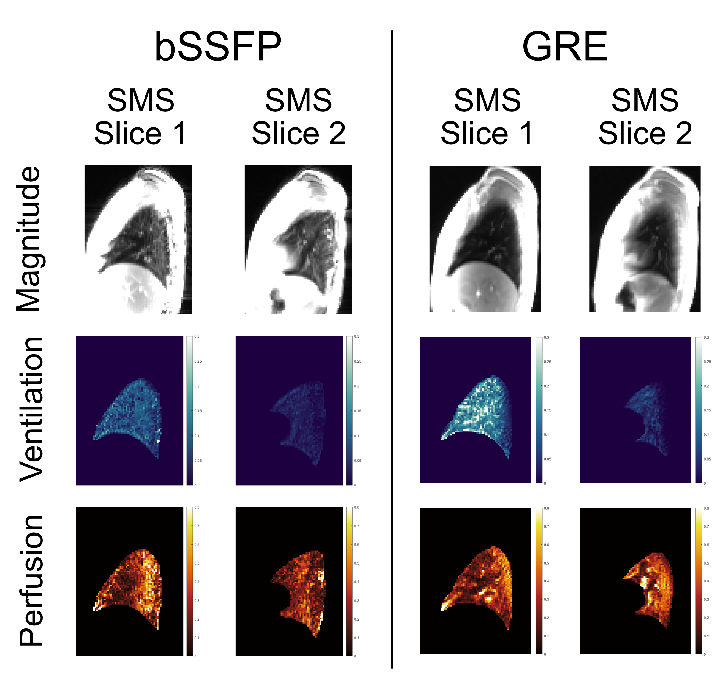
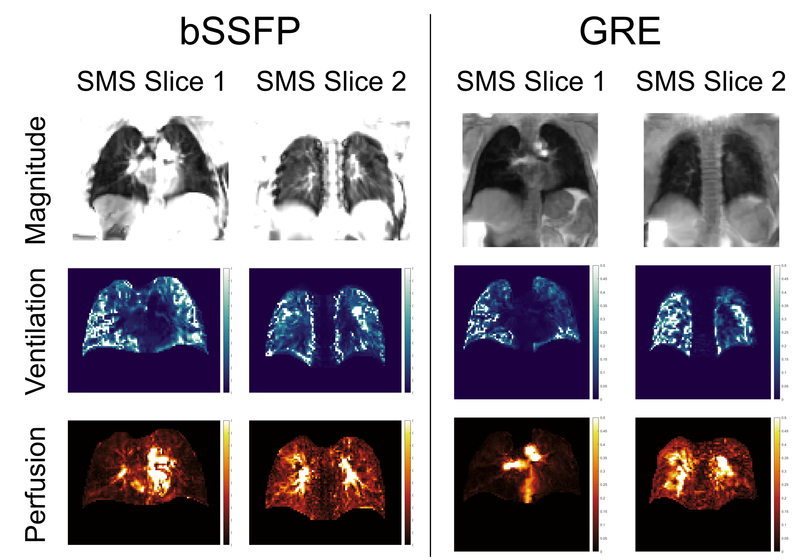
Figure 1 displays magnitude images, fractional ventilation, and normalized perfusion maps from a healthy volunteer, acquired in sagittal view using both bSSFP and GRE sequences with SMS. Both sequences exhibit similar performance, but GRE acquisitions demonstrate greater robustness against imaging artifacts.Similarly, Figure 2 presents magnitude images, fractional ventilation, and normalized perfusion maps from a different volunteer in coronal view, where SMS is utilized together with in-plane acceleration to improve acquisition rate. Here, we observe that bSSFP can better capture the effects stemming from cardiac pulsation, and provides improved visualization of smaller vascular structures, as evidenced by the perfusion maps.
Discussion & Conclusion
By acquiring multiple slices at the same time, SMS enables the observation of concurrent breathing dynamics. Consequently, it can be useful for investigating true breathing dynamics. In addition, when integrated with conventional acceleration methods, SMS has the potential to mitigate the signal-to-noise ratio (SNR) penalty resulting from extensive undersampling while preserving the overall scan time. However, further research is needed to explore the trade-offs between SNR loss due to undersampling and SMS acceleration, and to optimize the acquisition process.Regarding the measurements conducted with Volunteer 1, the acquisition rates were insufficient to accurately capture the cardiac pulsation. However, the aliased cardiac peaks differed from the respiratory peak, allowing us to distinguish between the two. In terms of acquisition orientation, the sagittal view is more effective at preserving inflow effects compared to the coronal view, resulting in improved perfusion map quality. Concerning the image quality derived from pulse sequences, our observations indicate that the bSSFP sequence did not yield better images despite its theoretical SNR advantage compared to GRE4. This observation is attributed to the utilization of GC-LOLA7 with a relatively high slice thickness to distance ratio in the presence of substantial off-resonance effects in the lungs11,12, which could potentially diminish the attainable SNR in bSSFP acquisitions.
In conclusion, we have showcased the application of SMS-based acquisitions for functional lung imaging at 1.5T using bSSFP and GRE pulse sequences. Although further investigations are warranted, our initial results highlight SMS as a promising alternative for dynamic lung acquisitions while maintaining scan efficiency.
Acknowledgements
This work was supported by Deutsche Forschungsgemeinschaft (grant number: DFG 397806429).References
1. Zapke M, Topf H-G, Zenker M, et al. Magnetic resonance lung function—a breakthrough for lung imaging and functional assessment? A phantom study and clinical trial. Respir Res. 2006;7:106.
2. Bauman G, Puderbach M, Deimling M, et al. Non-contrast-enhanced perfusion and ventilation assessment of the human lung by means of Fourier decomposition in proton MRI. Magn Reson Med. 2009;62:656–64.
3. Fischer A, Weick S, Ritter CO, et al. SElf-gated non-contrast- enhanced FUnctional lung imaging (SENCEFUL) using a quasi-random fast low-angle shot (FLASH) sequence and proton MRI. NMR Biomed. 2014;27:907–17.
4. Bauman G, Pusterla O, Bieri O. Functional lung imaging with transient spoiled gradient echo. Magn Reson Med. 2019;81:1915-1923. doi:10.1002/mrm.27535
5. Stäb D, Ritter CO, Breuer FA, Weng AM, Hahn D, Köstler H. CAIPIRINHA accelerated SSFP imaging. Magn. Reson. Med. 2011;65:157–164. doi: 10.1002/mrm.22600
6. Breuer FA, Blaimer M, Heidemann RM, Mueller MF, Griswold MA, Jakob PM. Controlled aliasing in parallel imaging results in higher acceleration (CAIPIRINHA) for multi-slice imaging. Magn Reson Med. 2005; 53: 684-691.
7. Stäb D, Speier P. Gradient-controlled local Larmor adjustment (GC-LOLA) for simultaneous multislice bSSFP imaging with improved banding behavior. Magn. Reson. Med. 2019;81:129–139 doi: 10.1002/mrm.27356.
8. Glover, G.H. (1991), Phase-offset multiplanar (POMP) volume imaging: A new technique. J. Magn. Reson. Imaging, 1: 457-461. https://doi.org/10.1002/jmri.1880010410
9. Chefd’Hotel C, Hermosillo G, Faugeras O (2001) A variational approach to multi-modal image matching. In: Proceedings IEEE Workshop on Variational and Level Set Methods in Computer Vision. IEEE Computer Soc, pp 21–28
10. Ilicak E, Ozdemir S, Zapp J, et al (2023) Dynamic mode decomposition of dynamic MRI for assessment of pulmonary ventilation and perfusion. Magn Reson Med 90:761–769. https://doi.org/10.1002/mrm.29656
11. Wild JM, Marshall H, Bock M, et al. MRI of the lung (1/3): methods. Insights Imaging. 2012;3:345–53.
12. Ilicak E, Ozdemir S, Schad LR, et al. Phase-cycled balanced SSFP imaging for non-contrast-enhanced functional lung imaging. Magn Reson Med. 2022;88:1764-1774. doi:10.1002/mrm.29302
Figures