2887
IVIM Combined with DCE-MRI Characterize Architectural Distortion Detected on Mammography: A Pilot Study1The First Affiliated Hospital of Dalian Medical University, Dalian, China, 2Dalian Medical University, Dalian, China, 3GE Healthcare, MR Research China, Beijing, Dalian, China
Synopsis
Keywords: fMRI Analysis, Breast
Motivation: Breast adenosis (AD) detection is crucial for early breast cancer diagnosis. Traditional mammography has limitations.
Goal(s): To enhance AD diagnosis using MRI and evaluate MRI's diagnostic value for AD detected in mammography. Assess the combined potential of dynamic contrast-enhanced MRI (DCE-MRI) and intravoxel incoherent motion (IVIM) sequences.
Approach: Collected data from AD-diagnosed patients who underwent breast MRI. Analyzed DCE-MRI and IVIM parameters, compared benign and malignant AD, and assessed their combined diagnostic efficiency.
Results: MRI exhibited high sensitivity (100%) and NPV (100%) for malignancy exclusion. DCE-MRI and IVIM combined yielded superior results with an AUC of 0.984.
Impact: This study demonstrates that combining DCE-MRI and IVIM significantly enhances the diagnostic accuracy of AD changes. It offers a non-invasive approach to differentiate benign from malignant lesions, reducing unnecessary invasive procedures and improving breast cancer management.
Introduction
At present, AD is recognized as the third most common imaging finding in breast cancer mammography, following only mass or calcification [1]. It is also a common presentation of nonpalpable breast cancer [2]. Detectiing AD early is crucial for improving prognosis. However, AD can closely resemble the normal appearance of overlapping breast tissue, making it challenging to evaluate. Breast MRI has emerged as a valuable supplemental tool for breast screening alongside mammography. Previous study has shown that dynamic contrast-enhanced (DCE-MRI) combined with apparent diffusion coefficient (ADC) is more reliable than mammography in distinguishing breast lesions that appear as primary AD on mammography [3]. Nonetheless, studies specifically focused on AD using MRI remain limited. The intravoxel incoherent motion (IVIM) model, a dual exponential diffusion-weighted imaging (DWI) technique, aims to describe both diffusion and microcirculation effects. Over the past decade, numerous studies have indicated that IVIM parameters are helpful for distinguishing malignant from benign breast lesions. In this study, we aimed to evaluate the diagnostic values of combining IVIM with DCE-MRI in patients with AD changes detected on mammography.Methods
Data were collected from patients who underwent breast MRI after being diagnosed with AD changes on mammography between April 2019 and February 2022. Two independent radiologists reviewed all MRI data, recorded DCE-MRI lesion characteristics based on the Breast Imaging Reporting and Data System (BI-RADS,2013), and measured IVIM parameters. The final outcome was determined using pathology results from biopsy or surgical excision or at least one year of clinical and radiological follow-up. Statistical analysis was performed using SPSS software (SPSS Version 19, SPSS Inc., Chicago, IL, USA). Independent-sample T-test or Mann-Whitney U test were employed to compare parameter difference between benign and malignant AD changes. Receiver operating characteristic (ROC) was used to evaluate the diagnostic performance of individual parameters and their combinations. A significance level of P<0 .05 was considered statistically significant.Results
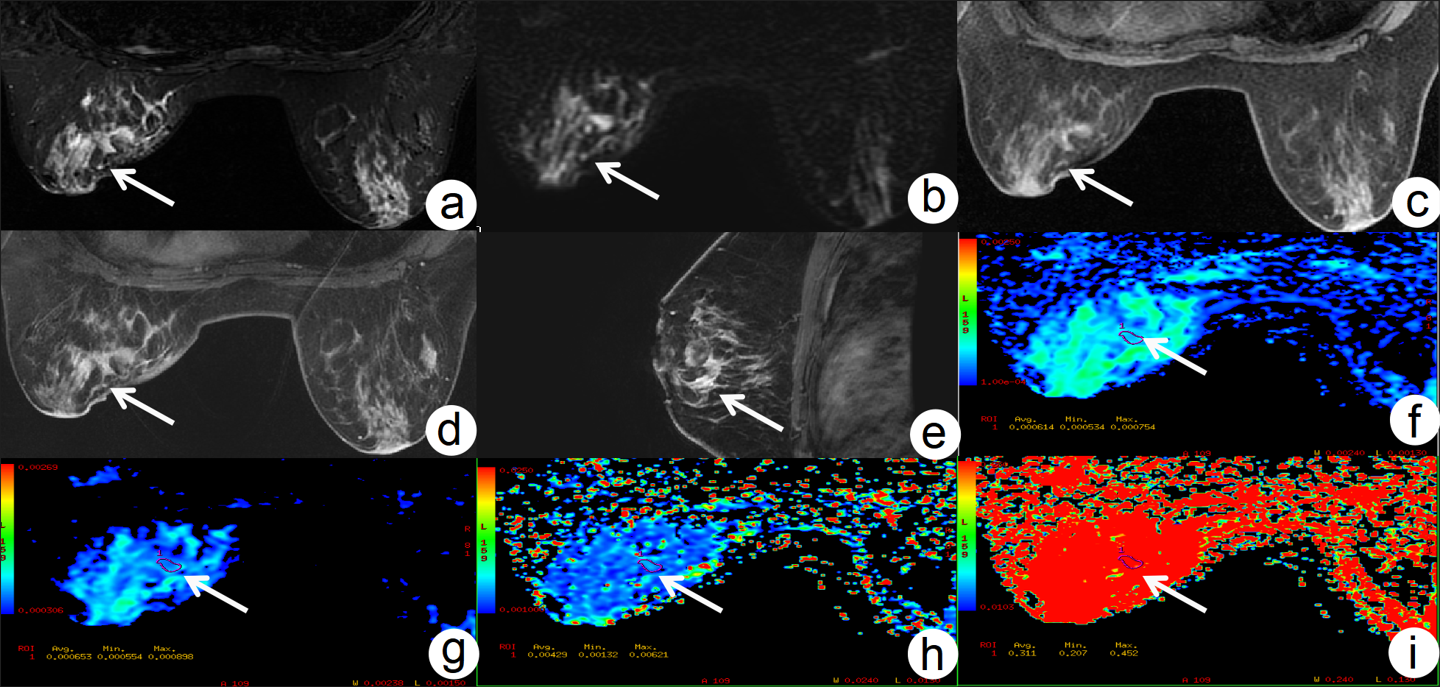
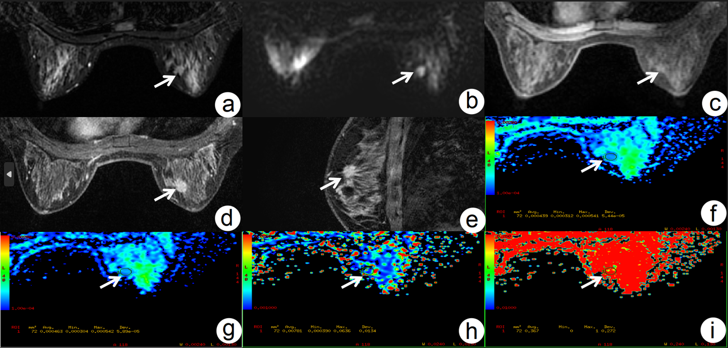
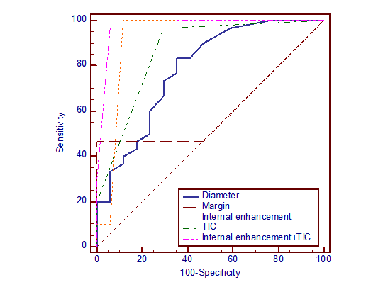
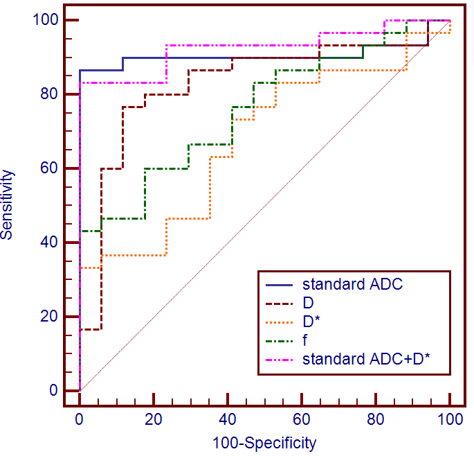
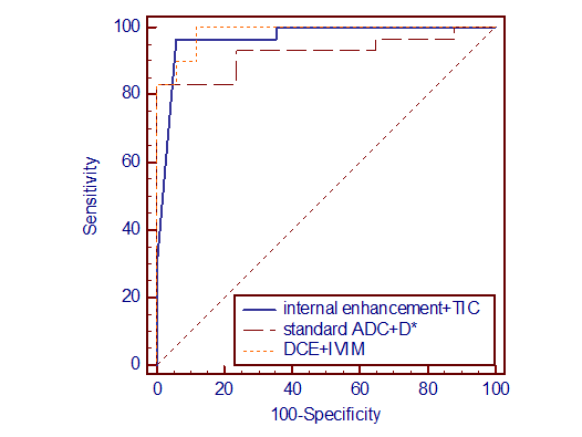
A total of 80 patients were included in this study. In 33 (41.25%) patients, no lesions were detected on MRI, resulting in negative findings. Among the remaining 47 (58.75%) patients with positive MRI findings, 17 (36.17%) had benign lesions, while 30 (63.83%) had malignant lesions(Figure 1-2). MRI demonstrated high sensitivity, specificity, negative predictive value(NPV), and positive predictive value (PPV) for distinguishing benign from malignant AD changes, with values of 100%, 92%, 100%, and 88.2%, respectively, and an overall accuracy of 95%. Regarding DCE-MRI characteristics, parameters such as fibrograndular tissue (FGT), background parenchymal enhancement (BPE), and enhancement mode did not show significant difference between benign and malignant cases. However, parameters such as lesion diameter, shape and margin of mass enhancement, distribution of non-mass enhancement(NME), internal enhancement, and time signal intensity curve(TIC) exhibited significant differences (all P<0.05). Among these, internal enhancement and TIC were identified as independent risk predictors, with a combined area under the curve (AUC) of 0.969(Figure 3). In terms of IVIM parameters, standard ADC, D, D*, and f values demonstrated significant differences between benign and malignant AD groups (all P<0.05), with AUC values of 0.908, 0.831, 0.680, and 0.761, respectively. Standard ADC and D* were identified as independent risk predictors, with a combined AUC of 0.925(Figure 4). Combined DCE-MRI and IVIM provided the highest AUC, reaching 0.984(Figure 5).Discussion and Conclusion
The findings of this preliminary study suggest that MRI can serve as a reliable problem-solving tool in patients with AD changes detected on mammography. MRI exhibited remarkably high sensitivity and NPV, making it a depandable means of excluding AD lesions complicated by malignancy. Regarding the differences for malignant tumors compared with benign lesions in IVIM results, these differences may be attributed to the dense cellularity and active proliferation of malignant tumor cells, leading to reduced extracellular space and hindered water molecules diffusion, resulting in decreased standard ADC and D values [4]. The decreased f value in malignant tumors may be due to the increased heterogeneous proliferation of malignant cells, narrowing of the extracellular space , and alterations in microvessels and capillary structures within the tumor, potentially influenced by the high b values [5] or TE and TR parameters [6]. An increased D* value may be attributed to microvascular perfusion, consistent with previous studies [4]. The combination of DCE-MRI and IVIM significantly improved sensitivity, reaching 100%, with an AUC of 0.984, surpassing the performance of DCE-MRI and IVIM alone. This underscores the advantages of a multi-modal magnetic resonance approach. In conclusion, the combination of IVIM and DCE-MRI exhibits high diagnostic efficacy in characterizing AD changes, providing a reliable non-invasive method to distinguish benign from malignancy lesions, ultimately reducing unnecessary biopsies and surgeries.Acknowledgements
This study was supported by the Natural Science Foundation of Liaoning Province (2020-MS-266), 2022 Teaching Reform of Continuing Education of Liaoning Adult Education Society (LCYJGZXYB22100), University-level teaching reform research general project of Dalian Medical University (DYLX21036), and 2022 General Project of "Peak Climbing Plan" of Dalian city key specialty of medicine(2022DF042).References
[1] R. Shaheen, C.A. Schimmelpenninck, L. Stoddart, H. Raymond, P.J. Slanetz, Spectrum of diseases presenting as architectural distortion on mammography: multimodality radiologic imaging with pathologic correlation, Semin Ultrasound CT MR, 32 (2011) 351-362.[2] Y. Goh, C.W. Chan, P. Pillay, H.S. Lee, H.B. Pan, B.H. Hung, S.T. Quek, C.P. Chou, Architecture distortion score (ADS) in malignancy risk stratification of architecture distortion on contrast-enhanced digital mammography, European radiology, 31 (2021) 2657-2666.[3] L. Si, R. Zhai, X. Liu, K. Yang, L. Wang, T. Jiang, MRI in the differential diagnosis of primary architectural distortion detected by mammography, Diagn Interv Radiol, 22 (2016) 141-150.[4] N. Meng, X.J. Wang, J. Sun, L. Huang, Z. Wang, K.Y. Wang, J. Wang, D.M. Han, M.Y. Wang, Comparative Study of Amide Proton Transfer-Weighted Imaging and Intravoxel Incoherent Motion Imaging in Breast Cancer Diagnosis and Evaluation, J Magn Reson Imaging, 52 (2020) 1175-1186.[5] T. Tamura, S. Usui, S. Murakami, K. Arihiro, Y. Akiyama, K. Naito, M. Akiyama, Biexponential Signal Attenuation Analysis of Diffusion-weighted Imaging of Breast, Magnetic resonance in medical sciences : MRMS : an official journal of Japan Society of Magnetic Resonance in Medicine, 9 (2010) 195-207.[6] S.X. Zhang, Q.J. Jia, Z.P. Zhang, C.H. Liang, W.B. Chen, Q.H. Qiu, H. Li, Intravoxel incoherent motion MRI: emerging applications for nasopharyngeal carcinoma at the primary site, European radiology, 24 (2014) 1998-2004.Figures