2886
Value of multi-parametric diffusion-weighted imaging in pathological grading of clear cell renal cell carcinoma1Tongji Hospital, Tongji Medical College, Huazhong University of Science and Technology, Wuhan, China, 2MR Research Collaboration Team, Siemens Healthineers Ltd., Chengdu, China
Synopsis
Keywords: Diffusion Modeling, Diffusion/other diffusion imaging techniques
Motivation: Accurate preoperative grading of clear cell renal cell carcinoma (ccRCC) is essential for treatment decisions, particularly in patients with comorbidities.
Goal(s): This study aims to investigate the utility of various diffusion-weighted imaging (DWI) models, including mono-exponential, intravoxel incoherent motion (IVIM), diffusion kurtosis imaging (DKI), and continuous time random walk (CTRW), in preoperative ccRCC grading to facilitate personalized treatment strategies.
Approach: We conducted MRI scans on 105 ccRCC patients with multi-b value DWI and employed machine learning for model construction for ccRCC grading.
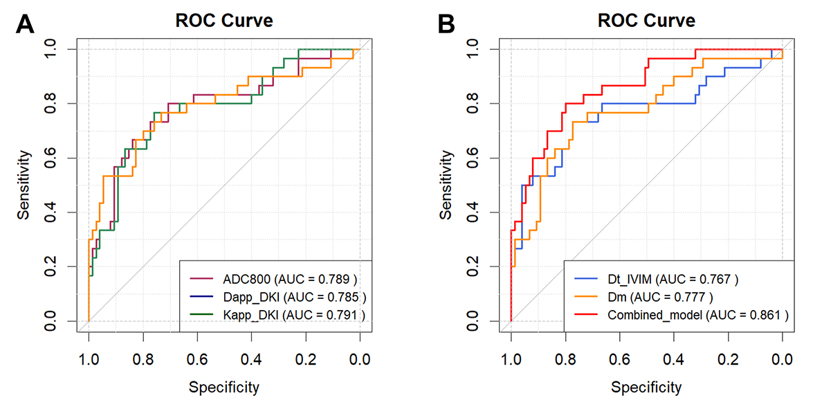
Results: This study demonstrates the protential of multi-parametric DWI models for accurate ccRCC grading, yield an AUC of 0.861.
Impact: This study has the potential to reshape the landscape of preoperative ccRCC grading, promote objectivity in treatment planning.
Introduction
Clear cell renal cell carcinoma (ccRCC) is the most prevalent histological subtype of renal cell carcinoma. This malignant renal tumor presents varying grades, wherein high-grade ccRCC is associated with heightened invasiveness and a less favorable clinical prognosis when compared to its low-grade counterpart. Hence, preoperative pathological grading plays a very important role in the treatment plan and prognosis, especially for patients with multiple comorbidities. In recent years, multiparametric MRI has been increasingly used in tumor diagnosis and tissue assessment. Diffusion-weighted imaging (DWI) technology provides valuable insights into the tumor microenvironment by quantifying the movement of water molecules within tissues, making it an effective tool for evaluating tumor characteristics. While traditional mono-exponential models are commonly employed in diagnosing ccRCC, research has revealed their limitations in fully encapsulating tissue microstructure complexity. This study aims to explore the efficacy of various diffusion models, including the mono-exponential, intravoxel incoherent motion (IVIM), diffusion kurtosis imaging (DKI), and continuous time random walk (CTRW) models, in preoperative pathological grading of ccRCC.Methods
In total of 105 patients with histologically confirmed ccRCC (75 low-grade and 30 high-grade) were recruited in this study. All patients underwent axial MRI scans at 3T (Magnetom Skyra, Siemens Healthcare, Erlangen, Germany) with an 18-element body matrix coil. DWI was obtained with 11 trace-weighted diffusion images, with b-values from 0 to 2000 s/mm2. Specific scan parameters were: TR/TE=7700/71 ms, FOV=288×125 mm, matrix=128×128, slice thickness=5 mm, bandwidth=1666Hz, b-values(averages): 01, 201, 501, 801, 1001, 2001, 5001, 8002, 10002, 15003, 20006. ccRCC lesions were manually delineated for region-of-interested based analysis. Diffusion parameters were generated from the following models: 1) apparent diffusion coefficient ADC from mono-exponential model using b0 and one high b value (800, 1000 or 1500 s/mm2); 2) diffusion coefficient Dt, perfusion fraction Fp and pseudo-diffusion coefficient Dp calculated from IVIM model with a segmented fitting using b-values from 0 to 1000 s/mm2; 3) mean kurtosis Kapp and mean diffusivity Dapp calculated from DKI model using all b values; and 4) CTRW model using S = S0·Eβ(-(b·Dm)α) [1]. Tumor size was measured on T2-weighted images. Pathological diagnosis and grading were conducted by experienced senior pathologists in accordance with the 2016 WHO classification criteria [2] based on postoperative pathology. WHO/ ISUP grades Ⅰ and Ⅱ were assigned to the low-grade group, and WHO/ ISUP grades Ⅲ and Ⅳ to the high-grade group. Using the least absolute shrinkage and selection operator (LASSO) regression for parameters selection and logistic regression for model construction. The area under the curve (AUC) was calculated to evaluate the predictive accuracy of diffusion parameters in ccRCC grading.Results
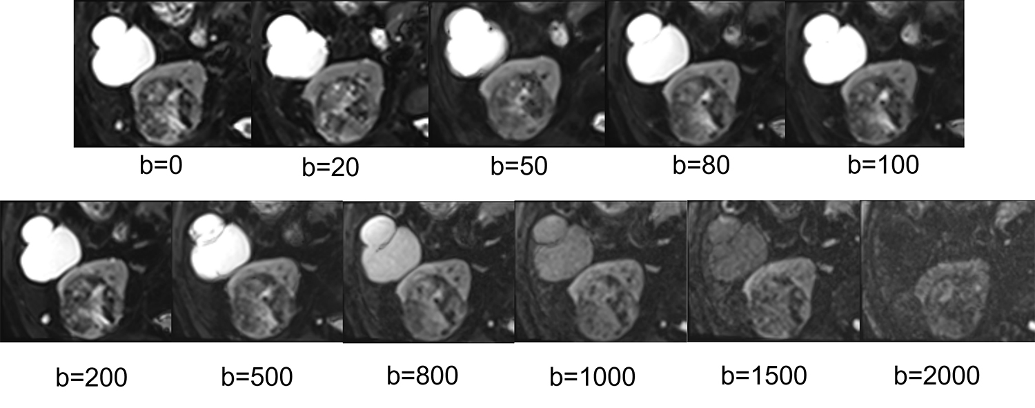
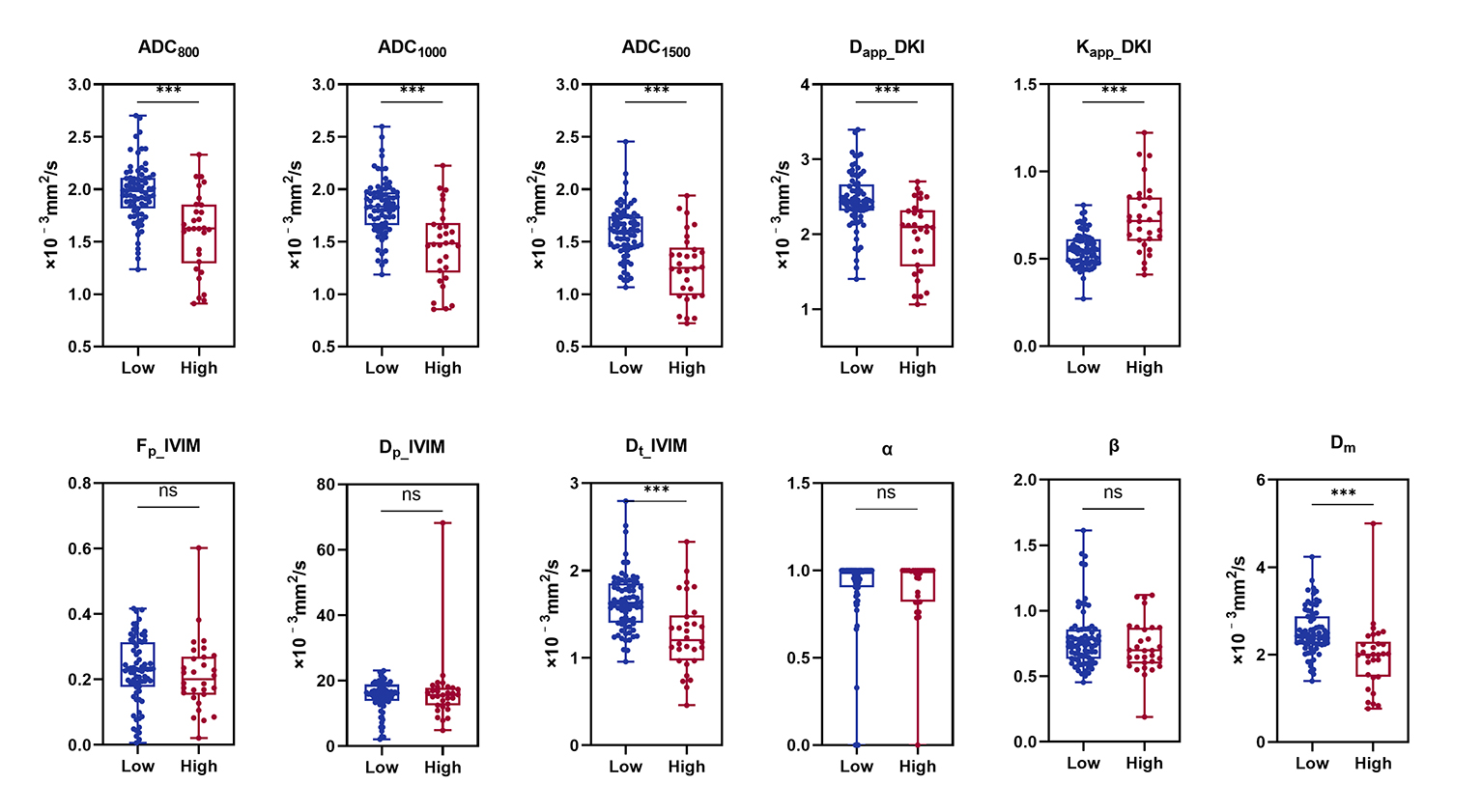
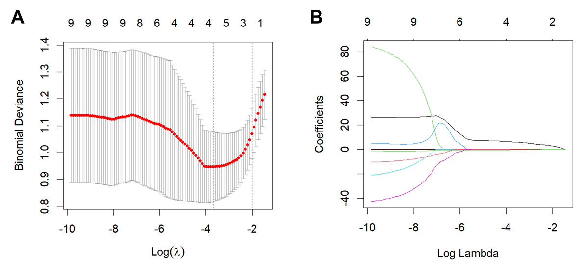
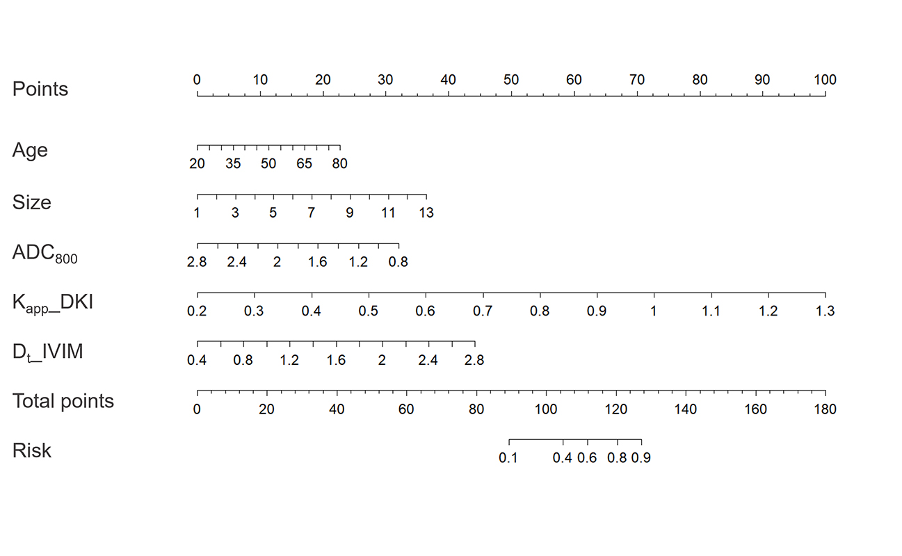
Figure 1 showed a typical multi-b-value DWI images of ccRCC case. Diffusion parameters calculated from different diffusion models for all 105 patients were summarized in figure 2. The low-grade lesion exhibited significantly lower values in ADC800, ADC1000, ADC1500, Dapp_DKI, Dt_IVIM, and Dm. While Kapp_DKI has typically been found to be higher in high-grade lesion. There were no significant differences in Dp_IVIM and Fp_IVIM between high-grade and low-grade ccRCC. Through the least absolute shrinkage and selection operator (LASSO) algorithm using five-fold cross-validation, five features (including age, tumor size, ADC800, Dt_IVIM, and Kapp_DKI) were finally selected. The process of LASSO analysis is shown in Figure 3. Logistic regression was then used to build a risk prediction model with an AUC of 0.861 (Figure 4), and the nomogram was plotted (Figure 5).Discussion
In conclusion, our study demonstrated the feasibility of the multi-parameter DWI model for preoperative prediction of ccRCC grading. By integrating multiple DWI parameters and clinical information such as age and tumor size, a risk prediction model can be established to achieve the best diagnostic efficiency.Acknowledgements
No acknowledgement found.References
1 Yang Q, Reutens DC, Vegh V (2022) Generalisation of continuous time random walk to anomalous diffusion MRI models with an age-related evaluation of human corpus callosum. Neuroimage 250:118903 2 Moch H, Cubilla AL, Humphrey PA, Reuter VE, Ulbright TM (2016) The 2016 WHO Classification of Tumours of the Urinary System and Male Genital Organs-Part A: Renal, Penile, and Testicular Tumours. Eur Urol 70:93-105Figures