2884
Value of intravoxel incoherent motion diffusion-weighted MRI in evaluating endometrial receptivity in women with polycystic ovary syndrome1The First Affiliated Hospital of Nanjing Medical University, 300 Guangzhou Road, Nanjing, China, 2MR Research Collaboration, Siemens Healthineers, Shanghai, China
Synopsis
Keywords: IVIM, Reproductive, polycystic ovary syndrome, vessels, hormonal levels
Motivation: This study evaluated endometrial receptivity in women with polycystic ovary syndrome (PCOS) impacting embryo implantation and pregnancy outcomes.
Goal(s): The primary objectives were to evaluate endometrial receptivity by measuring uterine junctional zone (UJZ) and myometrium parameters and to explore the correlation between these imaging parameters and hormonal levels.
Approach: Using intravoxel incoherent motion (IVIM), the study measured the quantitative parameters of the UJZ and myometrium in women with PCOS.
Results: Patients with PCOS had lower UJZ and myometrium f values than controls, indicating reduced perfusion affecting embryo implantation. A positive correlation with free thyroxine levels was observed.
Impact: Intravoxel incoherent motion imaging is vital in assessing endometrial receptivity. Patients with polycystic ovary syndrome had lower f values of the uterine junctional zone and myometrium than controls, suggesting less blood perfusion, which might be associated with poor pregnancy outcomes.
Introduction
Polycystic ovary syndrome (PCOS) is a common cause of female infertility, accounting for approximately 20%-40% of cases. Its main pathologic features are endocrine and metabolic abnormalities. Women with PCOS have poor endometrial receptivity, which increases the risk of adverse pregnancy outcomes1. Previous studies mainly assessed endometrial receptivity through ultrasound measurements of spiral and uterine artery blood flow parameters, but the accuracy was not high2. Intravoxel incoherent motion (IVIM) imaging is a quantitative imaging technique derived from diffusion-weighted imaging (DWI), which can noninvasively examine the diffusion of water molecules and perfusion of blood vessels in tissues3,4. This study aimed to evaluate the endometrial receptivity in patients with PCOS using IVIM quantitative parameters and analyze the correlation between imaging parameters and hormonal levels.Methods
This prospective study included 44 patients (aged 22-34 years) diagnosed with infertility at Jiangsu People's Hospital from April 2022 to July 2023, including 28 patients with PCOS and 16 patients with tubal factor infertility or unexplained infertility. All volunteers were in the luteal phase and underwent pelvic conventional sequences as well as IVIM and DWI sequences using a 3T MRI system scanner (MAGNETOM Skyra, Siemens Healthineers AG, Erlangen, Germany). All the participants also underwent serological examinations. The width of the junctional zone was measured on sagittal T2-weighted imaging (T2WI) images collected by Half-fourier Acquisition Single-shot Turbo spin Echo imaging (HASTE) technique. The IVIM applied 7 b values, including 0, 50, 100, 150, 200, 500, and 1000 s/mm2. Other parameters were as follows: repetition time = 5600ms; echo time = 71 ms; field of view = 250 × 250 mm2; slice thickness = 5.0 mm; voxel size = 1 × 1 × 5 mm3. The apparent diffusion coefficient(ADC), perfusion fraction (f), pure molecular diffusion coefficient (D), and perfusion-related diffusion coefficient (D) values were calculated using the FireVoxel software (https://firevoxel.org). Regions of interest (ROI) were determined according to axial T2WI images adjusting the measurement range based on the anatomical structure (Figure 1). Further, 2- to 3-mm-wide ROIs were delicately drawn on the uterine junctional zone (UJZ) and myometrium. The ADC value was calculated using the mono exponential model formula, S(b)/S(0) = exp(b × ADC). The D, D, and f were calculated using the biexponential model formula, S(b)/S(0) = f × exp(–b × D) + (l – f) × exp(–b × D).The f, D, D, and ADC values in the myometrium and UJZ of patients with PCOS and healthy controls were compared using Student t test and Mann–Whitney U test. The correlation between imaging parameters and clinical indexes of patients with PCOS was analyzed using Pearson and Spearman correlations. A P value <0.05 indicated a statistically significant difference.
Results
The f value of UJZ was substantially lower in patients with PCOS than in healthy controls [fpcos (16.062± 5.023)%, f control (26.802± 10.707)%, P <.001] (Figure 2). Also, the f value of the myometrium was lower in patients with PCOS than in healthy controls [fpcos (21.830 ± 6.530)%, fcontrol (29.051 ± 12.272)%, P =.045] (Figure 2). Remarkable differences were noted in anti-Mueller hormone levels between patients with PCOS and healthy controls [AMHpcos (10.834 ± 5.003) ng/mL, AMHcontrol (5.979 ± 2.412) ng/mL, P =.001]. In patients with PCOS, free thyroxine (FT4) level was positively correlated with the f value of UJZ and myometrium [r(UJZ ) = 0.405, P =.033; r(myometrium) = 0.432, P =.022] (Figure 3 and Table 1).Discussion
Studies have reported that reduced endometrial and subendometrial blood perfusion is not conducive to embryo implantation 2. In the IVIM parameter, the D value reflects the diffusion movement of water molecules, and the f value reflects the volume ratio of blood perfusion to the total diffusion effect3. Our study demonstrated that the f value of the UJZ and myometrium in patients with PCOS decreased, reflecting decreased subendometrial blood perfusion, possibly leading to endometrial receptivity damage in patients with PCOS. At the same time, the FT4 level was positively correlated with the junctional-zone f value. Previous studies demonstrated that thyroid hormone abnormalities affected fertilization and embryo implantation5,6. However, the pathophysiological mechanism underlying thyroid hormone abnormalities affecting the myometrium and junctional zone still needs further exploration.Conclusions
IVIM imaging can be used as an essential indicator of endometrial receptivity. The decrease in the f value of the junctional zone and myometrium in patients with PCOS suggests a decrease in blood flow perfusion in the subendometrial myometrium of PCOS, affecting the receptivity of the endometrium.Acknowledgements
No acknowledgement found.References
1. Lensen, S., Martins, W., Nastri, C., Sadler, L. & Farquhar, C. Pipelle for Pregnancy (PIP): study protocols for three randomised controlled trials. Trials17, 216 (2016).
2. Tong, R. et al. Analysis of the guidance value of 3D ultrasound in evaluating endometrial receptivity for frozen-thawed embryo transfer in patients with repeated implantation failure. Ann Transl Med 8, 944–944 (2020).
3. Le Bihan, D. What can we see with IVIM MRI? NeuroImage 187, 56–67 (2019).
4. Hu, Q. et al. Noninvasive assessment of endometrial fibrosis in patients with intravoxel incoherent motion MR imaging. Sci Rep 11, 12887 (2021).
5. Palomba, S., Colombo, C., Busnelli, A., Caserta, D. & Vitale, G. Polycystic ovary syndrome and thyroid disorder: a comprehensive narrative review of the literature. Front. Endocrinol. 14, 1251866 (2023).
6. Wu, Z. et al. Hashimoto’s thyroiditis impairs embryo implantation by compromising endometrial morphology and receptivity markers in euthyroid mice. Reprod Biol Endocrinol 17, 94 (2019).
Figures
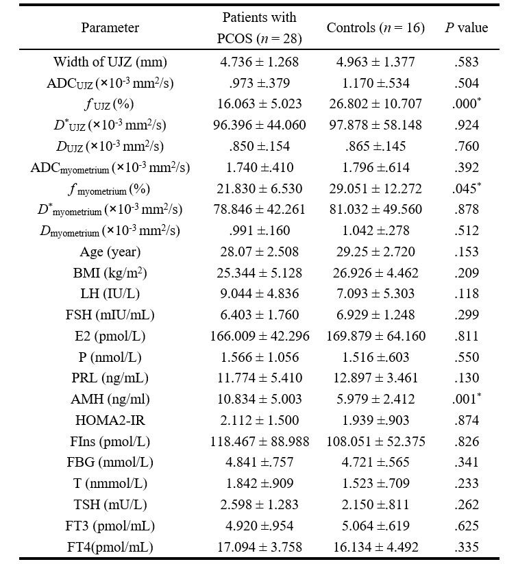
Table 1.Comparison of imaging parameters and hormonal levels between polycystic ovary patients and healthy controls
AMH, Anti-Müllerian hormone; BMI, body mass index; E2, estradiol; FBG, fasting blood glucose; FIns, fasting insulin; FSH, follicle-stimulating hormone; FT3, triiodothyronine; FT4, free thyroxine; HOMA2-IR, homeostatic model assessment index for insulin resistance [calculated by HOMA2 Calculator (v2.2.3)]; LH, luteinizing hormone; P, progesterone; PRL, prolactin; T, testosterone; TSH, thyroid-stimulating hormone.
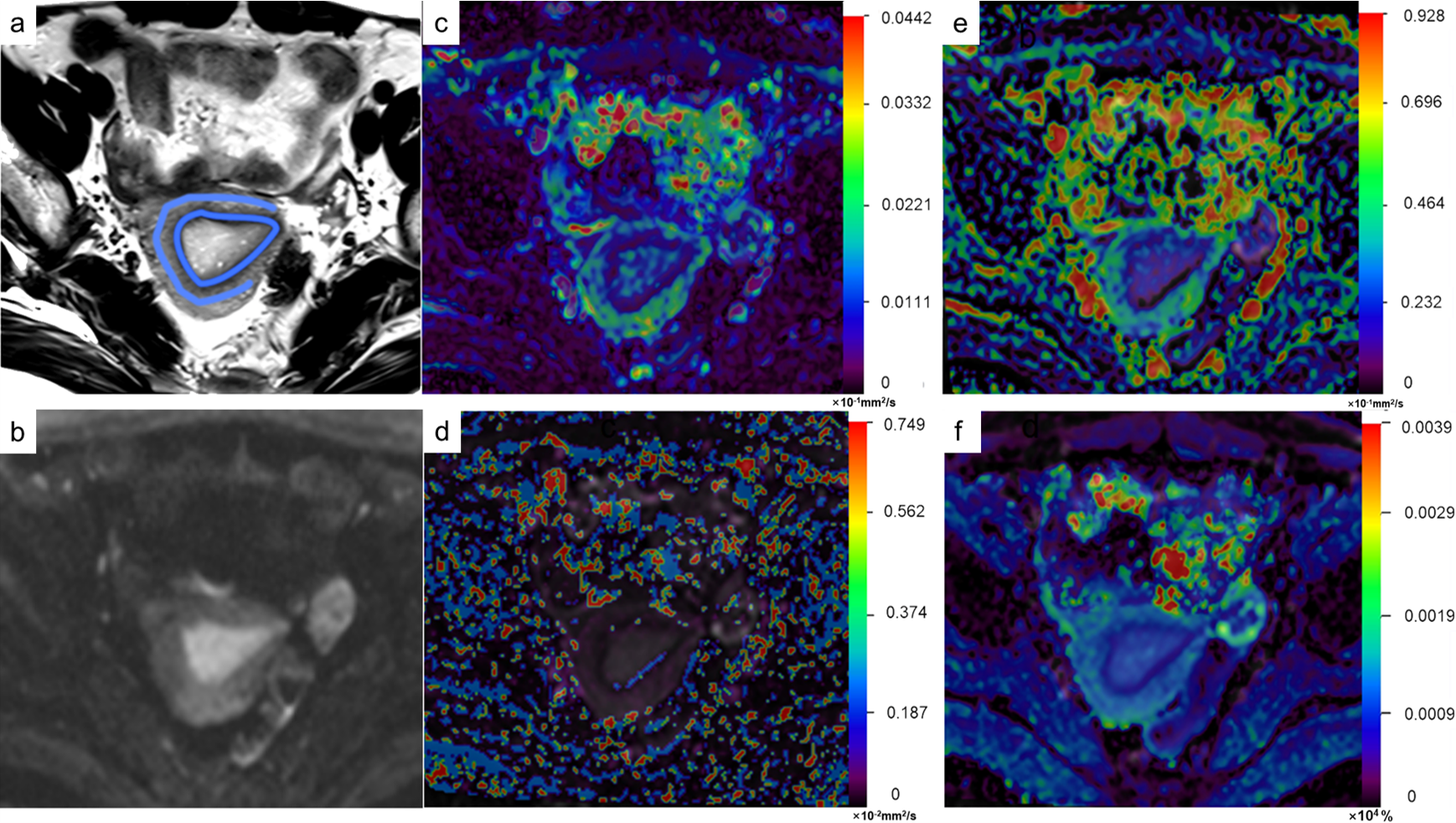
Figure 1. A patient with PCOS (age 29 years, body mass index 24.6). (a) Axial T2 image and (b) axial view of a diffusion-weighted image with b: 1000 s/mm2. (c) ADC pseudocolor plot and (d–f) pseudocolor maps of D*, D, and f generated for the IVIM biexponential model. ADC, Apparent diffusion coefficient; PCOS, polycystic ovary syndrome; IVIM, intravoxel incoherent motion.
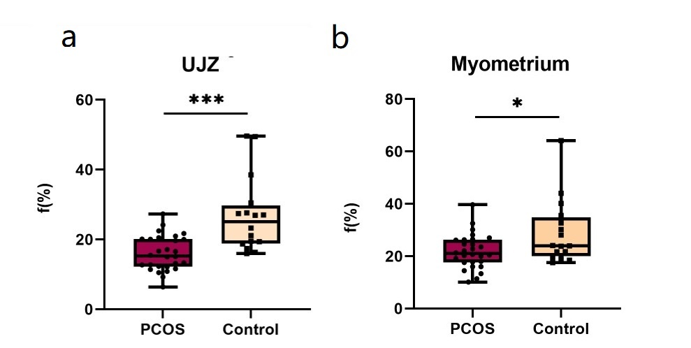
Figure 2. Box plot of considerable difference index.PCOS, polycystic ovary syndrome; UJZ, uterine junctional zone.
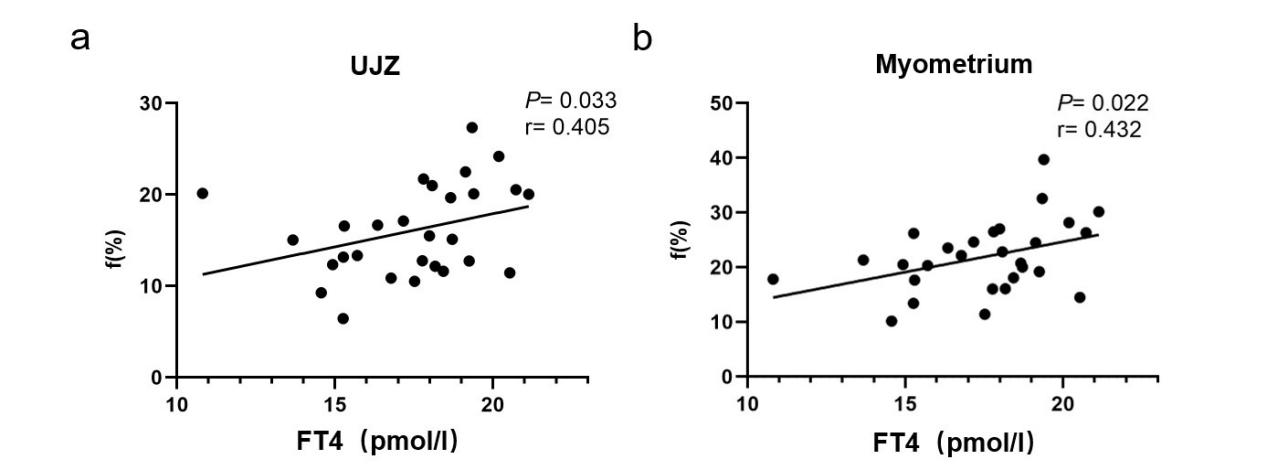
Figure 3. Correlations between considerable difference index and clinical indicators. FT4, free thyroxine; UJZ, uterine junctional zone.