2883
Sensitivity of IVIM in Detecting Acute Femoral Head Ischemia: A Comparison Between RESOLVE and Single Shot EPI Using An In Vivo Piglet Model1Veterinary Clinical Sciences, University of Minnesota, Saint Paul, MN, United States, 2Center for Magnetic Resonance Research, University of Minnesota, Minneapolis, MN, United States
Synopsis
Keywords: IVIM, Ischemia, perfusion, bone
Motivation: Intravoxel incoherent motion (IVIM) is a promising noninvasive technique to measure tissue diffusion and perfusion using a single multi-b-value diffusion-weighted imaging (DWI) acquisition.
Goal(s): To determine whether IVIM is sensitive to acute femoral head ischemia in the piglet model using RESOLVE and single-shot EPI.
Approach: 24 piglets underwent bilateral hip imaging using a 3T MRI before and after surgical induction of unilateral femoral head ischemia. IVIM and apparent diffusion coefficient (ADC) values were compared between the pre- and post-operative scans.
Results: IVIM is sensitive in detecting ischemia and subsequent injury to the femoral head. RESOLVE outperforms ssEPI in detecting changes in perfusion.
Impact: IVIM may be a clinically useful non-contrast-enhanced alternative to CE-MRI to measure femoral head ischemia and perfusion changes for detecting, staging, and monitoring LCPD and other forms of osteonecrosis of the femoral head.
Introduction
Legg-Calvé-Perthes disease (LCPD) is a childhood hip disorder characterized by interruption of blood supply to the developing femoral head1. Clinical evaluation of LCPD in the early, avascular stage involves assessment of perfusion changes in the developing femoral head using gadolinium-based contrast-enhanced MRI (CE-MRI) to confirm femoral head ischemia and measure the degree of femoral head involvement2. However, concerns about gadolinium deposition in the brain have limited the use of CE-MRI in children2, 3, prompting a need for non-contrast-enhanced approaches. Recently, IntraVoxel Incoherent Motion (IVIM), a technique that utilizes a series of diffusion-weighted images (DWI) to measure both tissue perfusion (perfusion fraction, f) and diffusion (diffusion coefficient, Ds)4, has been proposed as a non-contrast-enhanced alternative to CE-MRI to assess LCPD5. In particular, in an LCPD piglet model, IVIM perfusion fraction (f), obtained using RESOLVE DWI, decreased while IVIM diffusion (Ds) and apparent diffusion coefficient (ADC) increased in the secondary ossification center (SOC; the bone and bone marrow of the femoral epiphysis) one week after induction of femoral head ischemia. Inthe current study, we aimed to determine whether IVIM is sensitive to acute femoral head ischemia in a piglet model between 0 to 14 days post-operatively by comparing IVIM parameters between pre- and post-operative scans in the ischemic femoral heads using two sequences (RESOLVE and single-shot EPI, ssEPI). We hypothesized that the IVIM perfusion fraction (f) will decrease in the ischemic femoral heads immediately following the onset of ischemia and remain decreased over two weeks, whereas Ds and ADC will gradually increase with ischemic injury to the bone marrow.Methods
Animal Model and In Vivo 3T MRI: Our local IACUC approved this study. Bilateral hips of 24 six-week-old piglets were imaged in vivo at 3T MRI before the piglets underwent unilateral surgery to induce global femoral head ischemia by placing a ligature around the femoral neck and transecting the ligamentum teres6. The contralateral femoral head was unoperated. Piglets were then imaged a second time, using the same MRI protocol, at either 0, 1, 2, 4, 7, or 14 days post-operatively (n=4 piglets/timepoint). The MRI protocol included: (i) IVIM using RESOLVE and ssEPI DWI sequences; and (ii) subtraction CE-MRI to confirm the global femoral head ischemia. Acquisition parameters are summarized in Table 1.Data Analysis: Quantitative IVIM parameters (f and Ds) were calculated pixel-by-pixel using an Analytical Segmented approach (AS) 7,8, while ADC was calculated by fitting signals from all b values to a mono-exponential decay curve. A region of interest (ROI) constituting the SOC of the femoral head was drawn manually using RESOLVE b=0 diffusion-weighted images at a single middle slice. Median f, Ds, and ADC values were measured in each ROI, and the values were compared between the pre- and post-operative scans in the ischemic femoral heads. IVIM and ADC values from RESOLVE and ssEPI were also compared.
Results
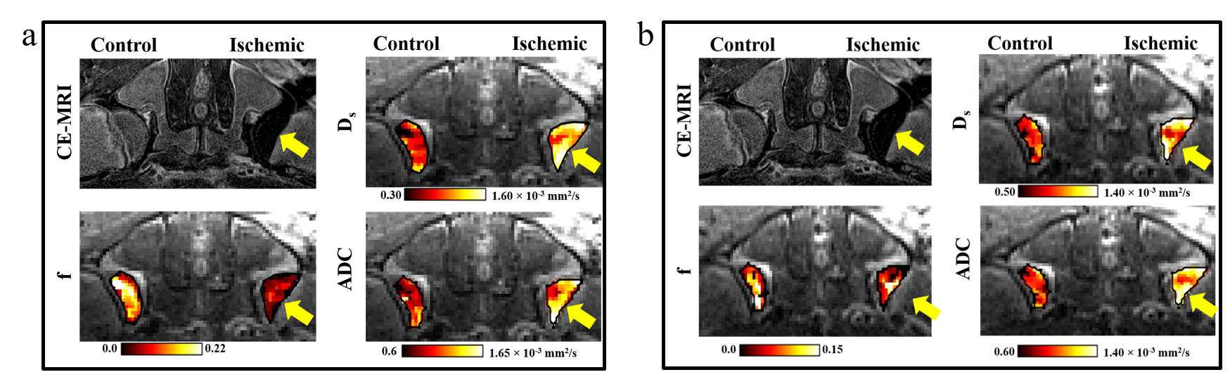
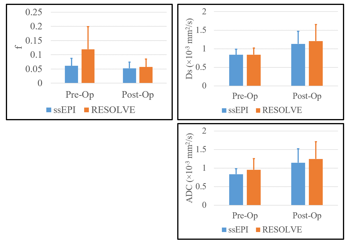
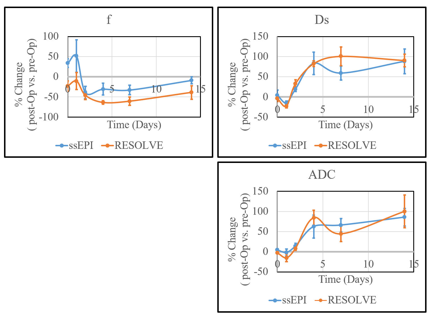
CE-MRI confirmed the global induction of ischemia in all 24 piglets post-operatively (Figure 1). Overall, f decreased while Ds and ADC increased in ischemic vs. control femoral heads (Figures 1 and 2). Pre-operatively, f values measured using RESOLVE were higher than those obtained using ssEPI, with no apparent differences in the measured Ds and ADC (Figure 2). Post-operatively, f, Ds, and ADC values obtained using RESOLVE and ssEPI were comparable. The f values from RESOLVE were consistently lower post- vs. pre-operatively from 0 to 14 days (Figure 3), while f values from ssEPI were increased before decreasing at 2 days post-operatively. The Ds and ADC values from RESOLVE and ssEPI initially decreased following the surgery, then gradually increased over the 14-day post-operative period (Figure 3).Discussion and Conclusion
Our findings demonstrate that IVIM parameters (f, Ds) and ADC are sensitive in detecting acute ischemia and subsequent injury to the femoral head without the need for a contrast agent. Consistently decreased f in the ischemic femoral heads confirms its potential to provide a non-contrast-enhanced alternative to assess ischemia and perfusion changes in bone. RESOLVE was more sensitive to perfusion changes than ssEPI, which could be attributed to its higher SNR and reduced sensitivity to artifacts. Thus, for IVIM measurement of bone perfusion, RESOLVE is preferred over ssEPI. The limitation of RESOLVE is its relatively long acquisition time. In conclusion, IVIM techniques represent potential non-contrast-enhanced alternatives to CE-MRI to measure femoral head ischemia and perfusion changes for detecting, staging, and monitoring LCPD and other forms of osteonecrosis of the femoral head.Acknowledgements
This project was supported by NIH grants R01AR081877, R56AR078315, and P41EB027061. We thank the staff of the University of Minnesota’s Clinical Investigation Center, Comparative Pathology Shared Resource, and Center for Magnetic Resonance Research for their assistance.References
1. Kim HK. Pathophysiology and new strategies for the treatment of Legg-Calve-Perthes disease. J Bone Joint Surg Am 2012; 94:659-669.
2. Laine JC, Martin BD, Novotny SA, Kelly DM. Role of Advanced Imaging in the Diagnosis and Management of Active Legg-Calvé-Perthes Disease. J Am Acad Orthop Surg. 2018 Aug 1;26(15):526-536.
3. Gulani V, Calamante F, Shellock FG, Kanal E, Reeder SB; International Society for Magnetic Resonance in Medicine. Gadolinium deposition in the brain: summary of evidence and recommendations. Lancet Neurol. 2017 Jul;16(7):564-570.
4. Le Bihan D, Breton E, Lallemand D, Aubin ML, Vignaud J, Laval-Jeantet M. Separation of diffusion and perfusion in intravoxel incoherent motion MR imaging. Radiology. 1988 Aug;168(2):497-505.
5. Buko EO, Bhave S, Tóth F, Johnson CP. IVIM Detects Bone Ischemia in a Piglet Model of Legg-Calvé-Perthes Disease. Proc. 31st Joint ISMRM-ESMRMB & ISMRT Annual Meeting 2022; No. 1598.
6. Kim HK, Su PH. Development of flattening and apparent fragmentation following ischemic necrosis of the capital femoral epiphysis in a piglet model. J Bone Joint Surg Am 2002; 84-A(8):1329-34.
7. Buko E, Zhang J, Ajala A, Hor PH, and Muthupillai R. An analytical segmented (AS) approach for extracting intravoxel incoherent motion (IVIM) model parameters. Proc. 26th ISMRM Annual Meeting 2018; No. 5361.
8. Buko EO, Ajala A, Zhang J, Hor PH, and Muthupillai R. An analytical segmented approach for extracting TE independent perfusion fraction in intravoxel incoherent motion (IVIM) MRI. In: Proceedings of the 28th Annual Meeting of ISMRM, Virtual. Abstract 4393.
Figures