2882
The impact of deep learning based image reconstruction on IVIM for benign and malignant pulmonary lesion discrimination1Department of Radiology, Second Affiliated Hospital of Naval Medical University, Shanghai, China, 2College of Health Sciences and Engineering, University of Shanghai for Science and Technology, Shanghai, China, 3MR Research,GE Healthcare, Beijing, China, 4Department of Thoracic Surgery, Second Affiliated Hospital of Naval Medical University, Shanghai, China
Synopsis
Keywords: IVIM, Lung, Deep learning reconstruction; Lung cancer; Magnetic resonance imaging; Diffusion weighted imaging; Intravoxel incoherent motion.
Motivation: DWI-based IVIM model provides information about tumor microenvironment and is reported useful in discriminating pulmonary lesions. However, lung DWI suffers low SNR and substantial susceptibility artifacts. Recently, a vendor-provided deep-learning reconstruction (DLR), relative to conventional reconstruction (ConR), is provided to improve image quality.
Goal(s): Investigate the impact of DLR on IVIM parameters for distinguishing malignant from benign pulmonary lesion.
Approach: DWI was acquired using FOCUS-MUSE to alleviate susceptibility artifacts and was reconstructed with DLR and ConR, separately. SNR and diagnostic performance of IVIM were compared.
Results: DLR significantly increased DWI image SNR and improved diagnostic performance of IVIM parameters for pulmonary lesion discrimination.
Impact: The application DLR would be beneficial for lung DWI in term of image quality and the performance of IVIM quantitative parameters for distinguishing benign from malignant pulmonary lesion.
Introduction
Lung cancer is the leading cause of cancer-related deaths worldwide [1]. Non-invasively characterizing lung cancer would be important for patient management. Diffusion-weighted imaging (DWI) is an advanced MRI technique which can reveal tissue microstructure by probing the motion process of water molecules [2-3]. DWI-based intravoxel incoherent motion (IVIM) model supplys information about both tissue diffusion and microcapillary perfusion components which are useful in discriminating tumors and monitoring and/or predicting efficacy of cancer treatments [3]. However, lung DWI commonly suffers low SNR and substantial susceptibility artifacts. This image quality of DWI may have impact on the quantitative parameters of IVIM for characterizing lung lesion. Recently, a vendor-provided deep learning reconstruction (DLR) technique (AIRTM ReconDL; GE Healthcare, USA) is commercially available and shows dramatically image quality improvement in comparison with conventional reconstruction (ConR) [4]. It had been applied to DWI for prostate and liver and resulted with increased SNR and image sharpness [5-6]. With these promising results, this study aimed at investigating the impact of DLR on lung DWI image quality and DWI-based IVIM quantitative parameters for pulmonary lesion discrimination. SNR and diagnostic performance were compared between ConR DWI-based and DLR DWI-based IVIM.Materials and Methods
Patients41 patients (30 malignant and 11 benign) were used in this study.
MRI Acquisition
All MRI experiments were performed at 3.0T scanner (SIGNA Premier; GE Healthcare, Milwaukee, USA) using a 21-channel AIRTM flexible coil (GE Healthcare). IVIM was scanned using FOCUS-MUSE sequence with TR/TE=2222-6316/55ms, FOV=32cm×16cm, matrix size=132×68, slice thickness=4 mm, number of shots=2, b values= 0/10/20/80/100/200/400/600/800/1000 s/mm2. FOCUS-MUSE is an improved EPI-based DWI which integrated the advantages of reduced FOV and multi-shot techniques. As a result, it is insensitive to susceptibility artifacts.
Data Analysis
DWI at b=1000 s/mm2 was used to compare the SNR of lesions between DLR and ConR. The SNR was defined as the ratio between the average signal intensity of lesion and the signal intensity standard derivation of background. IVIM quantitative parameters, including Dslow, Dfast and f, were calculated using the vendor-provided workstation (AW4.7; GE Healthcare). ROI of lesion was manually defined along the lesion boundary and avoid blood vessels and lung parenchyma. The average value of Dslow, Dfast and f within each ROI was separately extracted for further analysis.
Statistical Analysis
Paired Wilcoxon test was used to compare the SNR, Dslow, Dfast, and f between DLR DWI-based and ConR DWI-based IVIM. Mann-Whitney test were used to compare the differences between benign and malignant lesions. Receiver operating characteristic (ROC) analysis was used to assess the performance for differentiating malignant from benign pulmonary lesion. In this study, P<0.05 was considered statistically significant.
Results
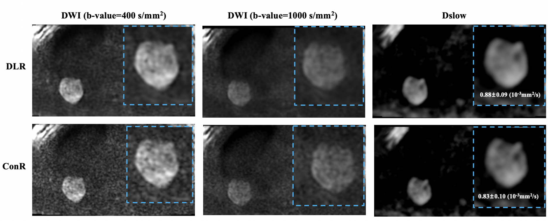
Figure 1 and Table 1 showed the SNR was significantly higher in the DLR DWI than in ConR DWI. The DLR DWI-based IVIM presented with significantly higher Dslow value than ConR DWI-based IVIM (Table 1 and Figure 1). No significant difference was detected in Dfast and f value between DLR and ConR DWI-based IVIM (Table 1).Table 2 demonstrated the f value was significantly higher in benign than in malignant for both ConR and DLR DWI-based IVIM. No significantly difference was observed in Dslow and Dfast between benign and malignant pulmonary lesions (Table2).
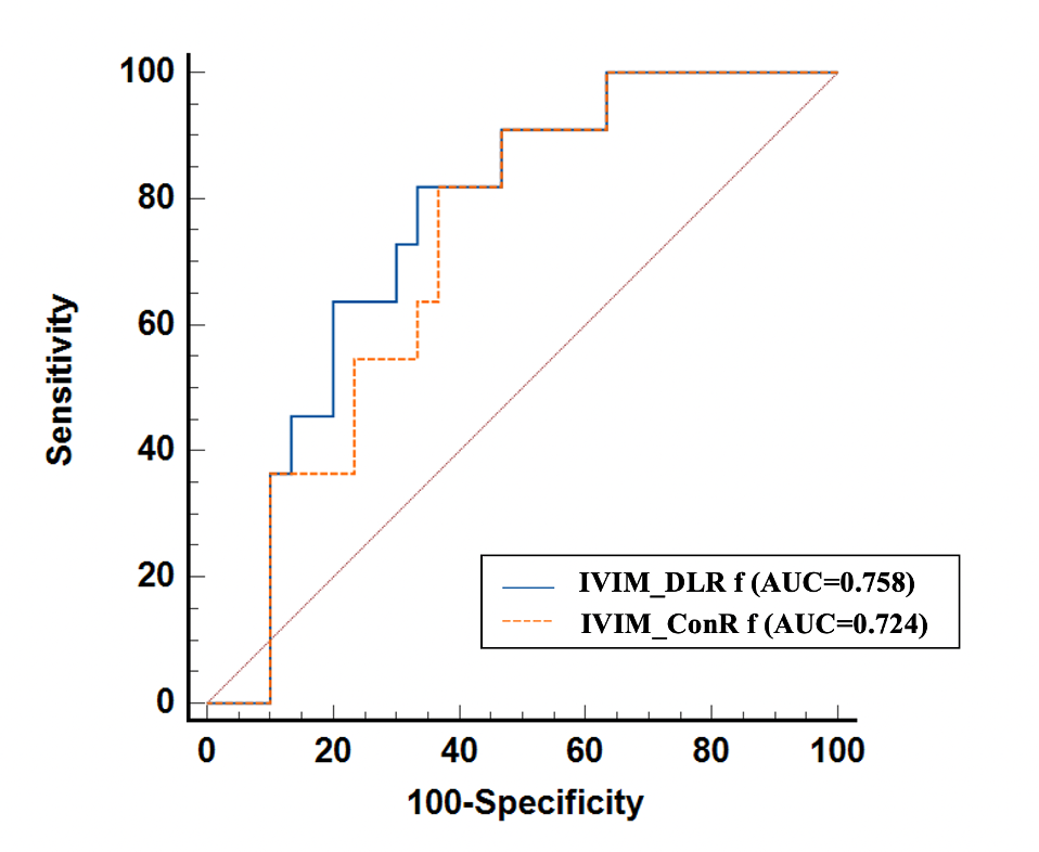
Figure 2 and Table 3 showed the f value calculated from both DLR and ConR DWI-based IVIM can significantly differentiating benign from malignant pulmonary lesion. The DLR DWI-based IVIM presented relatively better performance (AUC=0.758 and 0.724, respectively).
Discussion and Conclusion
This study investigated the impact of DLR on image quality of lung DWI and DWI-based IVIM quantitative parameters. The results showed DLR, relative to ConR, can significantly increase the SNR of lesion and can relatively improve the diagnostic performance of IVIM for distinguishing benign from malignant pulmonary lesion. The noise can act in a pernicious way specific to the quantification of DWI [2]. For example, lower SNR would result with underestimated apparent diffusion coefficient (ADC) [2]. Similarly, our results showed Dslow (i.e., true diffusion coefficient) were smaller in the lower SNR ConR DWI-based IVIM. The perfusion fraction parameter f derived from DLR DWI-based IVIM showed improved diagnostic performance. This may because the increased SNR allowed IVIM for more accurately characterizing the micro-organization of pulmonary lesion.In conclusion, DLR can increase SNR of lung DWI and can relatively improve the diagnostic performance of DWI-based IVIM for benign and malignant pulmonary lesion discrimination.
Acknowledgements
NoReferences
[1] Sung H, Ferlay J, Siegel RL, Laversanne M, Soerjomataram I, Jemal A, Bray F. Global Cancer Statistics 2020: GLOBOCAN Estimates of Incidence and Mortality Worldwide for 36 Cancers in 185 Countries. CA Cancer J Clin. 2021 May;71(3):209-249
[2] Mami Lima, Savannah C Partridge, Denis Le Bihan. Six DWI questions you always wanted to know but were afraid to ask: clinical relevance for breast diffusion MRI. Eur Radiol. 2020; 30(5):2561-2570.
[3] Masato Karayama, Nobuko Yoshizawa, Masataka Sugiyama, et al. Intravoxel incoherent motion magnetic resonance imaging for predicting the T long-term efficacy of immune checkpoint inhibitors in patients with non- small-cell lung cancer. Lung Cancer. 2020; 143:47-54.
[4] Lebel RM. Performance characterization of a novel deep learning-based MR image reconstruction pipeline. arxiv.org/abs/2008.06559.
[5] Kang-Lung Lee, Dimitri A Kessler, Simon Dezonie, et al. Assessment of deep learning-based reconstruction on T2-weighted and diffusion-weighted prostate MRI image quality. Eur J Radiol. 2023; 166:111017.
[6] Marta Zerunian, Francesco Paucciarelli, Damiano Caruso, et al. Artificial intelligence-based image quality enhancement in liver MRI: a quantitative and qualitative evaluation. Radiol Med. 2022; 127(10):1098-1105.
Figures