2881
Assessment of pathological prognostic factors in rectal cancer: comparison of DWI, IVIM, and DKI1Department of Radiology, Sichuan Provincial People's Hospital, University of Electronic Science and Technology of China, Chengdu, China, 2MR Research Collaboration, Siemens Healthineers, Chengdu, China, 3MRI clinical application, Customer Service Department, Siemens Digital Medical Technology Co., LTD, Shanghai, China
Synopsis
Keywords: IVIM, Diffusion/other diffusion imaging techniques
Motivation: The prognosis of rectal cancer (RC), depends on pathological prognostic factors. Despite the contributions of MRI, accurate preoperative prediction of these factors remains challenging.
Goal(s): Evaluate the values of DWI, IVIM, and DKI in predicting RC’s pathological prognostic factors.
Approach: In 162 rectal cancer patients, we compared DWI, IVIM, and DKI MRI techniques to predict EMVI, LNM, and histological differentiation, analyzing correlations with histological data.
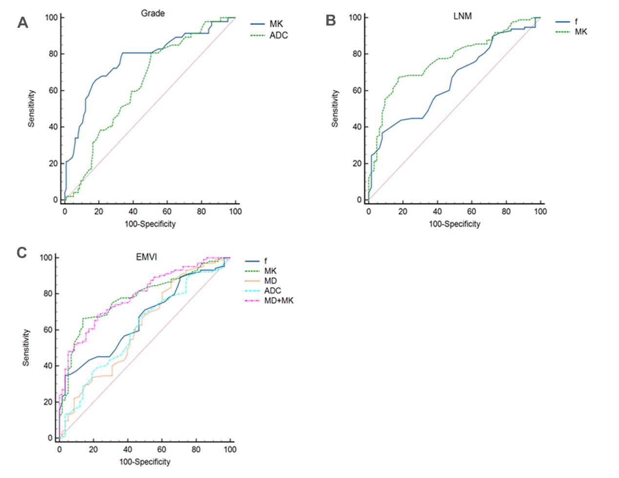
Results: MK was identified as a powerful predictor, especially for histological differentiation and LNM. The combine of MK+MD exhibited robust potential to predict EMVI, highlighting the diagnostic values of these models for rectal cancer prognosis.
Impact: This research enhances MRI's predictive capabilities for critical rectal cancer prognostic factors, potentially refining treatment planning and improving the prognosis of patients.
Introduction
Colorectal cancer, especially rectal cancer, is a major public health problem worldwide1. Its prognosis is significantly influenced by factors such as extramural vascular invasion (EMVI), lymph node metastasis (LNM), and histological differentiation2. Accurate preoperative prediction of these factors remains challenging, but is essential for effective treatment planning and improved the prognosis of patients. Magnetic resonance imaging (MRI), as a noninvasive method, has played an important role in the prediction of prognostic factors, but it haves some diagnostic limitations, such as low predictive accuracy, incomplete prediction of prognostic factors3. Despite the potential of advanced diffusion-weighted imaging (DWI) models such as intravoxel incoherent motion (IVIM) and diffusion kurtosis imaging (DKI) to provide more precise assessments, there has not been a thorough comparison of these crucial prognostic factors. This study aimed to evaluate the values of DWI, IVIM, and DKI in predicting EMVI, LNM, and histological differentiation among patients with rectal cancer.Methods
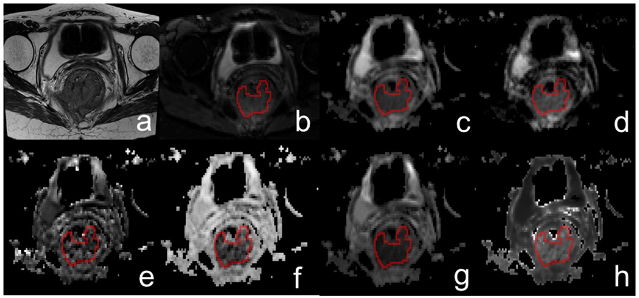
MR imaging: This study included 162 patients with rectal cancer who had undergone radical surgery. MRI was performed using a 3T MR scanner (MAGNETOM Vida; Siemens Healthineers, Erlangen, Germany) with a 30-channel coil. The MRI protocol consisted of sagittal, axial, and oblique coronal T2-weighted images and DWI. The axial DWI protocols were repetition time/echo time, 3100/99 ms; field of view, 226×226 mm2; matrix size, 110×110; slice thickness, 2 mm; intersection gap, 0.2 mm; and simultaneous multi-slice factor, 4. The DWI sequence duration was 6 minutes 21 seconds with eleven b-values (0, 50, 100, 200, 500, 800, 1000, 1500, 2000, 2300, and 2600 s/mm2 in three directions).Reconstruction & Segmentation: Data analysis involved computing mono-exponential DWI, IVIM, and DKI models from multi-b-value DWI data using an in-house postprocessing software (NeuDiLab) based on the open-resource tool DIPY (Diffusion Imaging in Python, https://dipy.org). The mappings included apparent diffusion coefficient (ADC) from the mono-exponential DWI; perfusion fraction (f), fast diffusion coefficient (D*) and slow diffusion coefficient (D) from IVIM; mean kurtosis (MK) and mean diffusion (MD) from DKI. A freehand region of interest (ROI) was manually placed on DWI images with b of 50 s/mm2, which was automatically copied to the corresponding ADC, D, D*, f, MD, and MK mappings. The ROIs were placed on the three different sections containing the largest tumor area, excluding any necrosis, vessels, or cysts within the lesion. EMVI, LNM, and histological differentiation were identified by analyzing the histology.
Statistical Analysis: Statistical analyses were conducted using SPSS version 26.0 and MedCalc version 16.8. The normal distribution continuous variables analyzed using independent samples t-test and one-way ANOVA. Univariable and multivariable logistic regression analyses were performed to identify independent risk factors. Diagnostic performances were assessed through receiver operating characteristic (ROC) curves, with areas under the curve (AUC) compared via the DeLong test. p-values < 0.05 were considered statistically significant.
Results
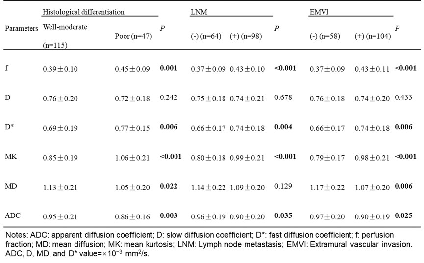
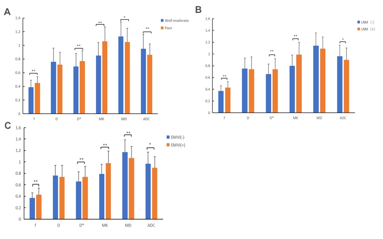
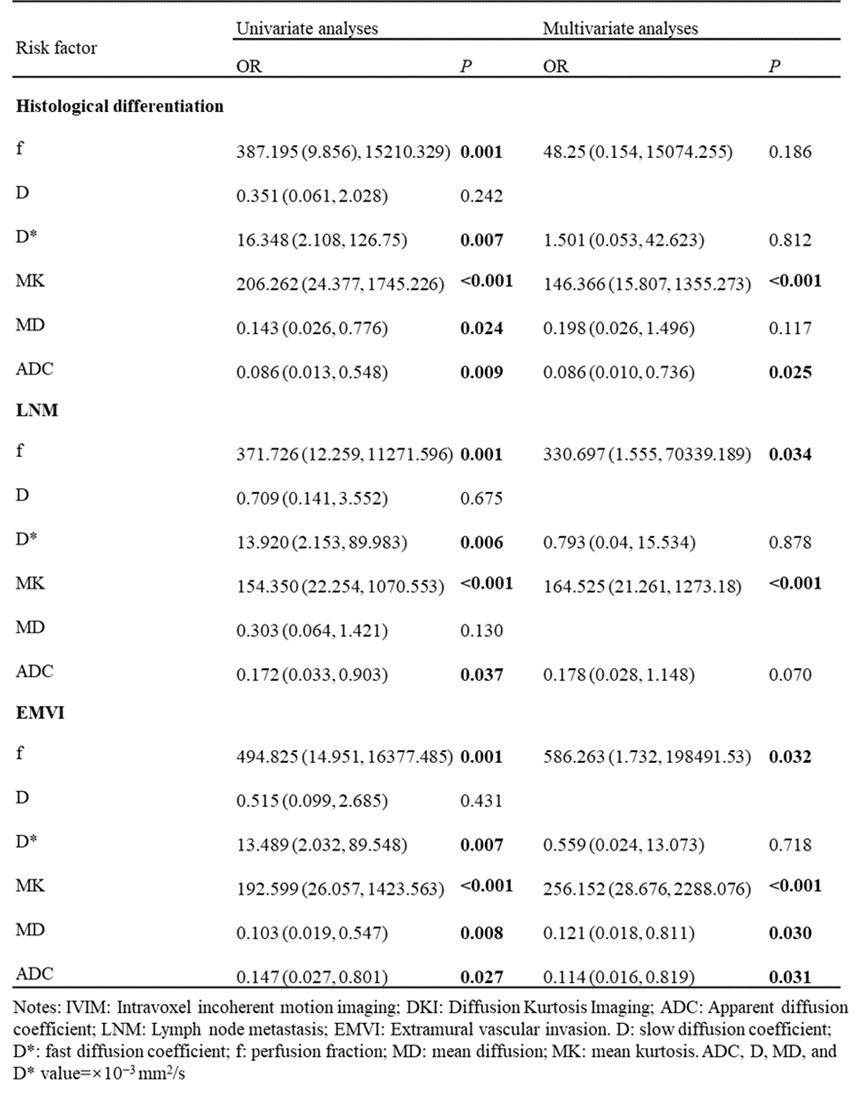
The mappings of various DWI models affecting rectal cancer prognosis revealed significant variation (Figure 1). For f, D*,MK,MD, and ADC mappings, significant differences were observed between well-moderate and poor histological differentiation and between EMVI (-) and EMVI (+) (all p<0.05) (Table 1 and Figure 2). Similarly, f, D*, MK, and ADC displayed significant difference between LNM (-) and LNM (+) (all p<0.05). In contrast, the D mapping did not show significant differences across prognostic factors. Multivariate analysis showed that MK and ADC were important indicators of histological differentiation; f and MK were significantly associated with LNM; and f, MK, MD, and ADC were significantly associated with EMVI (Table 2). Diagnostic assessments further emphasized the superiority of MK over other mappings in terms of predicting histological differentiation and LNM, whereas the combined MK+MD mappings displayed substantial diagnostic potential for EMVI (Figure 3).Discussion and Conclusion
Our findings demonstrate the superiority of MK over other mappings in terms of predicting histological differentiation and LNM. These results are consistent with recent literature highlighting the potential value of MK in tissue characterization4. MK reflects how water molecules interact with intracellular components and cell membranes; it represents the degree of diffusion property deviation from Gaussian behavior4. DKI model, especially MK mapping, may serve as imaging biomarkers for preoperative prediction of pathological prognostic factors in rectal cancer, helping to optimize the treatment planning and improved the prognosis of patients.Acknowledgements
No acknowledgement found.References
1. Asgeirsson T, Zhang S, Senagore AJ. Optimal follow-up to curative colon and rectal cancer surgery: how and for how long? Surg Oncol Clin N Am. 2021;19:861-873.
2. Kim JH, Beets GL, Kim MJ, et al. High-resolution MR imaging for nodal staging in rectal cancer: are there any criteria in addition to the size? Eur J Radiol. 2004;52:78-83.
3. Nerad E, Delli Pizzi A, Lambregts DMJ, et al. The Apparent Diffusion Coefficient (ADC) is a useful biomarker in predicting metastatic colon cancer using the ADC-value of the primary tumor. PLoS One. 2019;14:e0211830.
4. Jensen JH, Helpern JA. MRI quantification of non-Gaussian water diffusion by kurtosis analysis. NMR Biomed. 2021;23:698-710.
Figures