2880
Predicting estrogen receptor factor expression based on tumor habitats in patients with malignant breast cancer: a feasibility study1Department of Radiology, The Fourth Hospital of Hebei Medical University, Shijiazhuang, China, 2MR Research Collaboration, Siemens Healthineers Ltd., Beijing, China, 3East China Normal University, Shanghai, China
Synopsis
Keywords: IVIM, Radiomics, IVIM, MRI, habitats, breast tumor, estrogen receptor
Motivation: Immunohistochemistry remains the gold standard for diagnosing the estrogen receptor (ER) factor in breast cancer. However, the non-invasive IVIM radiomics method offers a meaningful alternative for predicting ER expression.
Goal(s): The objective of this study is to assess the feasibility and performance of a prediction model based on tumors habitats in predicting ER expression in malignant breast cancer patients.
Approach: We developed and evaluated a predictive model that utilizes tumors habitats to diagnose ER expression.
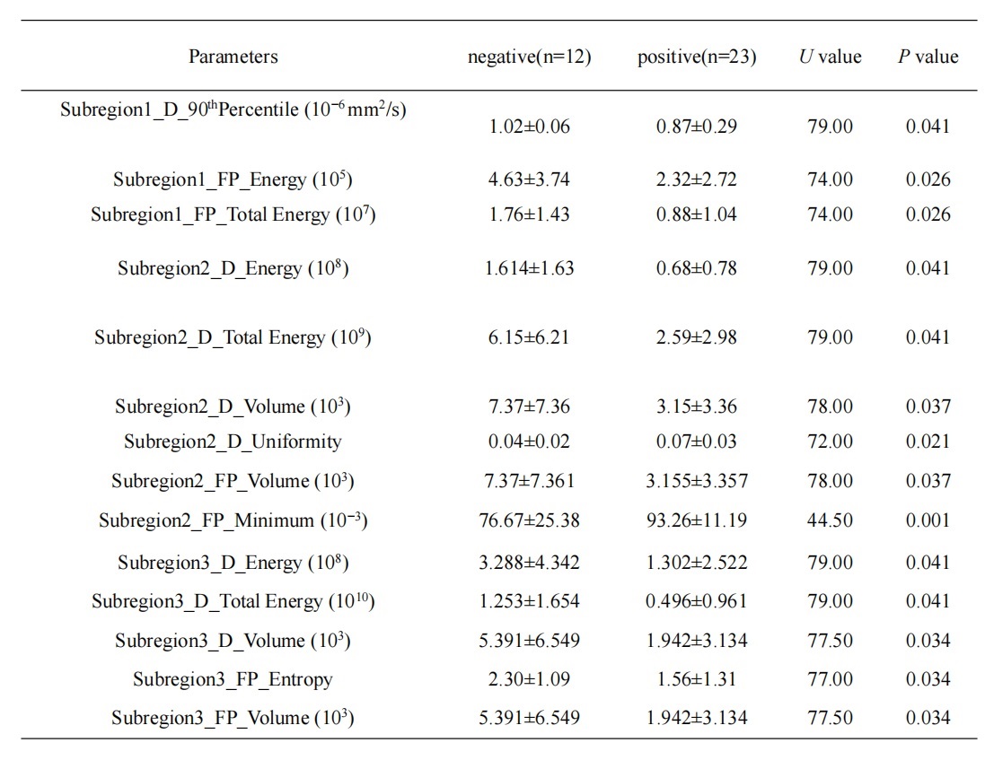
Results: The IVIM indicators can be employed for tumor subregions’ segmentation to hypoxia. The model demonstrates effective prediction capabilities for determining the probability of ER-positive in breast cancer.
Impact: Our research highlights the excellent performance of the predictive model based on tumors habitats in predicting ER expression in breast cancer. The model can aid clinicians in making more informed treatment decisions and subsequently improve patient outcomes.
Introduction
Breast cancer (BC) is responsible for approximately 30% of female cancer cases worldwide, with a mortality-to-incidence ratio of 15% 1. Research indicates that the overexpression of estrogen metabolites is associated with the development of BC, and the expression of the estrogen receptor (ER) serves as an important indicator of prognosis, with ER-positive expression suggesting a better prognosis 2. Tumor heterogeneity, which is directly proportional to malignancy, is closely linked to tumor habitats, including perfusion and oxygen-supply levels 3. Intravoxel incoherent motion (IVIM) can assess water molecule diffusion (D) to reflect tumor cellularity, as well as evaluate tumor microvascular perfusion fraction levels (FP). A novel data-driven method for partitioning tumor subregions based on IVIM, known as habitats, can better represent tumor heterogeneity and improve prediction accuracy. In this study, we aim to explore the application of radiomics features extracted from these imaging-defined tumor subregions to predict ER factor expression in malignant BC.Methods
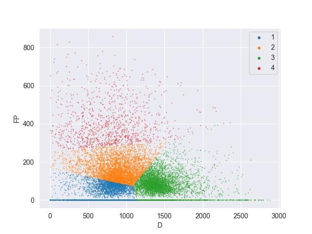
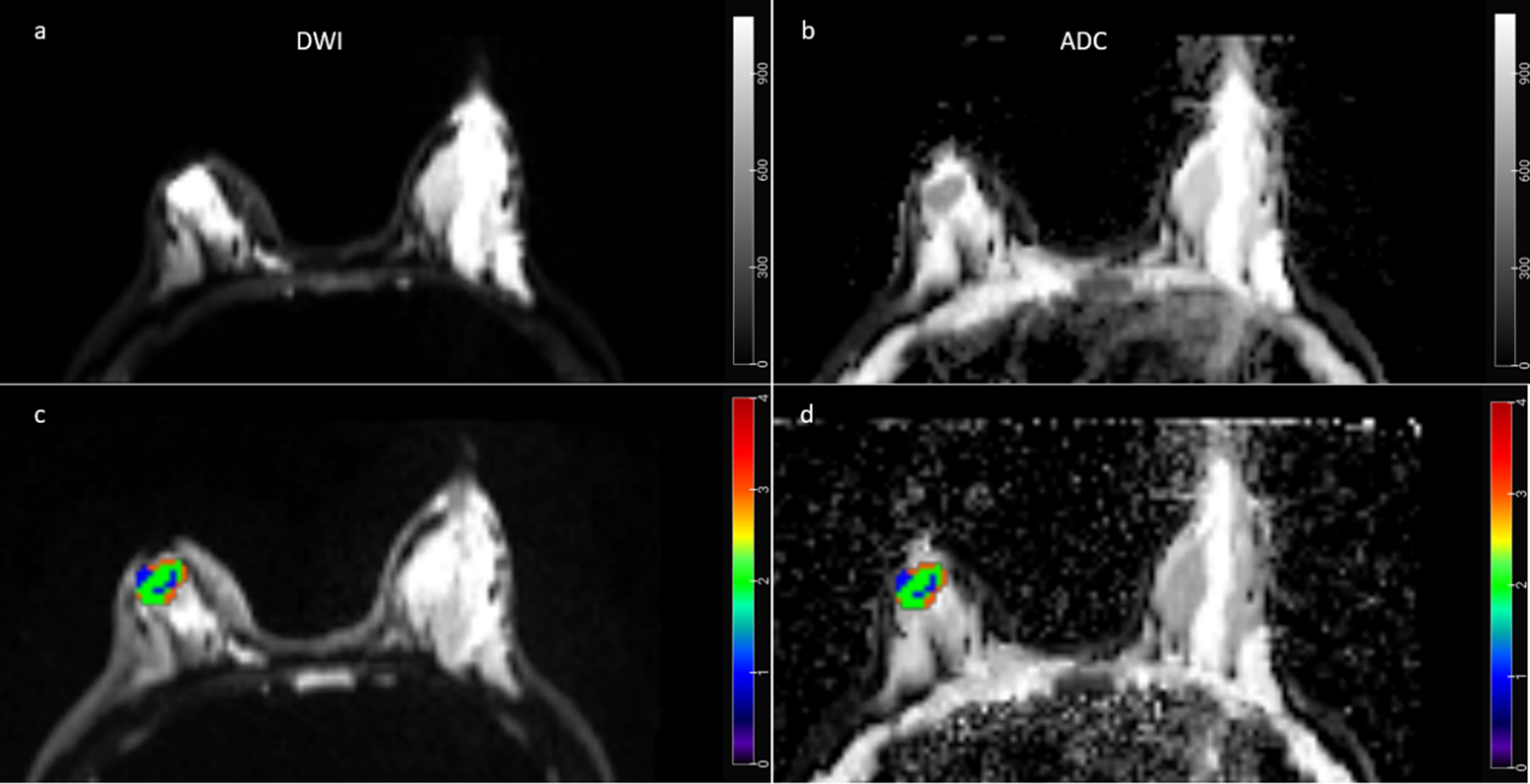
This study included 35 patients diagnosed with breast malignant invasive ductal carcinoma confirmed by surgery and pathology. Of these, 12 patients had an ER-negative factor and 23 patients had an ER-positive factor. All patients underwent breast MRI on a 3T MR scanner (MAGNETOM Vida, Siemens Healthineers, Erlangen, Germany). IVIM data was acquired using a single-shot spin-echo echo planar imaging (SE EPI) sequence with 9 b values ranging from 0 to 1200 s/mm2. The IVIM matrices (D, D*, and FP) were calculated with a research software (MR Body Diffusion Toolbox v1.4.0, Siemens Healthineers, Germany), and an ROI encompassing the maximum tumor level on b0 images was manually outlined using 3D Slicer software (https://www.slicer.org/,version 5.2.2). Utilizing these three metrics from each voxel, consensus clustering (K means) was employed to partition the tumor into habitats with varying cellularity and perfusion levels (subregion1: DlowFPlow, subregion2: DmoderateFPmoderate, subregion3: DhighFPlow, and subregion4: DlowFPhigh) using in-house development software nnFAE. First-order radiomics features were extracted for all four subregions. The mean ± standard deviation was used to express all features, and differences between the two groups were assessed using the nonparametric Mann-Whitney U Test. Logistic regression analysis was conducted to establish a predictive model using features with significant differences, and the predictive performance of the model was assessed using the area under the receiver operating characteristic curve (AUC).Results
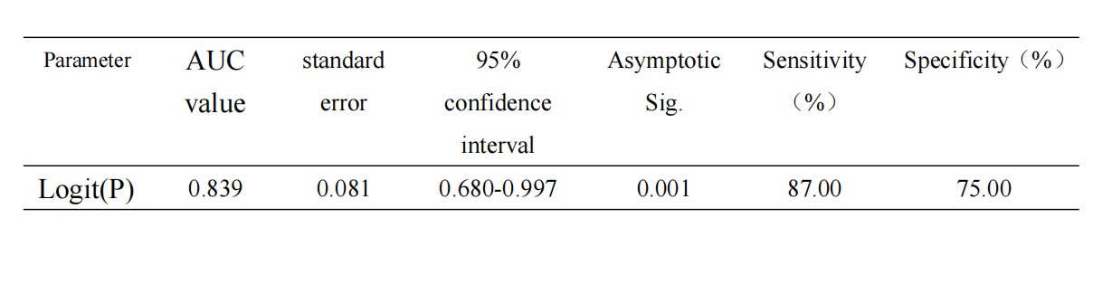
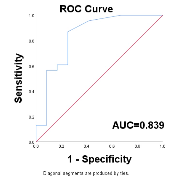
Table 1 demonstrates significant differences in the features of DlowFPlow, DmoderateFPmoderate, and DhighFPlow subregions between the ER-negative and ER-positive groups (P < 0.05). The logistic regression model based on these features exhibited excellent predictive performance for ER status, with an AUC of 0.839, sensitivity of 0.870, and specificity of 0.750 (Table 2, Figure 1).Discussion and conclusion
Our research demonstrates the feasibility of using IVIM-based habitat analysis to predict ER expression in BC. The results indicate that IVIM metrics (D and FP), which reflect tumor cellularity and vascularity, can be employed to divide tumors into subregions using clustering methods. Significant differences exist in the features of DlowFPlow, DmoderateFPmoderate, and DhighFPlow subregions, representing hypoperfusion and hypercellularity regions called hypoxia, between ER-negative and ER-positive BC. The uniformity of D and the minimum of FP in the DmoderateFPmoderate subregion were higher in the ER-positive group, suggesting greater intratumor heterogeneity in the ER-negative group. Additionally, the 90th percentile of D, the energy and total energy of FP in the DlowFPlow subregion, as well as the volume of FP, energy, total energy, and volume of D in the DmoderateFPmoderate subregion, were higher in the ER-negative group. This may indicate that ER-negative tumors are more aggressive, exhibit reduced microcirculation perfusion, lack oxygen supply and nutrients, and are more likely to degrade3-4. Therefore, to some extent, this logistic regression model based on features of habitats can predict ER-positive malignant BC, evaluate tumor prognosis, and guide clinical treatment decisions.Acknowledgements
No acknowledgement found.References
1. Siegel RL, Miller KD, Jemal A. Cancer statistics, 2020. CA Cancer J Clin 2020; 70: 7–30.
2. Wu JR, Zhao Y, Zhou XP, et al. Estrogen receptor 1 and progesterone receptor are distinct biomarkers and prognostic factors in estrogen receptor-positive breast cancer: Evidence from a bioinformatic analysis. Biomed Pharmacother. 2020;121:109647.
3. Mo T, Brandal SHB, Köhn-Luque A, et al. Quantification of Tumor Hypoxia through Unsupervised Modelling of Consumption and Supply Hypoxia MR Imaging in Breast Cancer. Cancers (Basel). 2022 Mar 4;14(5):1326.
4. Jalnefjord O, Montelius M, Arvidsson J, et al. Data-driven identification of tumor subregions based on intravoxel incoherent motion reveals association with proliferative activity. Magn Reson Med. 2019;82(4):1480-1490.
Figures