2879
Potential Diagnostic Efficiency of Intravoxel Incoherent Motion-based Virtual Magnetic Resonance Elastography in Pulmonary Neoplasms1Radiology, The First Affiliated Hospital of Xi'an Jiaotong University, Xi'an, China
Synopsis
Keywords: IVIM, Cancer, Stiffness
Motivation: Exploring non-invasive techniques with the potential of replacing invasive pathological analysis of pulmonary neoplasms remains urgent.
Goal(s): To investigate the feasibility of IVIM-based vMRE to provide quantitative estimates of tissue stiffness in pulmonary neoplasms and to verify the diagnostic performance in distinguishing neoplasm property.
Approach: sADC and virtual stiffness values of neoplasm were extracted, and the diagnostic performance of vMRE in distinguishing benign and malignant and detailed pathological type were explored.
Results: Virtual stiffness values of malignant neoplasms were significantly higher than benign ones. Subsequent sub-type analyses showed adenocarcinoma and squamous cell carcinoma were mostly stiffer than non-specific benign neoplasms.
Impact: IVIM-based vMRE has good feasibility in reflecting stiffness of pulmonary neoplasms. Virtual stiffness and sADC values showed significant difference between malignant and benign lesions.The application of non-invasive vMRE is promising as a new method for clinical diagnosis.
Introduction
Lung cancer is the most common malignancy and the leading cause of cancer-related death [1]. Exploring non-invasive techniques with the potential of replacing invasive pathological analysis of pulmonary neoplasm remains urgent.Stiffness is an intrinsic material property of the tissue. IVIM-based virtual MRE (vMRE) is first proposed in the evaluation of liver fibrosis, which is a novel method with potential application for the non-invasive assessment of tissue stiffness and without the limitation of specialized hardware and expertise of traditional MRE [2]. vMRE reveals a strong correlation between tissue water diffusivity and tissue elasticity in liver and has been extended to other diseases of liver and brain [3-6]. Therefore, we hypothesized that vMRE could judge the nature of pulmonary neoplasms. This study was to explore the capability of vMRE in the characterization of pulmonary neoplasms through quantitative sADC and virtual stiffness values.
Methods
This study was approved by the ethical committee of the First Affiliated Hospital of Xi’an Jiaotong University. Pulmonary neoplasm patients underwent CT guided percutaneous lung biopsy between April 2018 and March 2020 were enrolled. IVIM was performed on a 3T MRI system (GE Healthcare) with an 8-channel body flex coil before biopsy. Images were collected with each of the following b values: 0, 10, 15, 20, 25, 50, 80, 150, 300, 500 and 800 s/mm2. IVIM images of the lower key b-value (LKb = 150 s/mm2) and the higher key b-value (HKb = 800s/mm2) were used to estimate the virtual stiffness. Virtual stiffness (kPa) = α·ln (SLKb/SLKb) + β. The scaling (α) and the shift (β) factors were set to -9.8 and 14 according to the calibration studies [2]. Regions of interest (ROI) were placed on three contiguous axial slices, in which the tumors had maximum diameters, and to avoid necrotic, bleeding, or inhomogeneous areas.Results
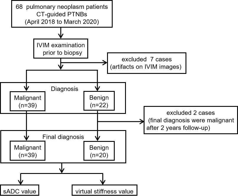
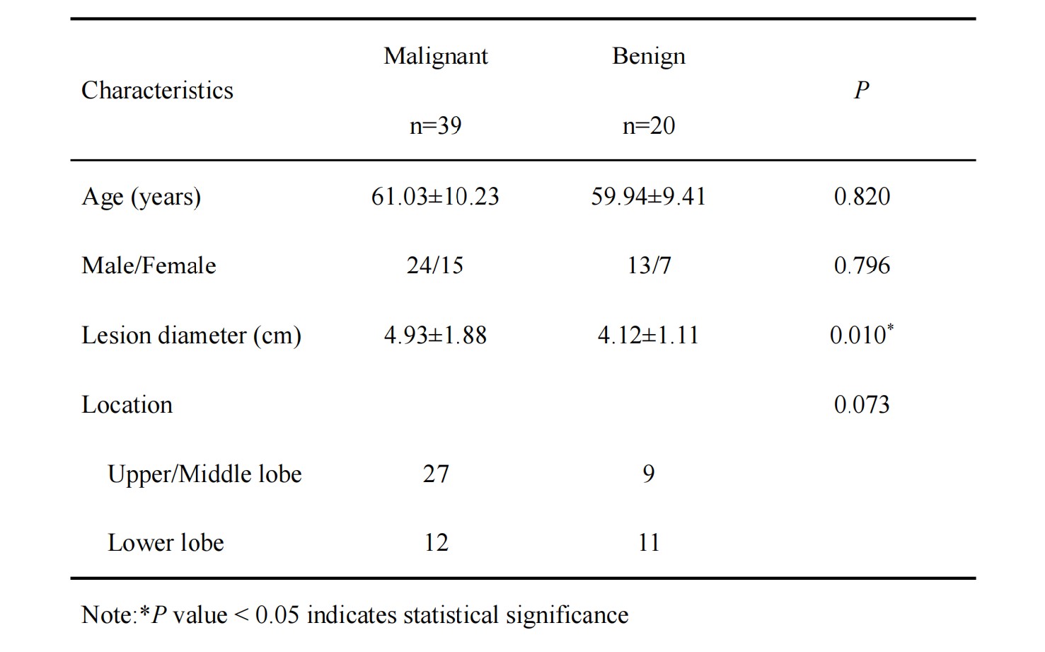
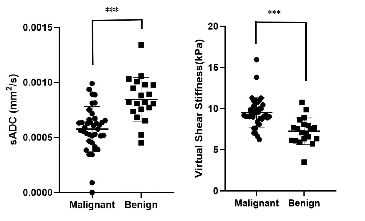
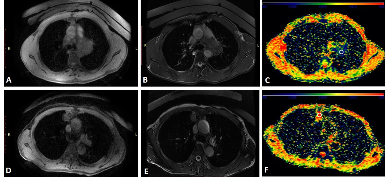
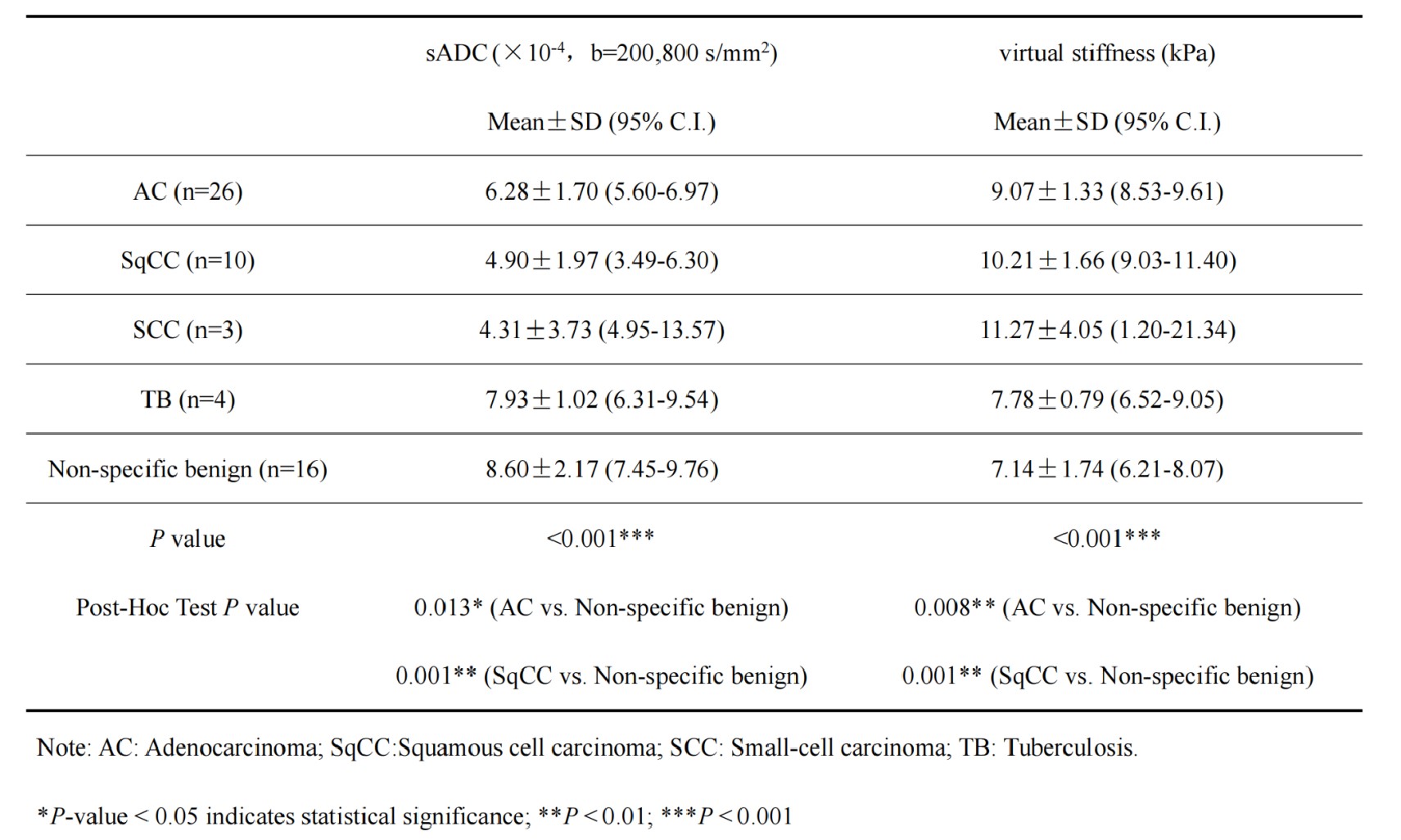
We ultimately enrolled 59 participants (Figure 1). The malignant group (39/59) included patients with adenocarcinoma, squamous cell carcinoma and small-cell carcinoma, whereas the benign group (20/59) included tuberculosis and non-specific benign (Figure 2). Compared to benign neoplasms, malignant ones had significantly lower sADC and higher virtual stiffness value (P < 0.001) (Figure 3). The cut-off value of sADC was 6.78 ×10⁻⁴ s/mm2, while that for the virtual stiffness value was 8.08 kPa. FIESTA and T2FSE images together with sADC maps are presented in Figure 4. Sub-type analyses showed sADC values of adenocarcinoma and squamous cell carcinoma groups were significantly lower than non-specific benign group (P = 0.013 and 0.001, respectively). Additionally, virtual stiffness values showed the opposite trend (P = 0.008 and 0.001, respectively) (Figure 5).Discussion
vMRE was first developed for liver fibrosis assessments, suggesting a strong correlation between liver elastic properties and tissue microstructure [2]. This correlation was due to the elastic properties of tissues are linked to the layout of elementary tissue components (i.e., cells, fibers, stroma) to which diffusion MR imaging was exquisitely sensitive [7]. In this study, vMRE for evaluation of pulmonary neoplasm was proposed to have the potential to indicate stiffness and distinguish property. We present malignant neoplasms possessed significantly lower sADC and higher mean virtual stiffness values compared to benign neoplasms.The reason for this may because increased stiffness of cancer is caused by matrix deposition and remodeling, which can activate signaling pathways that promote proliferation, invasiveness, and metastasis [8,9]. When normal tissue architecture is disrupted by cancer growth and invasion, microarchitecture is altered [10]. Increased deposition and cross-linking of the extracellular matrix lead to matrix stiffening. Additionally, some collagen fibers are under tension due to cell contraction or local expansion caused by tumor growth [11]. These tensile stresses increase the stiffness of collagen network, which further activates the focal adhesion contractility of cancer-associated fibroblasts in their vicinity, leading to a vicious cycle of matrix deposition and stiffening [12]. Hence, vMRE may potentially provide a better understanding of microstructural tissue changes and enable fast and detailed evaluation of tumor stiffness.
Sub-type analyses showed sADC and virtual stiffness values of adenocarcinoma and squamous cell carcinoma were significantly different from non-specific benign. However, no correlation was found in small cell carcinoma and tuberculosis. As small cell carcinoma has a faster proliferation rate and reduced extracellular space, which results in a significant reduction of water diffusion in the tumor, it should theoretically present a higher stiffness. Our negative result may due to the small sample size.
Conclusion
Our findings provide evidence that malignant neoplasms have significantly lower sADC and higher virtual stiffness values than benign ones. Non-invasive vMRE is promising as a new method for clinical diagnosis.Acknowledgements
This study was supported by National Natural Science Foundation of China (No. 82202998), Natural Science Basic Research Program of Shaanxi Province (2021JQ-389, 2022JQ-884), Clinical Research Award of the First Affiliated Hospital of Xi'an Jiaotong University of China (XJTU1AF2021CRF-015) and National Natural Science Foundation of China (No. 82272073).References
[1]. Chen W, Zheng R, Baade PD, Zhang S, Zeng H, Bray F, et al. Cancer statistics in China, 2015. CA Cancer J Clin 2016;66:115-132
[2]. Le Bihan D, Ichikawa S, Motosugi U. Diffusion and Intravoxel Incoherent Motion MR Imaging-based Virtual Elastography: A Hypothesis-generating Study in the Liver. Radiology 2017;285:609-619
[3]. Ota T, Hori M, Le Bihan D, Fukui H, Onishi H, Nakamoto A, et al. Diffusion-Based Virtual MR Elastography of the Liver: Can It Be Extended beyond Liver Fibrosis? J Clin Med 2021;10
[4]. Hanniman E, Costa AF, Bowen CV, Abdolell M, Stueck A, McLeod M, et al. Prospective Evaluation of Virtual MR Elastography With Diffusion-Weighted Imaging in Subjects With Nonalcoholic Fatty Liver Disease. J Magn Reson Imaging 2022;56:1448-1456
[5]. Lagerstrand K, Gaedes N, Eriksson S, Farahmand D, De Coursey E, Johansson G, et al. Virtual magnetic resonance elastography has the feasibility to evaluate preoperative pituitary adenoma consistency. Pituitary 2021;24:530-541
[6]. Aunan-Diop JS, Andersen MCS, Friismose AI, Halle B, Pedersen CB, Mussmann B, et al. Virtual magnetic resonance elastography predicts the intraoperative consistency of meningiomas. J Neuroradiol 2022
[7]. Alkalay RN, Burstein D, Westin CF, Meier D, Hackney DB. MR diffusion is sensitive to mechanical loading in human intervertebral disks ex vivo. J Magn Reson Imaging 2015;41:654-664
[8]. Nia HT, Munn LL, Jain RK. Physical traits of cancer. Science 2020;370
[9]. Guo H, Zhang T, Yu Y, Xu F. Cancer Physical Hallmarks as New Targets for Improved Immunotherapy. Trends Cell Biol 2021;31:520-524
[10]. Le Bihan D. What can we see with IVIM MRI? Neuroimage 2019;187:56-67
[11]. Samuel MS, Lopez JI, McGhee EJ, Croft DR, Strachan D, Timpson P, et al. Actomyosin-mediated cellular tension drives increased tissue stiffness and beta-catenin activation to induce epidermal hyperplasia and tumor growth. Cancer Cell 2011;19:776-791
[12]. Choquet D, Felsenfeld DP, Sheetz MP. Extracellular matrix rigidity causes strengthening of integrin-cytoskeleton linkages. Cell 1997;88:39-48
Figures