2878
Evaluation of PD-1/PD-L1 Expression in Hepatocellular Carcinoma Using Intravoxel Incoherent Motion: A Diagnostic Perspective1Department of Radiology, The First Affiliated Hospital of Guangxi Medical University,, Naning, China, 2MR Research Collaboration, Siemens Healthineers Ltd., Wuhan, China, 3MR Application Predevelopment, Siemens Healthcare GmbH, Erlangen, Germany
Synopsis
Keywords: IVIM, Diffusion/other diffusion imaging techniques, PD-1/PD-L1
Motivation: In clinical practice of hepatocellular carcinoma, there are lacking in preoperative and pre-medication results regarding immunosuppressive checkpoint inhibitors PD-1 and PD-L1.
Goal(s): This study aimed to evaluate the expression of PD-1 and PD-L1 using intravoxel incoherent motion (IVIM) before surgery and immunotherapy.
Approach: Immunohistochemistry staining was performed on liver cancer tissue to analyze the correlation between immunohistochemistry results and derived parameters (D, D*, and f) from IVIM and ADC, allowing for the assessment of their diagnostic value.
Results: The parameters D and D* from IVIM had statistically significant differences between the expression of PD-1 and PD-L1.
Impact: The results of this study may have an impact on clinical decision-making in the future and demonstrate a favorable outcome for scientific research in the immune microenvironment.
Introduction
Hepatocellular carcinoma (HCC) poses a serious threat to the lives and health of people worldwide1. Precision medicine and improvement in the tumor microenvironment have become current focuses in treating liver cancer, aiming to achieve improved prognoses for patients. However, currently available immune checkpoint inhibitors lack the evaluation of patients' immune checkpoint expression and immune status before medication. Therefore, the noninvasive prediction of the expression of immune checkpoints and immune status would greatly help in the precision treatment of HCC. The application of intravoxel incoherent motion (IVIM) in predicting the immune microenvironment has not been explored yet. This study aimed to evaluate the expression of immunosuppressive checkpoints using IVIM in preoperative and pre-immunotherapy patients.Methods
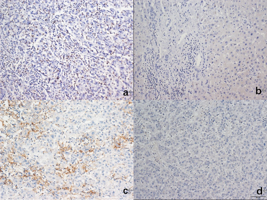
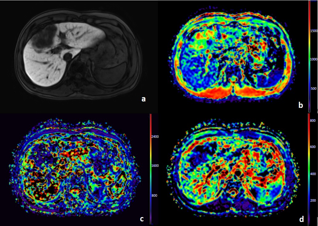
The inclusion criteria were as follows: (1) Patients with HCC receiving no intervention, radiotherapy, targeted therapy, or other treatments prior to surgery; (2) patients who underwent MRI protocol 2 weeks before surgery; and (3) patients who underwent surgical resection and obtained postoperative pathologic confirmation of HCC. Finally, 74 patients with primary HCC were included. All patients underwent preoperative examination using a 3T MRI scanner (MAGNETOM Prisma, Siemens Healthcare, Erlangen, Germany). A research integrated shimming (iShim) diffusion-weighted imaging (DWI) sequence with multiple b values was performed using free-breathing scans2. Eight b values (number of excitations) were acquired in 3 orthogonal directions: 0 (1), 20 (1), 50 (1), 100 (1), 150 (1), 200 (1), 600 (1), and 1000 (2) s/mm2. Specific scanning parameters were as follows: TR/TE= 4900/57 milliseconds; FOV= 380 × 261 mm2; matrix size= 128 × 88; slice thickness= 5 mm; slice gap= 1 mm; parallel imaging acceleration factor = 2; diffusion scheme = monopolar; and bandwidth = 2442 Hz/pixel. PD-1 and PD-L1 immunohistochemical staining was performed on tumor specimens resected from the 74 patients. The apparent diffusion coefficient (ADC) map using b = 0 and 1000 and IVIM-derived parameters of molecular diffusion coefficient (D), perfusion fraction (f), and perfusion-related diffusion coefficient (D*) from IVIM using all 8 b values were calculated. The remarkable differences in 10th, mean, and 90th parameter values were evaluated between PD-1-positive (n = 36) and PD-1-negative groups (n = 38), as well as between PD-L1-positive (n = 38) and PD-L1-negative (n = 36) groups using the t test. P <.05 indicated statistically significant differences.Results
For PD-1 expression, no statistically significant difference was observed in the ADC value (P >.05). However, statistically significant differences were noted in the 10th value of IVIM-D, as well as the mean values of IVIM-D and IVIM-D* (P<.05) between PD-1-positive (PD-1+) and PD-1-negative groups (PD-1–), as illustrated in Table 1. The combined area under the curve (AUC) of these non-Gaussian model diffusion parameters with substantial differences was 0.746, as depicted in Figure 1a.Additionally, for PD-L1 expression, no statistically significant difference was observed in the ADC value (P >.05). However, statistically significant differences were noted in the 10th and mean value of IVIM-D* (P<.05) between PD-L1-positive (PD-L1+) and PD-L1-negative groups (PD-L1–), (Table 2). The combined AUC of these 3 parameters was 0.654, as illustrated in Figure 1b. Figures 2 and 3 represent immunohistochemical staining and MRI images, respectively.
Discussion
This study explored the feasibility of DWI in predicting the tumor immune microenvironment preoperatively. The results revealed that D* could differentiate the expression of PD-1 and PD-L1. However, the ADC value displayed no statistically significant difference between any immune-related markers. Previous studies demonstrated that ADC values might not fully reflect the diffusion characteristics of water molecules in tissues3-5. In contrast, the IVIM model provided an improved description of the diffusion characteristics of water molecules in tumors6,7. Our study also verified the value of IVIM. The IVIM-D* value usually reflects the speed of capillary flow8. In this study, both PD-1 and PD-L1 positivity displayed a remarkable decrease. This was likely due to the close relationship between PD-1 positivity and immune infiltration in tumors. The increased tissue viscosity of immune cell infiltration might cover the perfusion of tumor capillaries9. The D* value of patients with PD-L1 positivity was considerably lower than that of patients with PD-L1 negativity, indicating that microvascular perfusion might be lower in patients with PD-L1 positivity. In the tumor microenvironment, distinct interactions existed between the immune and vascular microenvironments. A more accurate understanding of the motion patterns of water molecules within the tumor and the microenvironment of tumor tissue can be achieved using the IVIM model.Conclusion
The evaluation of immune checkpoint expression using IVIM preoperatively and before immunotherapy may contribute to clinical decision-making in the futureAcknowledgements
This study was supported by the National Natural Science Foundation of China (No. 82060310).References
[1] Sung H, Ferlay J, Siegel R L, et al. Global Cancer Statistics 2020: GLOBOCAN Estimates of Incidence and Mortality Worldwide for 36 Cancers in 185 Countries [J]. CA Cancer J Clin, 2021, 71(3): 209-49.
[2] Stemmer A , Kiefer B,. Combination of integrated slice-specific dynamic shimming and pixel-wise unwarping of residual EPI distortions [J]. Proc Intl Soc Mag Reson Med. 23 (2015).
[3] Shankar S, Kalra N, Bhatia A, et al. Role of Diffusion Weighted Imaging (DWI) for Hepatocellular Carcinoma (HCC) Detection and its Grading on 3T MRI: A Prospective Study [J]. Journal of Clinical and Experimental Hepatology, 2016, 6(4): 303-10.
[4] Kuai Z-X, Sang X-Q, Yao Y-F, et al. Evaluation of non-monoexponential diffusion models for hepatocellular carcinoma using b values up to 2000 s/mm2 : A short-term repeatability study [J]. Journal of Magnetic Resonance Imaging : JMRI, 2019, 50(1): 297-304.
[5] Koh D-M, Collins D J. Diffusion-weighted MRI in the body: applications and challenges in oncology [J]. AJR American Journal of Roentgenology, 2007, 188(6): 1622-35.
[6] Jensen J H, Helpern J A. MRI quantification of non-Gaussian water diffusion by kurtosis analysis [J]. NMR In Biomedicine, 2010, 23(7): 698-710.
[7] Le Bihan D. What can we see with IVIM MRI? [J]. Neuroimage, 2019, 187: 56-67.
[8] Lee H-J, Rha S Y, Chung Y E, et al. Tumor perfusion-related parameter of diffusion-weighted magnetic resonance imaging: correlation with histological microvessel density [J]. Magnetic Resonance In Medicine, 2014, 71(4): 1554-8.
[9] Swartz J E, Driessen J P, Van Kempen P M W, et al. Influence of tumor and microenvironment characteristics on diffusion-weighted imaging in oropharyngeal carcinoma: A pilot study [J]. Oral Oncology, 2018, 77.
Figures
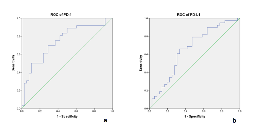
Figure 1. (a) ROC curve of the combined 10th values of IVIM-D, as well as the mean values of IVIM-D and IVIM-D* for PD-1. (b) ROC curve of the combined 10th and mean values of IVIM- D* for PD-L1.
IVIM, intravoxel incoherent motion; ROC, receiver operating characteristic.

Table 1. Univariate analysis of PD-1 with MRI diffusion parameters (ADC, IVIM-D: ×10-6 mm2/s; IVIM-D*: ×10-5 mm2/s)
ADC, Apparent diffusion coefficient