2877
The value of IVIM-based tumor habitats in predicting the lymph node metastasis of breast cancer1Department of Radiology, The Fourth Hospital of Hebei Medical University, Shijiazhuang, China, 2MR Research Collaboration, Siemens Healthineers Ltd., Beijing, China, 3East China Normal University, Shanghai, China, 4MR Application Predevelopment, Siemens Healthineers AG, Erlangen, Germany
Synopsis
Keywords: IVIM, Diffusion/other diffusion imaging techniques, IVIM,MRI, breast tumor, Lymph node metastasis, habitats
Motivation: Biopsy is usually used to diagnose lymph node metastasis in breast cancer patients. Our study evaluated the clinical significance of a non-invasive method.
Goal(s): Our aim was to explore the performance of a prediction model using tumor habitats. It predicted lymph node metastasis in patients with malignant breast cancer.
Approach: We developed a predictive model of tumor habitats. It used the data of intravoxel incoherent motion (IVIM) and diagnosed lymph node metastasis.
Results: The IVIM indicators can segment whole tumor into four subregions, which were related to hypoxic condition. The model can effectively predict lymph node metastasis of breast cancer patients.
Impact: The predictive model elucidates the features of habitats. Its performance is good for the diagnosis of lymph node metastasis in patients with breast cancer, helping clinicians make better decisions and recommend appropriate treatment options. Thus, patient outcomes are improved.
Introduction
The prognosis of breast cancer is characterized by the involvement of the axillary lymph nodes 1. Internal microangiogenesis leads to the development and metastasis of a tumor. The higher the degree of malignancy in tumors, the faster would be the invasion and spread of tumor tissues and the higher would be the heterogeneity of tumors. However, tumor perfusion is often the cause of heterogeneity within the tumor. For example, hypoxic areas develop due to an insufficient supply of microcirculation within the lesion 2. The technique of intravoxel incoherent motion (IVIM) is used to evaluate the diffusion of water molecules in tissues (D) and to decipher the cellularity of tumors. We evaluated the levels of microvascular perfusion fraction in tumors (FP). A new data-driven method, which reflected the heterogeneity of tumors, was used for partitioning the tumor subregion and for estimating the tumor microenvironment, that is, the habitats. Our aim was to explore the application of first-order radiomics features, which were extracted with an imaging-defined technique, based on tumor subregions. We predicted the lymph node metastasis in patients with malignant breast cancer.Methods
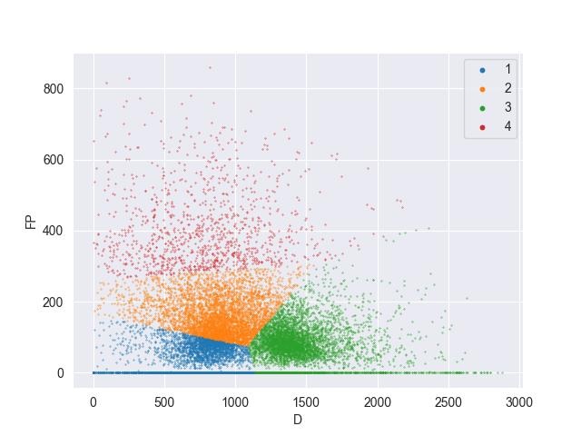
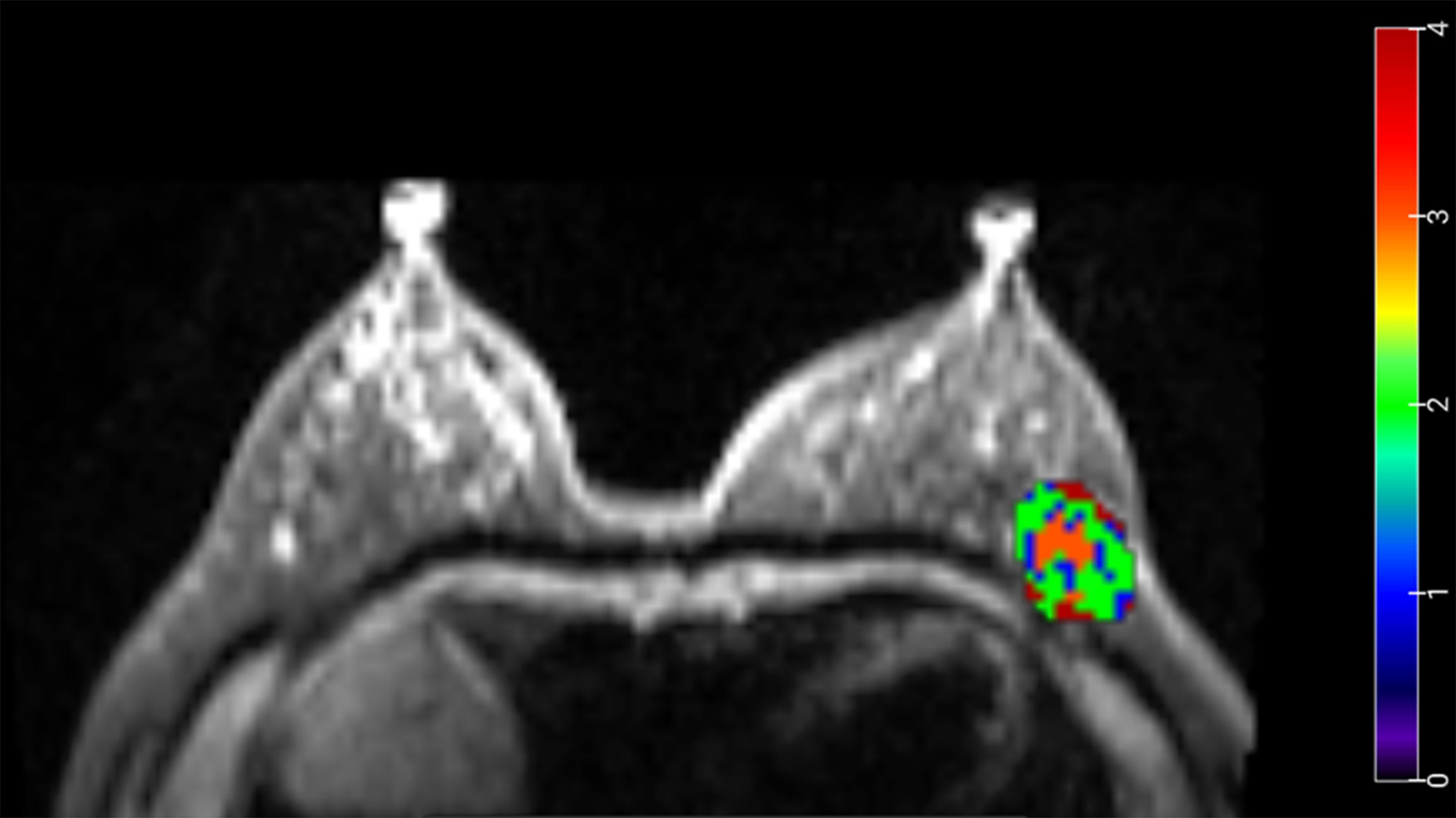
Thirty-five patients of malignant breast cancer were diagnosed with invasive ductal carcinoma by performing surgery and pathology. This included 18 lymph node non-metastasis patients and 17 lymph node metastasis patients. A MRI scan was performed on the breasts of all the patients by using a 3T MR scanner (MAGNETOM Vida, Siemens Healthineers, Erlangen, Germany). The IVIM data was acquired with a single-shot spin-echo echo planar imaging (SE EPI) sequence, with 9 b values of 0, 20, 40, 80, 120, 200, 400, 800, 1200 s/mm2. TR = 6000 ms, TE = 61 ms, slice thickness = 4.5 mm, slice gap = 0.9 mm, FOV = 340×180 mm, matrix = 128 x 128. The matrices of IVIM (D, D* and FP) were calculated with a research software (MR Body Diffusion Toolbox v1.4.0, Siemens Healthineers, Germany). The region of interest (ROI) included the whole tumor on b0 images, and it was manually outlined with a 3D Slicer software (https://www.slicer.org/,version 5.2.2). These three metrics of IVIM were located in each voxel. We performed the consensus clustering (K means) technique. We divided the tumor into habitats of different cellularity and perfusion levels: subregion 1 with low D and low FP (DlowFPlow), subregion 2 with moderate D and moderate FP (DmoderateFPmoderate), subregion 3 with high D and low FP (DhighFPlow), and subregion4 with low D and high FP (DlowFPhigh). The analyses were done with an in-house development software named nnFAE. We extracted the first-order radiomics features of all the four subregions and estimated their mean ± standard deviation. The differences between the two groups were determined with the nonparametric Mann-Whitney U Test. A logistic regression test was performed, and a predictive model was established. It was based on indicators, with significant differences. The predictive performance was assessed by estimating the area under the receiver operating characteristic curve (AUC).Results
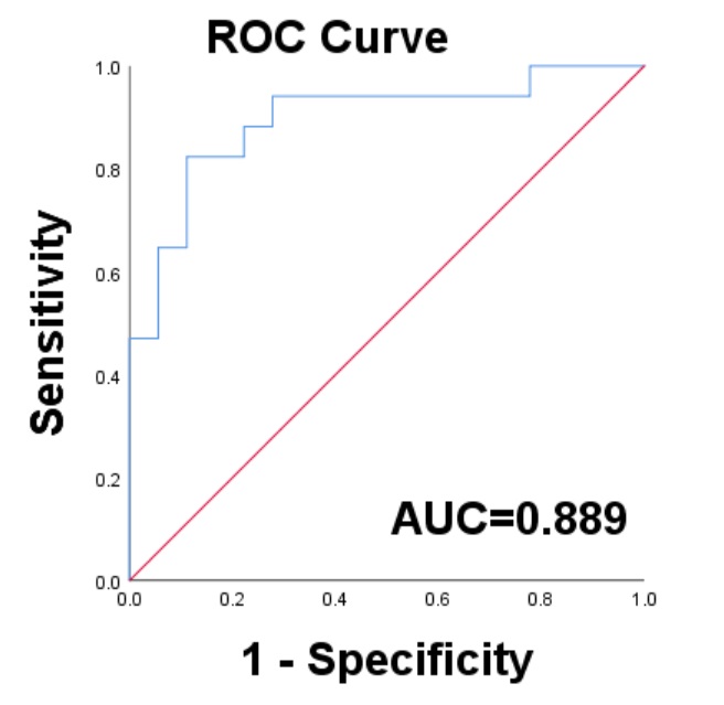
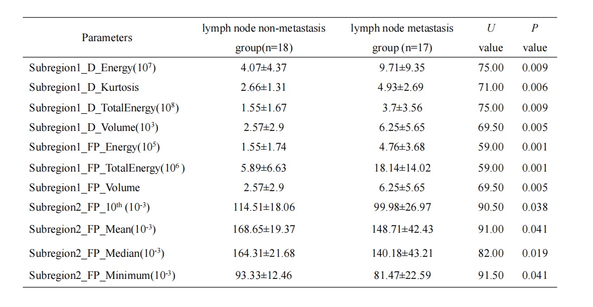
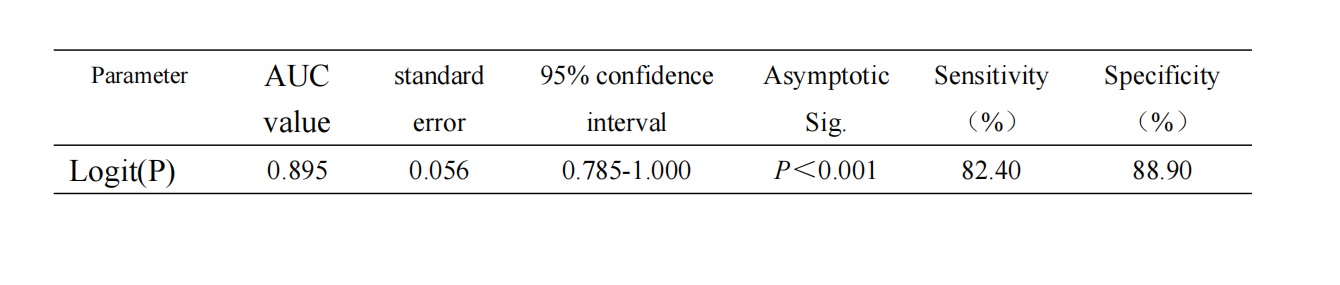
Table 1 shows that significant differences exist in the features of DlowFPlow and DmoderateFPmoderate subregions of the lymph node metastasis group and the non-metastasis group (P<0.05). The logistic regression model was based on these features. It accurately predicted the metastasis of lymph nodes, with the following parameters: an AUC of 0.895, sensitivity of 0.824, and specificity of 0.889 (Table 2, Figure 1).Discussion and conclusion
We evaluated whether IVIM based habitat analysis could be used to predict lymph node metastasis of breast cancer patients. The IVIM metrics (D and FP) reflect the cellularity and the vascularity of a tumor. We found that these could segregate the tumor into different subregions using a clustering method. DlowFPlow and DmoderateFPmoderate subregions represent the hypoperfusion and hypercellularity regions, which constitute the hypoxia. In the DlowFPlow subregion, we observed that the kurtosis, the energy, and the volume of D were higher for the metastatic group. Therefore, the intra-tumor heterogeneity was higher in the metastatic group. In the DmoderateFPmoderate subregion, we observed that the mean, the median, the minimum and 10th Percentile of FP were lower for the metastatic group. These findings imply that with a lower degree of microcirculation and perfusion in tumors, there is an increased lack of oxygen supply and nutrients, and higher tendency of the tumor to degrade and metastasize3,4. The logistic regression model, which was based on the features of habitats, has good performance in predicting in predicting the metastasis of malignant lymph nodes in cancer tissue, which is beneficial for evaluating the prognosis of tumors.Acknowledgements
The authors want to thank these guarantors of this entire study. They also thank all of the researchers who have assisted in data analysis/interpretation.References
1.McDonald ES, Clark AS, Tchou J, et al. Clinical Diagnosis and Management of Breast Cancer. J Nucl Med. 2016;57 Suppl 1:9S-16S. doi: 10.2967/jnumed.115.157834.
2.Mo T, Brandal SHB, Köhn-Luque A, et al. Quantification of Tumor Hypoxia through Unsupervised Modelling of Consumption and Supply Hypoxia MR Imaging in Breast Cancer. Cancers (Basel). 2022 ;14(5):1326. doi: 10.3390/cancers14051326.
3.Mo T, Brandal SHB, Köhn-Luque A, et al. Quantification of Tumor Hypoxia through Unsupervised Modelling of Consumption and Supply Hypoxia MR Imaging in Breast Cancer. Cancers (Basel). 2022 ;14(5):1326. doi: 10.3390/cancers14051326.
4.Jalnefjord O, Montelius M, Arvidsson J, et al Data-driven identification of tumor subregions based on intravoxel incoherent motion reveals association with proliferative activity. Magn Reson Med. 2019 ;82(4):1480-1490. doi: 10.1002/mrm.27820. Epub 2019 May 13.
Figures