2876
Assessment of mesenchymal stromal cells promoting liver regeneration via intravoxel incoherent motion diffusion-weighted imaging1Department of Radiology, Tianjin First Central Hospital, Tian Jin, China
Synopsis
Keywords: fMRI Analysis, Diffusion/other diffusion imaging techniques, intravoxel incoherent motion
Motivation: Scholars are proposing the use of mesenchymal stem cells (MSCs) to promote liver regeneration and prevent postoperative liver function failure; however, no method currently exists for evaluating their effectiveness in vivo.
Goal(s): We assessed whether intravoxel incoherent motion (IVIM) could be used to assess changes in liver regeneration in rats after partial hepatectomy.
Approach: Rats undergoing stem cell therapy underwent imaging examinations at baseline and postoperative days 1, 2, 3, 5, 7, and 14, and were compared with untreated rats.
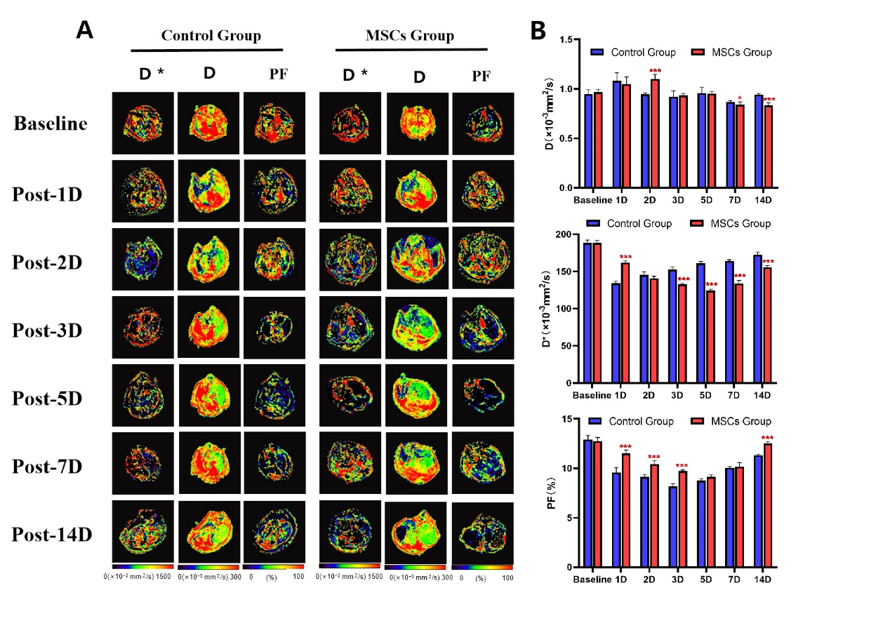
Results: Liver D, D*, and PF differed significantly between control rats and those receiving MSCs at all time points.
Impact: Mesenchymal stem cells can promote liver regeneration by inducing cell hypertrophy and extending the proliferation period of liver cells. IVIM parameters enable monitoring changes in liver blood perfusion after mesenchymal stem cell therapy and show correlations with pathological indicators.
Assessment of mesenchymal stromal cells promoting liver regeneration via intravoxel incoherent motion diffusion-weighted imaging
Introduction: The liver possesses a strong regenerative capacity, and ensuring a certain future liver remnant volume is critical for successful surgery. Inadequate future liver remnant volume can result in insufficient liver regeneration, leading to liver function failure[1,2]. Previous studies have indicated that intravenous administration of mesenchymal stem cells (MSCs) after partial hepatectomy can promote liver regeneration[3,4]. However, the regenerative process following MSC injection remains unclear. In clinical practice, changes in liver volume and liver function recovery are often used to assess post-hepatectomy liver regeneration, but this approach underestimates the risk of liver function failure[5]. Therefore, this study was conducted to investigate the liver regeneration differences between control and MSC-injected rats after partial hepatectomy. Intravoxel incoherent motion (IVIM) diffusion-weighted imaging was used to monitor liver regeneration. The study’s secondary purpose was to determine whether IVIM could be used to evaluate the effect of MSC therapy.Methods: All liver MRI examinations were performed on a 3T MRI scanner (MAGNETOM Prisma, Siemens Healthcare, Erlangen, Germany) with an eight-channel rat-specific coil (Chenguang, Shanghai, China). Seventy rats were randomly divided into the control and treatment groups, with 35 rats per group. All rats underwent a 70% major hepatectomy. Rats in each group were further divided into seven subgroups based on time points (preoperative baseline and postoperative days 1, 2, 3, 5, 7, and 14), with 5 rats per subgroup. During surgery, rats in the treatment group received 2×10⁶ MSCs via the portal vein, and the control group received an equivalent volume of phosphate-buffered saline. Liver IVIM imaging was performed on each group at the corresponding time points. Subsequently, the rats were euthanized, and liver tissue and venous blood were collected to measure liver function assay parameters and the Ki-67 proliferation index of liver cells. IVIM images were acquired using a single-shot echo-planar imaging sequence with repetition time (TR)/echo time (TE): 2300/74 ms, field of view: 120 mm × 98 mm, slice thickness: 3 mm, matrix: 120 × 98, and reconstructed voxel size: 0.5 mm × 0.5 mm × 3 mm. Ten b-values (0, 10, 20, 30, 50, 75, 100, 300, 500, and 800 s/mm2) were applied in three diffusion directions, with an acquisition time of 7 min 12 s. The IVIM-derived parameters, including the true diffusion coefficient (D), pseudodiffusion coefficient (D*) and perfusion fraction (PF), were obtained via a research post-processing software (MR Body Diffusion Toolbox, Siemens Healthcare, Erlangen, Germany). Repeated-measures analysis of variance was used to compare the IVIM-derived parameters in each group at the given time points. Multivariate analysis of variance was used to compare the parameters between both groups at each time point. Correlations between IVIM parameters and histological and blood biochemical indices were analyzed by Spearman analysis. The significance threshold was set at P<0.05.
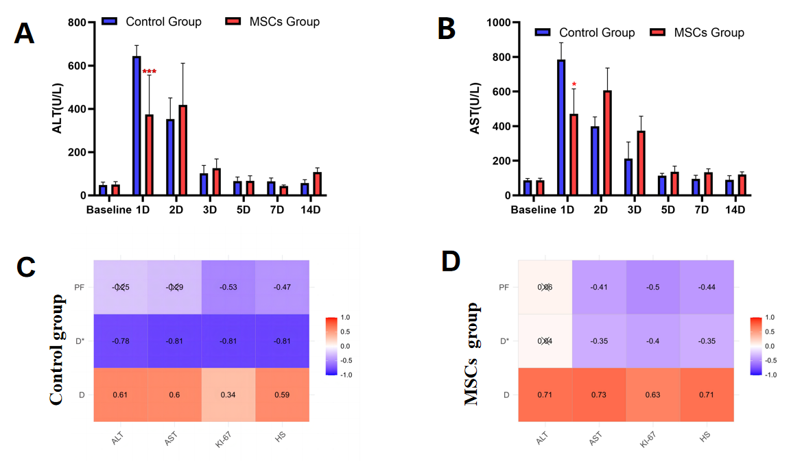
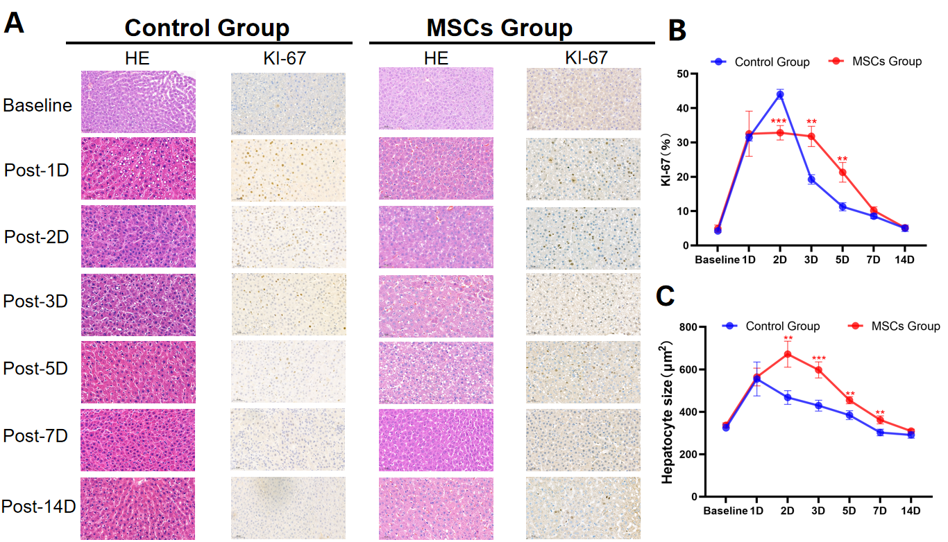
Results: The liver parenchymal D, D*, and PF values differed significantly between the control and treatment groups (P<0.05). In both groups, liver parenchymal D values initially increased after surgery, then decreased. D values differed significantly on postoperative days 2, 7, and 14 (P<0.05). The liver parenchymal D* and PF values in both groups initially decreased after surgery, then increased. The treatment group exhibited slower decreases in D* and PF values than did the control group, and the D* values did not recover to baseline levels on postoperative day 14 (P<0.05; Figure 1). Alanine transaminase (ALT) and aspartate aminotransferase (AST) values in both groups initially increased after surgery, then gradually decreased. On postoperative day 1, ALT and AST values were significantly reduced in the treatment group compared with those of the control group (P<0.05; Figure 2). Both the Ki-67 proliferation index and liver cell size initially increased, then decreased. On postoperative day 2, the treatment group had a lower Ki-67 proliferation index than did the control group, whereas on postoperative days 3 and 5, the Ki-67 proliferation index was higher in the treatment group. Liver cells were larger in the treatment group than in the control group from postoperative days 2–14 (Figure 3). Correlations were observed among the liver parenchymal D, D*, and PF; AST levels, the Ki-67 proliferation index of the liver cells, and liver cell size in the treatment group, with |r| values ranging from 0.35–0.71 (P<0.05; Figure 2).
Conclusion: MSCs can promote liver regeneration by inducing cell hypertrophy and extending the proliferation period of liver cells. Additionally, IVIM parameters allow monitoring changes in liver blood perfusion following MSC therapy, showing a correlation with pathological indicators. IVIM might be used as a non-invasive tool for evaluating the liver regenerative process and MSC therapy.
Acknowledgements
No acknowledgement found.References
1.de Graaf W, Bennink RJ, Heger M, et al. Quantitative assessment of hepatic function during liver regeneration in a standardized rat model. J Nucl Med. 2011;52(2):294-302.
2.Kele PG, de Boer M, van der Jagt EJ, Lisman T, Porte RJ. Early hepatic regeneration index and completeness of regeneration at 6 months after partial hepatectomy. Br J Surg. 2012;99(8):1113-1119.
3.Jun JH, Kim JY, Choi JH, Lim JY, Kim K, Kim GJ. Exosomes from Placenta-Derived Mesenchymal Stem Cells Are Involved in Liver Regeneration in Hepatic Failure Induced by Bile Duct Ligation. Stem Cells Int. 2020;2020:5485738. Published 2020 Oct 9.
4.Li DL, He XH, Zhang SA, Fang J, Chen FS, Fan JJ. Bone marrow-derived mesenchymal stem cells promote hepatic regeneration after partial hepatectomy in rats. Pathobiology. 2013;80(5):228-234.
5.Ribero D, Amisano M, Bertuzzo F, et al. Measured versus estimated total liver volume to preoperatively assess the adequacy of the future liver remnant: which method should we use?. Ann Surg. 2013;258(5):801-807.
Figures