2875
A comparison of four non-Gaussian diffusion models combined with whole-tumor histogram analysis for evaluating cervical cancer1Department of Radiology, Sun Yat-Sen Memorial Hospital, Sun Yat-Sen University, No. 107 Yanjiang Roa, GuangZhou, China, 2MR Research Collaboration, Siemens Healthineers Ltd., Beijing, China, 3MR Research Collaboration, Siemens Healthineers, Guangzhou, China
Synopsis
Keywords: DWI/DTI/DKI, Diffusion/other diffusion imaging techniques
Motivation: Non-Gaussian diffusion models can effectively characterize the microstructure of tissues.
Goal(s): To investigate the potential predictive value of multiple non-Gaussian diffusion models for assessing cervical cancer (CC).
Approach: Diffusion parameters derived from continuous-time random-walk (CTRW), diffusion-kurtosis imaging (DKI), fractional order calculus (FROC) and intravoxel incoherent motion (IVIM) were calculated. The most significant histogram features selected by univariate analysis and multivariate logistic regression were used to establish predictive models. The predictive performance was evaluated by receiver operating characteristic (ROC) analyses.
Results: The combination of multiple non-Gaussian diffusion models and whole-tumor histogram analysis could distinguish pathological types and differentiation degree in CC.
Impact: Predicting pathological types and differentiation degree of cervical cancer is crucial for appropriate treatment and prognosis. The use of multiple non-Gaussian diffusion models combined with whole-tumor histogram analysis offers a precise and non-invasive solution to this clinical issue.
Introduction
In recent years, DWI-derived Gaussian diffusion model has been used to differentiate subtypes and grades of cervical cancer (CC), but showing an unsatisfactory performance1, 2. Non-Gaussian diffusion models can capture more complex non-Gaussian diffusion processes, thereby allowing for a more effective reflection of microstructure3. Several non-Gaussian diffusion models, based on DWI at high b values, such as continuous-time random-walk (CTRW), diffusion-kurtosis imaging (DKI), fractional order calculus (FROC) and intravoxel incoherent motion (IVIM), have been employed to evaluate pathological characteristics in other tumors4, 5. Nevertheless, to our knowledge, no studies have compared the performance of these advanced models in assessing CC. Therefore, the purpose of this study was to investigate the potential predictive value of CTRW, DKI, FROC and IVIM, in combination with histogram analysis, for predicting pathological types and differentiation degree of CC.Methods
The study enrolled 89 women who had been diagnosed with CC and underwent DWI examinations. Participants with incomplete pathological information, poor image quality or rare pathological types were excluded. MR examinations were performed on a 3T MR scanner (MAGNETOM Vida; Siemens Healthcare, Erlangen, Germany). The DWI data were acquired with 16 b-values of 0, 10, 20, 50, 80, 100, 150, 200, 400, 500, 800, 1200, 1500, 2000, 3000 and 4000 s/mm2 by the spin-echo echo-planar imaging (SE-EPI) sequence with fat saturation. The other parameters were as follows: repetition time/echo time = 2,500/84 ms, filed of view = 248 × 248 mm2, matrix size = 124 × 124, slice thickness/gap = 4 mm/0.8 mm, flip angle = 180°, and acquisition time = 3 min and 56 s. The four diffusion models (CTRW, DKI, FROC, IVIM) were generated using the NeuDiLab software rooted in DIPY (Diffusion Imaging In Python, https://dipy.org/). Histogram features were extracted from the segmentation of the whole tumor on each diffusion metric, then the parameters with statistically significant differences were selected by Mann-Whitney U test, and the logistic regression models were established for each individual diffusion models as well as for the combined diffusion models. Receiver operating characteristic (ROC) curves and area under the curves (AUCs) were used to evaluate the predictive performance.Results
The combined model, which incorporated the CTRW, DKI, FROC, and IVIM diffusion models, showed superior diagnostic performance in distinguishing cervical squamous cell cancer (SCC) from cervical adenocarcinoma (CA), with a significant higher AUC (0.836 vs 0.664, 0.642, 0.651, 0.649, respectively; p < 0.05). Regarding the differentiation degree of tumor, the combined model exhibited better predictive performance compared to the DKI model. However, no significant differences were observed in the AUCs among individual models and the combined model (AUC, 0.809, 0.697, 0.747, 0.756, and 0.839, respectively; p > 0.05)Discussion
This study conducted a preliminary feasibility analysis of multiple non-Gaussian diffusion models to evaluate CC. Previous studies have preliminarily applied DKI, FROC and IVIM to assess different subtypes or grades of CC6-11. Nevertheless, the value of CTRW in assessing CC has not been explored, and the comparative performance of CTRW, DKI, FROC, IVIM diffusion models in predicting pathological types and differentiation degree of CC remains unknown. In our study, we applied an extended version of DWI with multi-b value and high b value (a total of 16 b values, ranging from 0 to 4000 s/mm2) in one scan sequence to generate four non-Gaussian diffusion models (CTRW, DKI, FROC and IVIM). The results indicated that these non-Gaussian diffusion models, individually and in combination, can be used to predict pathological types and differentiation degree of CC. The combined model showed the highest predictive performance, indicating the potential of whole-tumor histogram analysis using multiple non-Gaussian diffusion models at high b values in informing future therapeutic strategies for CC. Notably, among the four individual models, both the CTRW model showed the most significant value in predicting pathological types and differentiation degree of CC. This may be attributed to the sensitivity of CTRW diffusion model to alterations of tumor microstructure and heterogeneity, allowing for a more accurate depiction of tumor microstructure and a robust association with pathological findings12. In addition, out of the four individual non-Gaussian diffusion models, DKI model exhibited the poorest predictive performance. This finding was consistent with a prior study that found no significant added value of DKI compared to conventional DWI in evaluating CC13. These results suggested that the DKI model is not adequately reliable for assessing CC, likely due to the susceptibility of kurtosis quantification to noise, motion, and artifacts14.Conclusion
Whole-tumor histogram analysis of multiple non-Gaussian diffusion models is a promising approach for differentiating different pathological types of cervical cancer and their degree of differentiation.Acknowledgements
The authors would like to thank all patients who have participated in this study, and all the investigators who have assisted in data collection.
References
1. Liu, Y., et al., Radiomics analysis of apparent diffusion coefficient in cervical cancer: A preliminary study on histological grade evaluation. J Magn Reson Imaging, 2019. 49(1): p. 280-290.
2. Kuang, F., et al., The value of apparent diffusion coefficient in the assessment of cervical cancer. Eur Radiol, 2013. 23(4): p. 1050-8.
3. Otikovs, M., et al., Diffusivity in breast malignancies analyzed for b > 1000 s/mm(2) at 1 mm in-plane resolutions: Insight from Gaussian and non-Gaussian behaviors. J Magn Reson Imaging, 2021. 53(6): p. 1913-1925.
4. Xu, J., et al., Incorporating multiple magnetic resonance diffusion models to differentiate low- and high-grade adult gliomas: a machine learning approach. Quant Imaging Med Surg, 2022. 12(11): p. 5171-5183.
5. Li, C., et al., Preoperative prediction of VETC in hepatocellular carcinoma using non-Gaussian diffusion-weighted imaging at high b values: a pilot study. Front Oncol, 2023. 13: p. 1167209.
6. Zhang, Q., et al., Whole-tumor texture model based on diffusion kurtosis imaging for assessing cervical cancer: a preliminary study. Eur Radiol, 2021. 31(8): p. 5576-5585.
7. Hou, M., et al., Comparative analysis of the value of amide proton transfer-weighted imaging and diffusion kurtosis imaging in evaluating the histological grade of cervical squamous carcinoma. BMC Cancer, 2022. 22(1): p. 87.
8. Becker, A.S., et al., Assessment of Cervical Cancer with a Parameter-Free Intravoxel Incoherent Motion Imaging Algorithm. Korean J Radiol, 2017. 18(3): p. 510-518.
9. Winfield, J.M., et al., Separation of type and grade in cervical tumours using non-mono-exponential models of diffusion-weighted MRI. Eur Radiol, 2017. 27(2): p. 627-636.
10. Li, B., et al., The utility of APT and IVIM in the diagnosis and differentiation of squamous cell carcinoma of the cervix: A pilot study. Magn Reson Imaging, 2019. 63: p. 105-113.
11. Zhang, A., et al., Value of non-Gaussian diffusion imaging with a fractional order calculus model combined with conventional MRI for differentiating histological types of cervical cancer. Magn Reson Imaging, 2022. 93: p. 181-188.
12. Zhong, Z., et al., High-Spatial-Resolution Diffusion MRI in Parkinson Disease: Lateral Asymmetry of the Substantia Nigra. Radiology, 2019. 291(1): p. 149-157.
13. Wang, M., et al., Diffusion Kurtosis Imaging in the Assessment of Cervical Carcinoma. Acad Radiol, 2020. 27(5): p. e94-e101.
14. Jensen, J.H. and J.A. Helpern, MRI quantification of non-Gaussian water diffusion by kurtosis analysis. NMR Biomed, 2010. 23(7): p. 698-710.
Figures
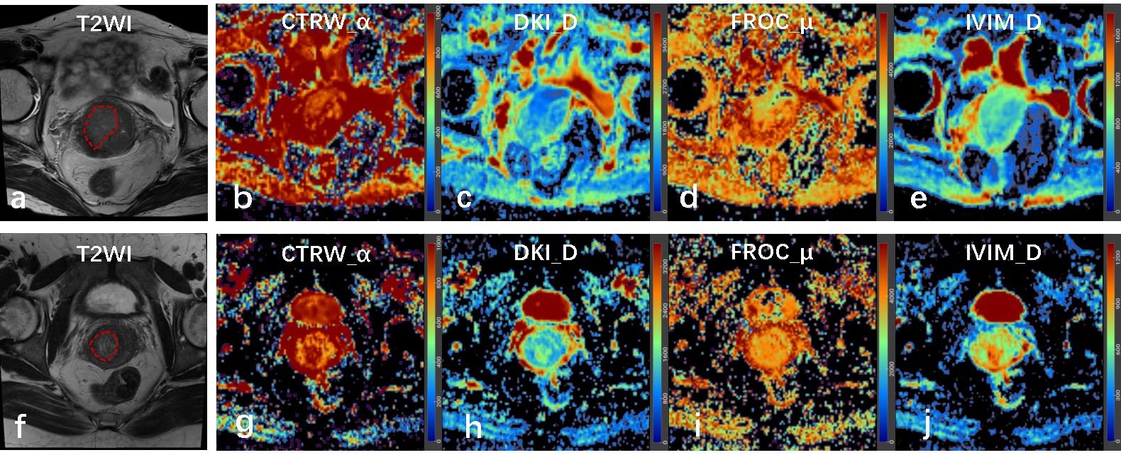
Fig. 1 a-e Images of a 64-year-old female with moderately differentiated SCC. f-j Images of a 40-year-old female with moderately differentiated CA. In these images, a and f are conventional T2-weighted images, the red lines show the lesions included in the volume of interest (VOI); b-e/g-j are pseudo-colorized images showing the CTRW_α (b/g), DKI_D (c/h), FROC_µ (d/i), IVIM_D (e/j) maps derived from continuous-time random-walk (CTRW), diffusional kurtosis imaging (DKI), fractional order calculus (FROC), intravoxel incoherent motion (IVIM), respectively.
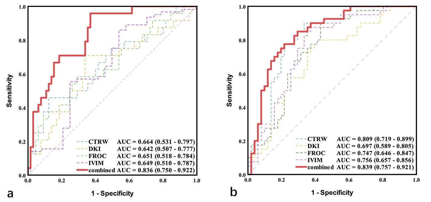
Fig. 2 shows the areas under the receiver operating characteristic curve (AUCs) and 95% CIs (in parentheses) for CTRW, DKI, FROC, IVIM and combined model. The histogram logistical models predict (a) pathological types and (b) differentiation degree in cervical cancer.
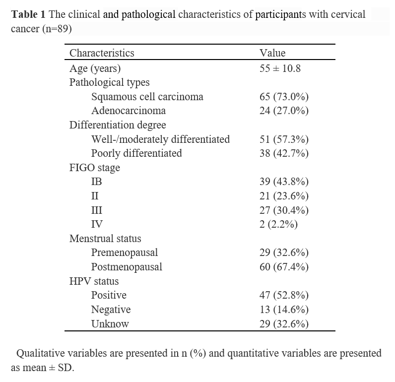
Table 1 The clinical and pathological characteristics of participants with cervical cancer (n=89)
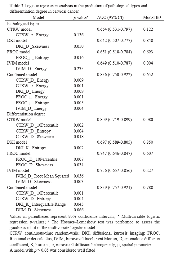
Table 2 Logistic regression analysis in the prediction of pathological types and differentiation degree in cervical cancer
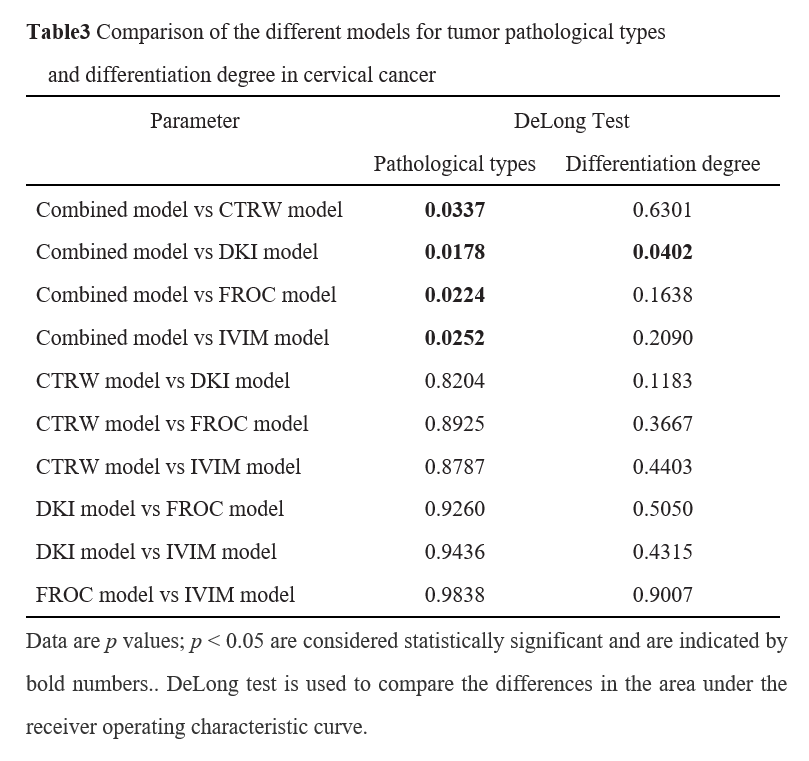
Table3 Comparison of the different models for tumor pathological types and differentiation degree in cervical cancer