2874
Effectiveness of IVIM/DWI MRI parameters in evaluating anti-PD-1 antibody treatment response in xenograft models in vivo1Faculty of Medicine, Kyoto University, Kyoto City, Kyoto, Japan, 2Department of Diagnostic Imaging and Nuclear Medicine, Graduate School of Medicine, Kyoto University, Kyoto City, Kyoto, Japan, 3iACT, Kyoto University Hospital, Kyoto City, Kyoto, Japan, 4Department of Radiology, Kobe City Medical Center General Hospital, Kobe City, Hyogo, Japan, 5Department of Systems Science, Graduate School of Informatics, Kyoto University, Kyoto City, Kyoto, Japan, 6Human Brain Research Center, Graduate School of Medicine, Kyoto University, Kyoto City, Kyoto, Japan, 7NeuroSpin, CEA Paris-Saclay Center, Gif-sur-Yvette, Essonne, France
Synopsis
Keywords: IVIM, Cancer
Motivation: Although intravoxel incoherent motion (IVIM) parameters are garnering attention as a DWI analytical method, their effectiveness in evaluating the immunotherapy efficacy is still largely unvalued.
Goal(s): Our goal was to assess if IVIM/DWI parameters reflect tissue changes subsequent to anti-PD-1 antibody treatment in vivo.
Approach: Colon (CT26) and breast (4T1) cancer xenograft murine models received the treatment and afterward MR study.
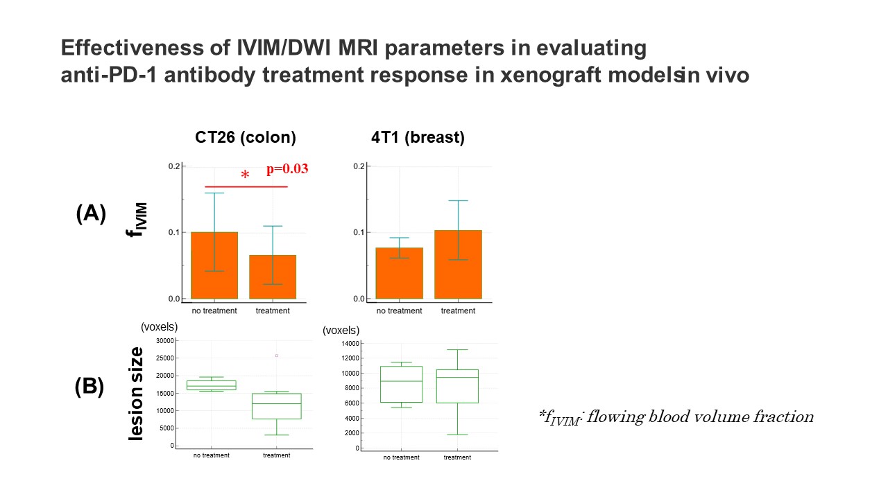
Results: fIVIM (flowing blood volume fraction) was significantly lower in the treated tissue in the CT26 model, while no change was observed in the 4T1 model. Also, there was no difference in the lesion size in both models.
Impact: The difference of fIVIM (flowing blood volume fraction) parameter and lesion size between two anti-PD-1 antibody-treated xenograft models might reflect a different level of treatment efficacy, which may indicate the IVIM potential to observe tissue heterogeneity.
Introduction
The concept of intravoxel incoherent motion (IVIM), encompassing diffusion-weighted imaging (DWI), is sensitive not only on water diffusion in tissues, providing non-invasively localized information on tissue microstructure, but also on blood microcirculation (microperfusion). Perfusion-driven IVIM effects result from blood flow in pseudo-randomly oriented capillary segments, hence depending on the local density of capillaries and small vessels segments.Although IVIM MRI has gradually proven its usefulness in evaluating oncologic tissue characteristics such as cellularity through its diffusion component and microvascularization through its perfusion component1, investigations on its potential to evaluate treatment efficacy are still lacking, particularly in the field of immunotherapy, such as anti-PD-1 antibody therapy. Anti-PD-1 antibody therapy is a general term for antibody pharmaceuticals that block the PD-1 receptor on T-cells, inhibiting immune suppression signals2. It is now considered one of the most successful cancer immunotherapies. However, it also presents challenges, such as high costs and varying individual efficacy2,3.
Therefore, in this study, we aimed to assess the potential of IVIM and non-Gaussian DWI parameters and whether they are sensitive to anti-PD-1 antibody therapy efficacy in colon and breast cancer xenograft models.
Methods
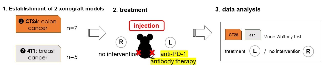
Tumor models:Seven and five BALB/c mice were subcutaneously injected with 1×10⁶ CT26 and 4T1 cells, respectively, in each hind limbs. 7 days after the injection, they started to receive treatment on one side, the other side remaining untreated as a control: the left side was treated with the subcutaneous injection of anti-PD-1 antibody, while the right side received no treatment. The details are shown in Figure1.
Image acquisition:
MR study was carried out on them after the injection in vivo. The imaging was conducted on a 7T MRI scanner (Bruker, Germany). The SE-EPI acquisition parameters were set as follows: resolution 250×250μm2, matrix size 100×100, field of view 25×25 mm2, slice thickness 1.5 mm, TE=57ms, TR=2500 ms, 8 averages, 4 segments. IVIM/DW images were acquired using a diffusion time of 27.6ms and 19 b values (7-4105 s/mm2). The acquisition time was 22 min 40 sec. fIVIM, D* as well as ADCo and K values were estimated using the combined IVIM/non-Gaussian diffusion kurtosis model4.
Statistical analysis:
MRI data analysis was performed using a code developed in Matlab (Mathworks, Natick, MA). Diffusion parameters were compared between the treated and untreated side of each xenograft model using Mann-Whitney test.
Results
We observed significantly lower fIVIM values on the treated tumor side compared to the untreated side in the CT26 model (p=0.03). The lesion size (estimated from the lesion voxels count) also tended to be smaller, although the difference was not statistically significant. In the 4T1 group, there was no significant difference in fIVIM values or lesion size (Fig.2). All other diffusion parameters, ADCo and K, did not show significance between the treatment and control groups.Discussion
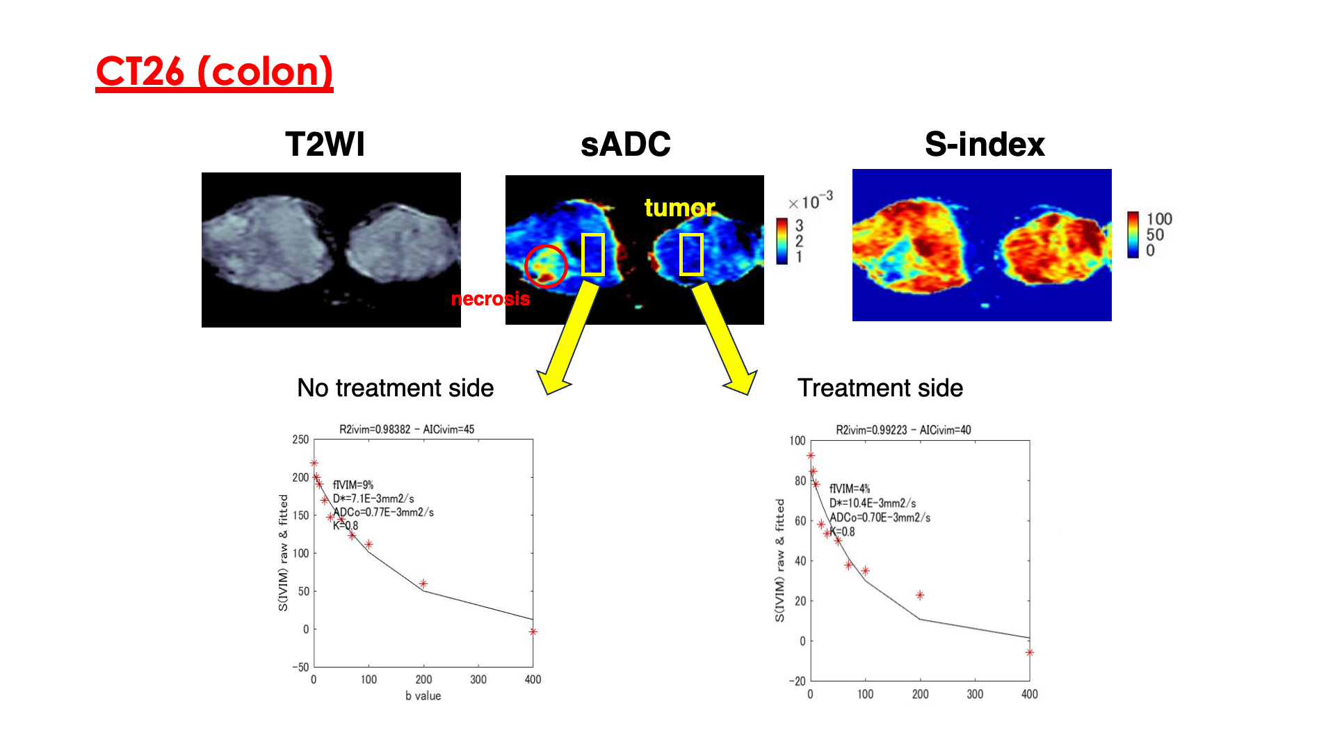
A decrease of fIVIM, representing the flowing blood volume fraction, indicates a decrease in microperfusion in the treated tissue in the CT26 group. However, there was no significant difference in fIVIM in the 4T1 group. The lesion size tended to be smaller in CT26, while there was not a significant difference in the 4T1 group. These differences, depending on the tumor type, might reflect different levels of treatment efficiency on tumors. Interestingly, the absence of changes in the diffusion parameters suggest that at this stage of treatment no change in tissue microstructure has yet occurred, besides changes in microcirculation. The relationship between microvessel density and PD-1 treatment remains poorly investigated with widespread results. IVIM MRI has the potential to provide such information non-invasively and on the form of maps (Fig.3), an essential tool to take into account tumor heterogeneities.Conclusion
fIVIM from IVIM MRI may have the potential to non-invasively reflect tissue microvascular structural changes induced by anti-PD-1 antibody treatment in vivo. Our results which need to be confirmed with a larger cohort of subjects already suggest the need for further in-depth research into the link of IVIM parameters and underlying tissue microvascularization, and their clinical relevance to evaluate immunotherapeutic efficacy in cancers.Acknowledgements
We express our gratitude to Ms. Setsuko Inoue for her invaluable technical support in conducting the animal experiments. This study was supported by AMED grant (23he0422025j0002).References
1. Le Bihan D. What can we see with IVIM MRI? NeuroImage. 2019;187:56–67.
2. Chamoto K, et al. Current issues and perspectives in PD-1 blockade cancer immunotherapy. Int J Clin Oncol. 2020;25(5):790-800.
3. Chiang C, et al. Cost-effectiveness of Pembrolizumab as a Second-Line Therapy for Hepatocellular Carcinoma. JAMA Netw Open. 2021;4(1):e2033761.
4. Someya Y, et al. Quantitative non- Gaussian diffusion and intravoxel incoherent motion magnetic resonance imaging: differentiation of malignant and benign breast lesions. Invest Radiol 2015;50:205–211.
Figures