2869
Multi-tissue constrained spherical deconvolution with spherical mean density estimation1Biomedical Engineering, School of Biomedical Engineering and Imaging Sciences, King's College London, London, United Kingdom, 2Centre for the Developing Brain, School of Biomedical Engineering and Imaging Sciences, King's College London, London, United Kingdom
Synopsis
Keywords: Diffusion Reconstruction, Diffusion/other diffusion imaging techniques
Motivation: Multi-tissue constrained spherical deconvolution (MT-CSD) average densities depend on the local fibre arrangement, as it relies on the non-negativity of the fibre orientation density functions (fODF).
Goal(s): We investigate whether MT-CSD can be performed without influence of the orientation structure of fibre ODF.
Approach: Using the spherical mean of each b-value shell to factor out the angular structure, we rely purely on the different tissue types' b-value dependence, with the angular part of the fODF estimated subsequently .
Results: The method produces plausible, but subtly different results from 'regular' MT-CSD, which appear to be less affected by fibre arrangement. It is also substantially faster.
Impact: The ability to decompose the signal based purely on its spherical mean b-value dependence may provide more reliable density estimates, though this needs further investigation. The computational efficiency of this approach may also make it more suitable for real-time use.
Introduction
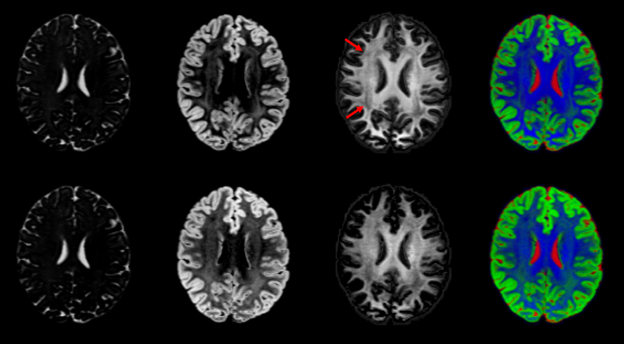
Multi-tissue constrained spherical deconvolution [1] is widely used to analyse multi-shell diffusion MRI data, and provides a decomposition of the signal into a set of orientation density functions (ODF). Typically, these correspond to a 'WM-like' fibre ODF, and scalar 'GM-like' and 'CSF-like' densities with no orientation structure. The use of a non-negativity constraint on the fibre ODF introduces a dependence of the estimated densities on the exact orientation structure of the fibre ODF, which can be observed in the data (Figure 1).Here, we investigate the possibility of performing MT-CSD using the spherical means of each b-value shell only as a first step to estimate the mean densities for each constituent, and subsequently estimate the remaining angular part of the fibre ODF while holding the mean densities fixed.
Methods
MT-CSD is typically performed in the spherical harmonic (SH) domain, and involves solving a least-squares problem with inequality constraints:$$
f' = \min_f || Hf - s ||^2 \quad \textrm{s.t.} \quad Af \geq 0
$$
where $$$f$$$ is the vector of all coefficients for all tissue types, $$$s$$$ is the vector of all measurements across all b-values, and $$$H$$$ performs the spherical convolution, summation of the contributions from all the tissue types, and mapping to the orientations and b-values acquired. The matrix $$$A$$$ maps each set of coefficients in $$$f$$$ to their corresponding amplitudes along a dense set of orientations.
The spherical mean of each b-value shell has been shown to remove any influence from the angular structure of an ODF [2]. If the different tissue types have sufficiently distinct b-values dependencies, it should be possible to perform the decomposition based on per-shell mean signal. We can therefore partition $$$H$$$ & $$$f$$$ into their spherical mean terms ($$$l=0$$$ SH coefficients) and angular terms ($$$l>0$$$ SH coefficients):
$$
f_0' = \min_{f_0}|| \begin{pmatrix} H_0 & H_1 \end{pmatrix} \begin{pmatrix} f_0 \\ f_1 \end{pmatrix} - s ||^2 \quad \textrm{s.t.} \quad \begin{pmatrix} A_0 & A_1 \end{pmatrix} \begin{pmatrix} f_0 \\ f_1 \end{pmatrix} \geq 0
$$
Where $$$f_0$$$ and $$$f_1$$$ correspond to the $$$l=0$$$ and $$$l>0$$$ SH coefficients respectively, and likewise for the matrices $$$H$$$ & $$$A$$$. This can be written as:
$$
f_0' = \min_{f_0}|| H_0 f_0 - (s - H_1 f_1) ||^2 \quad \textrm{s.t.} \quad A_0 f_0 \geq 0
$$
and solved readily using the same inequality-constrained least-squares solver as previously.
Having estimated the mean densities, these can be held fixed as the orientation structure of the fibre ODF is recovered. This step can be performed using a non-negativity regulariser rather than a hard constraint (as used in the original implementation of CSD [3]), which has the advantage of being much more computationally expedient, and also allows for slightly higher angular resolution by allowing some negativity in the fibre ODF (some ringing is expected due to the truncation of the SH series):
$$
f_1' = \min_{f_1} || H_1 f_1 - (s - H_0 f_0) ||^2 + \lambda || A_0 f_0 + A_1 f_1 ||_+^2
$$
where the norm of the regulariser $$$|| \cdot ||_+^2$$$ is computed using only those directions where the amplitudes are negative (see [3] for details).
This approach was implemented within the MRtrix3 framework and tested on an example subject from the Human Connectome Project (2×90 directions per shell, b = 1000, 2000, 3000 s/mm², 1.25mm isotropic resolution) [4].
Results & Discussion
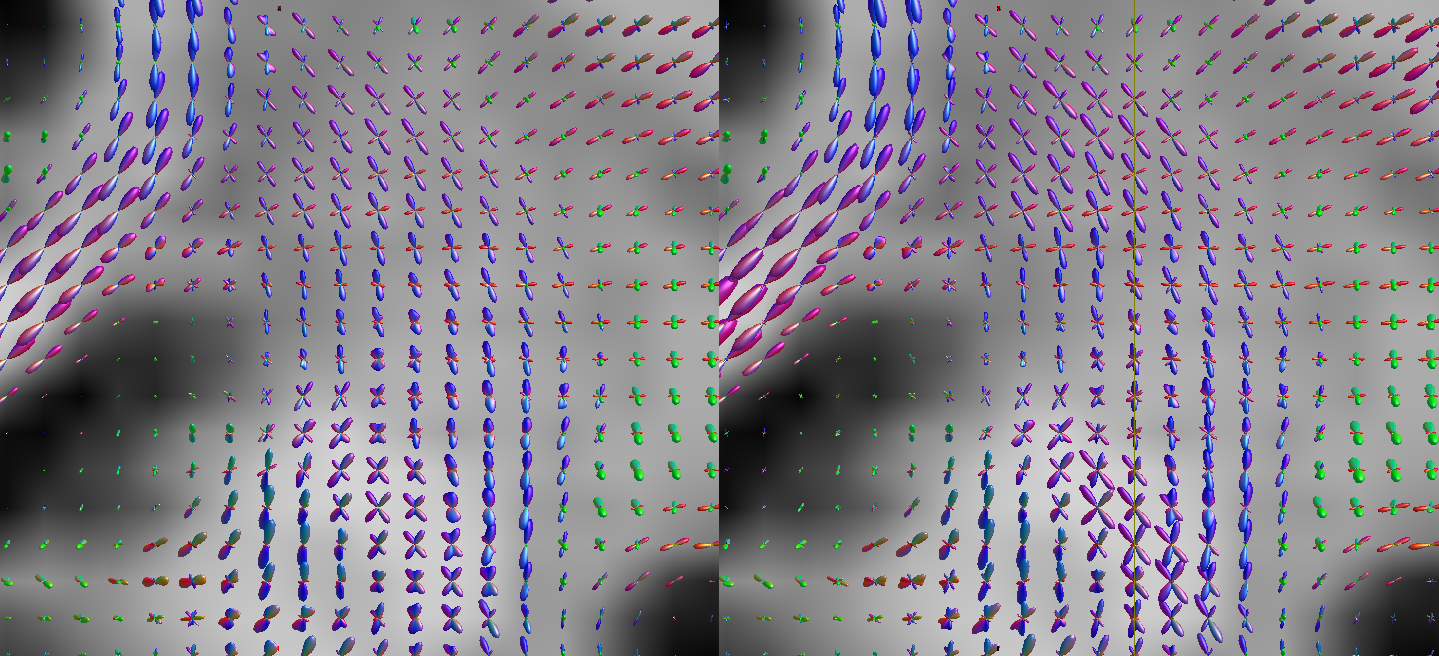
The results obtained using the proposed modification are similar but not identical to those obtained using regular MT-CSD. The mean densities suggest that there is indeed a reduction in the effects of the fibre configurations (red arrows in Figure 1), though there is also a clear increase in the estimated GM-like density within white matter regions, along with a notable reduction the estimated WM-like densities in cortical regions. However, the orientation structure of the fibre ODF is comparable to regular MT-CSD, if anything with improved angular resolution, at the cost of some negativity in the fibre ODFs (Figure 2).In terms of computational performance, processing time was 2m10s for MT-CSD compared to 11s for the proposed modification.
Conclusion
The proposed approach offers some advantages in terms of computation, invariance of the estimated densities to the angular structure of the fibre ODF, and improved angular resolution of the fibre ODF. It does however seem to produce higher GM-like densities in white matter regions, and some loss of WM-like density in cortical regions compared to MT-CSD, which warrants some caution and further investigation.Acknowledgements
This work was supported by EPSRC grant EP/W030411/1 (MRtrix project), ERC grant agreement no. 319456 (dHCP project), by core funding from the Wellcome/EPSRC Centre for Medical Engineering [WT203148/Z/16/Z] and by the National Institute for Health Research (NIHR) Clinical Research Facility based at Guy’s and St Thomas’ NHS Foundation Trust and King’s College London. The views expressed are those of the author(s) and not necessarily those of the NHS, the NIHR or the Department of Health and Social CareReferences
[1] B. Jeurissen, J.-D. Tournier, T. Dhollander, A. Connelly, and J. Sijbers, ‘Multi-tissue constrained spherical deconvolution for improved analysis of multi-shell diffusion MRI data’, Neuroimage, vol. 103, pp. 411–426, Dec. 2014, doi: 10.1016/j.neuroimage.2014.07.061.
[2] E. Kaden, F. Kruggel, D.C. Alexander, ‘Quantitative mapping of the per‐axon diffusion coefficients in brain white matter’, Magnetic Resonance in Medicine, vol. 75, no. 4, pp. 1752–1763, May 2015, doi: 10.1002/mrm.25734.
[3] J.-D. Tournier, F. Calamante, and A. Connelly, ‘Robust determination of the fibre orientation distribution in diffusion MRI: Non-negativity constrained super-resolved spherical deconvolution’, NeuroImage, vol. 35, no. 4, pp. 1459–1472, May 2007, doi: 10.1016/j.neuroimage.2007.02.016.
[4] S. N. Sotiropoulos et al., ‘Advances in diffusion MRI acquisition and processing in the Human Connectome Project.’, NeuroImage, vol. 80, pp. 125–43, Oct. 2013, doi: 10.1016/j.neuroimage.2013.05.057.
Figures