2868
Investigating Postmortem Neurite Orientation Dispersion and Density Imaging Parameters and Their Influences on Deceased Human Brains1Institute of Forensic Medicine, Department of Biomedical Engineering, University of Basel, BASEL, Switzerland, 2Institute of Forensic Medicine, Health Department Basel-Stadt, Basel, Switzerland, 3Department of Neurology, Medical University of Graz, Austria, Graz, Austria
Synopsis
Keywords: Microstructure, Ex-Vivo Applications, NODDI
Motivation: Neurite orientation dispersion and density imaging (NODDI) is expected to advance our still incomplete understanding of diffusion properties in postmortem brains.
Goal(s): We examined NODDI parameters in deceased human brains in situ and investigated their associations with postmortem interval (PMI), age at death and core temperature.
Approach: Ten subjects underwent postmortem in situ brain MRI, enabling NODDI analysis. Correlations between NODDI parameters and external factors were assessed.
Results: The results revealed higher NODDI parameters in deceased compared to living subjects. Longer PMIs were associated with increased fractional intracellular volume (FICVF) and orientation dispersion index (ODI) values, while higher temperatures had the opposite effect.
Impact: Our study expands the understanding of postmortem brain microstructure. NODDI's potential in deceased brain analysis and its relation to postmortem interval and temperature pave the way for further research with applications in diagnostics and forensics.
Introduction
Neurite orientation dispersion and density imaging (NODDI) is a powerful technique for assessing the microstructural parameters fractional intracellular volume (FICVF), isotropic water fraction (FISO), orientation dispersion index (ODI) and isotropic restriction fraction (IRFRAC) 1,2. Although NODDI could be beneficial in the noninvasive examination of deceased, its application in postmortem studies is sparse and does not yet exist for postmortem in situ human brains. Therefore, we examined NODDI parameters of deceased human brains in situ and their associations with the external factors postmortem interval (PMI), age at death and core temperature in this study.Materials and Methods
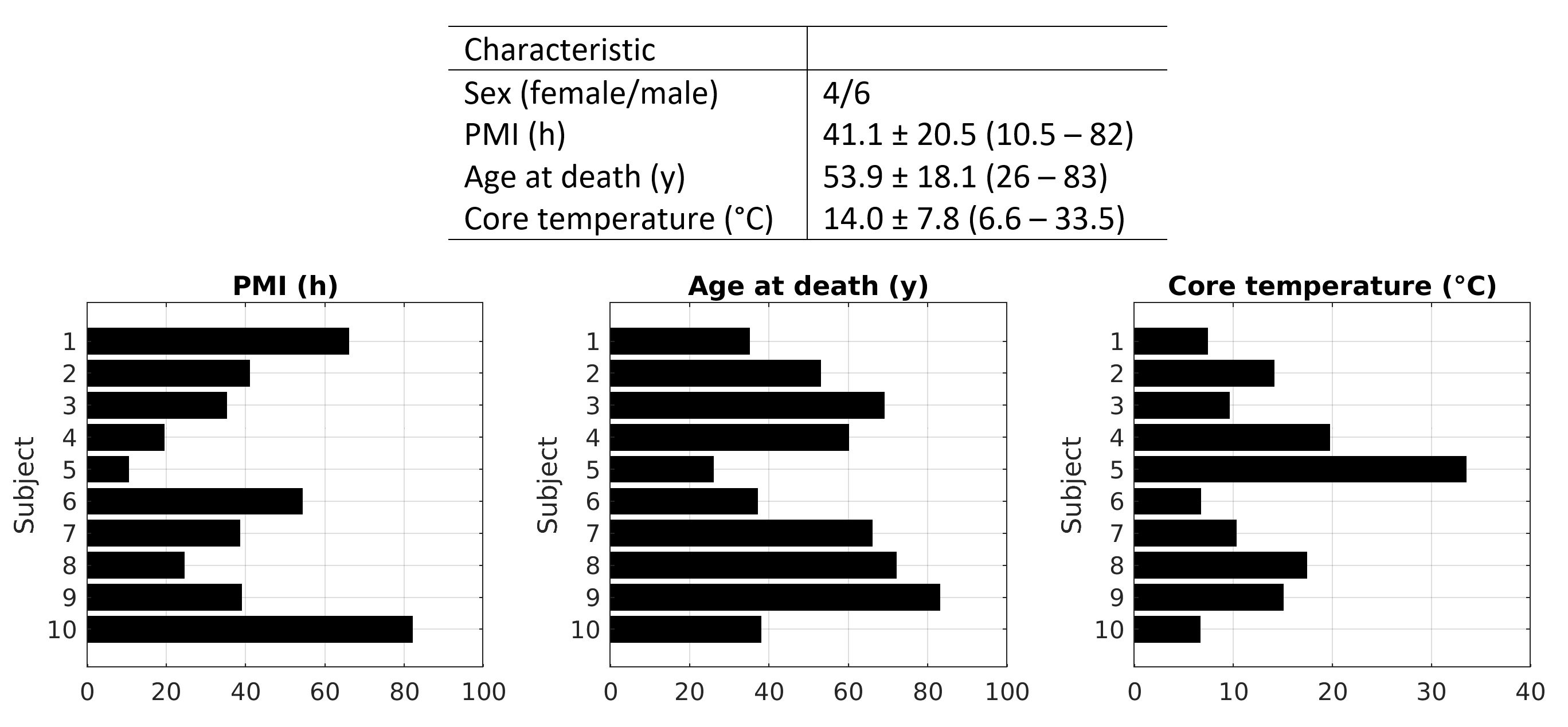
10 deceased subjects with recorded PMI, age at death and core temperature measured directly before the MRI scans were included (Figure 1). Postmortem in situ brain MRIs were performed using a 3 T MRI scanner (Magnetom Prisma, Siemens Healthineers, Erlangen, Germany). A diffusion-weighted single-shot-echo-planar imaging sequence with 64 isotropically distributed directions was acquired at b-values of 2000 s/mm2 and 6000 s/mm2. After eddy current correction, the NODDI analysis was carried out on one central axial brain slice by using the NODDI MATLAB toolbox v1.0.5 1 on MATLAB R2018 and MATLAB R2023b (The MathWorks, Inc., Natick, MA, United States). Due to postmortem data, the model WatsonSHStickTortIsoVIsoDot_B0 was applied. Automatic segmentation of white matter (WM) and gray matter (GM) was achieved on brain extracted b0 volumes using FSL 6.0.0 (FMRIB Software Library, Analysis Group 3). Mean values for FICVF, FISO, ODI and IRFRAC were calculated for each segmented tissue. The Pearson’s and Spearman’s correlations, respectively, between the mean NODDI parameters and PMI, age at death as well as core temperature were assessed.Results
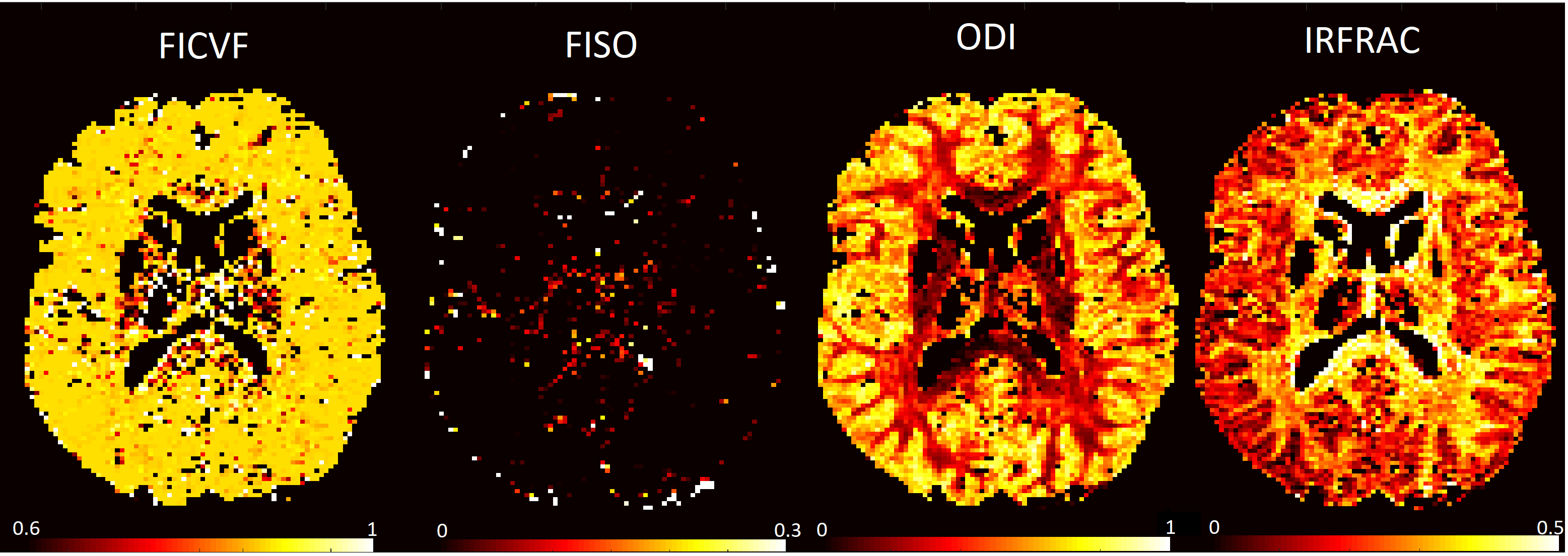
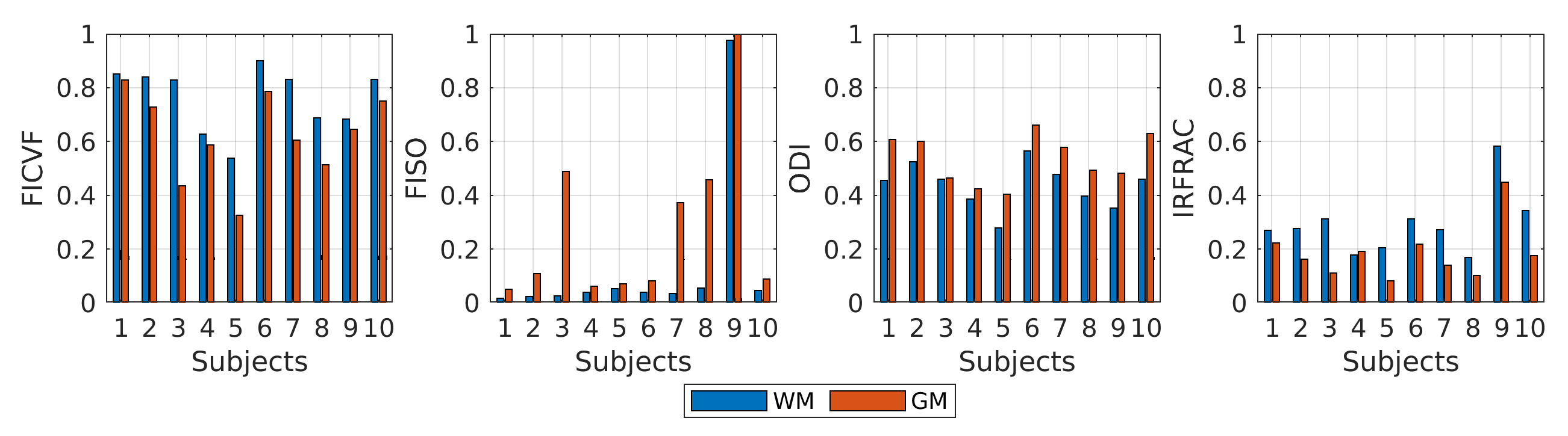
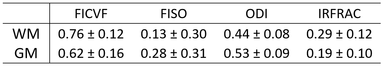
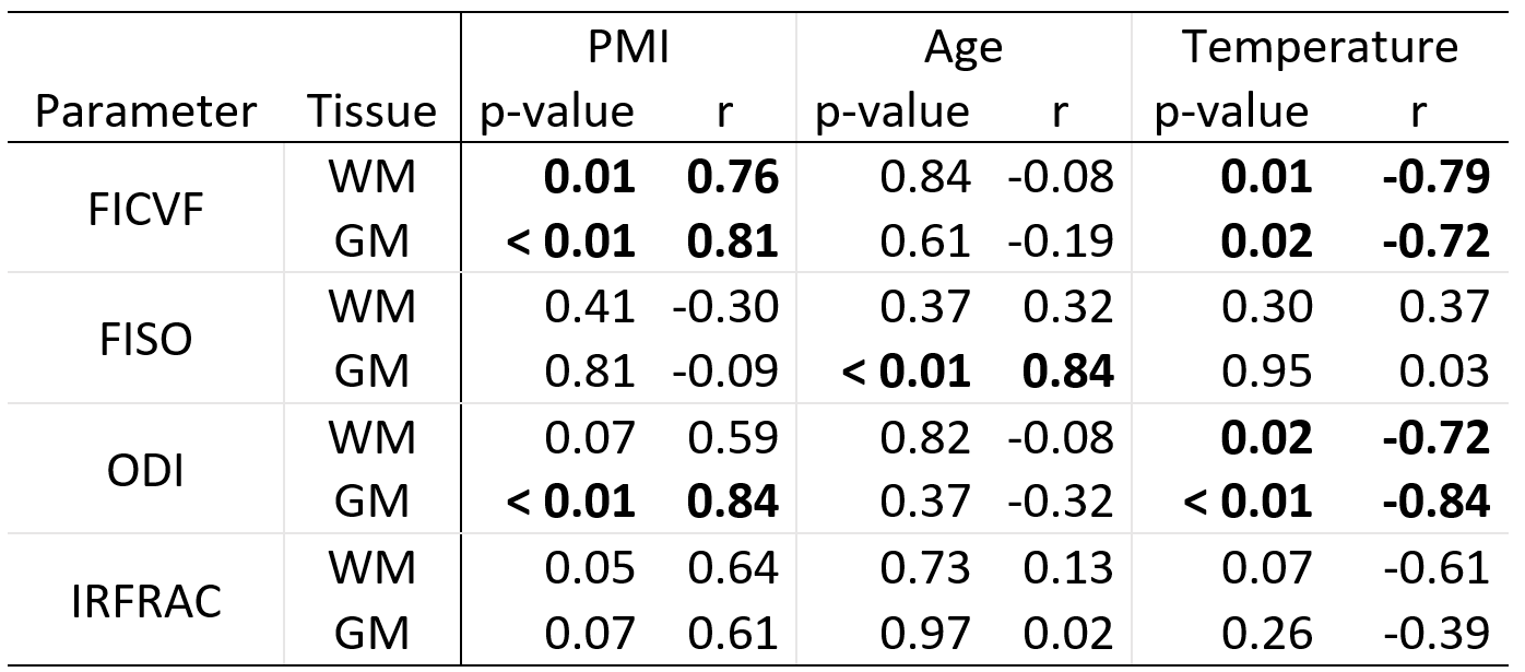
NODDI parameter maps for an exemplary deceased are presented in Figure 2. The bar plots in Figure 3 display the individual NODDI parameters in WM and GM for each subject. Table 1 summarizes the mean NODDI parameter values and their respective standard deviations for all deceased. Statistically significant correlations were observed between PMI and FICVF in both WM and GM, between PMI and ODI in GM, between age at death and FISO in GM and between core temperature and FICVF as well as ODI in WM and GM (Table 2).Discussion
Postmortem NODDI parameters in deceased brains exhibited higher values compared to healthy living subjects with a substantial increase in FICVF and FISO and a smaller increase in ODI 4,5. These findings align with a study involving mice brains in vivo and ex vivo (i.e. fixated) 6. However, the in situ nature of our MRI scans excluded influences of fixation in our study. Longer PMIs correlated with higher FICVF and ODI values as the progressive degradation associated with longer PMI results in cellular swelling and axonal disorganization 7,8. Age related changes in FISO in GM were consistent with previous findings in healthy living patients 9,10. Participant #3 of our study had an excessive brain atrophy (a natural process of brain shrinkage affecting mainly the GM), which was displayed in the exceptional high FISO value in GM. Temperature effects on diffusivity were consistent with the Einstein derivation of the Brownian motion and previous postmortem in situ brain MRI studies 11,12. In our study, FICVF and ODI were temperature dependent, pointing out the necessity to perform temperature corrections. In the case of subject #9 with Parkinson’s disease, increased FISO and IRFRAC values were observed due to sparse tissue structure 13. This mirrors the findings of a study on subjects with Parkinson’s diseases and healthy controls 14.Conclusion
Our study reveals that longer PMIs lead to increased FICVF and ODI values, while higher temperatures have the opposite effect. By definition, FICVF should distinguish vasogenic and cytotoxic brain edema as the intracellular volume fraction is only increased in cytotoxic edema. However, multiple factors influence NODDI parameters postmortem, necessitating a large and balanced sample size in the future to reliably identify individual influences.Acknowledgements
No acknowledgement found.References
1. Zhang H (2012). NODDI: Practical in vivo neurite orientation dispersion and density imaging of the human brain | Elsevier Enhanced Reader. 10.1016/j.neuroimage.2012.03.072.
2. Kleinnijenhuis M, Zhang H, Wiedermann D, Küsters B, Norris DG, and van Cappellen van Walsum A (2013). Detailed laminar characteristics of the human neocortex revealed by NODDI and histology. In.
3. Jenkinson M, Beckmann CF, Behrens TEJ, Woolrich MW, and Smith SM (2012). FSL. NeuroImage 62, 782–790. 10.1016/j.neuroimage.2011.09.015.
4. Andica C, Kamagata K, Hayashi T, Hagiwara A, Uchida W, Saito Y, Kamiya K, Fujita S, Akashi T, Wada A, et al. (2020). Scan–rescan and inter-vendor reproducibility of neurite orientation dispersion and density imaging metrics. Neuroradiology 62, 483–494. 10.1007/s00234-019-02350-6.
5. Chung AW, Seunarine KK, and Clark CA (2016). NODDI reproducibility and variability with magnetic field strength: A comparison between 1.5 T and 3 T. Human Brain Mapping 37, 4550–4565. 10.1002/hbm.23328.
6. Vinh To X, Kurniawan ND, Cumming P, and Nasrallah FA (2023). A cross-comparative analysis of in vivo versus ex vivo MRI indices in a mouse model of concussion. Brain Research 1820, 148562. 10.1016/j.brainres.2023.148562.
7. D’Arceuil H, and de Crespigny A (2007). The effects of brain tissue decomposition on diffusion tensor imaging and tractography. Neuroimage 36, 64–68. 10.1016/j.neuroimage.2007.02.039.
8. Shepherd TM, Flint JJ, Thelwall PE, Stanisz GJ, Mareci TH, Yachnis AT, and Blackband SJ (2009). Postmortem interval alters the water relaxation and diffusion properties of rat nervous tissue--implications for MRI studies of human autopsy samples. Neuroimage 44, 820–826. 10.1016/j.neuroimage.2008.09.054.
9. Merluzzi AP, Dean III DC, Adluru N, Suryawanshi GS, Okonkwo OC, Oh JM, Hermann BP, Sager MA, Asthana S, Zhang H, et al. (2016). Age-dependent differences in brain tissue microstructure assessed with neurite orientation dispersion and density imaging. Neurobiology of Aging 43, 79–88. 10.1016/j.neurobiolaging.2016.03.026.
10. Billiet T, Vandenbulcke M, Mädler B, Peeters R, Dhollander T, Zhang H, Deprez S, Van den Bergh BRH, Sunaert S, and Emsell L (2015). Age-related microstructural differences quantified using myelin water imaging and advanced diffusion MRI. Neurobiology of Aging 36, 2107–2121. 10.1016/j.neurobiolaging.2015.02.029.
11. Einstein A (1926). Investigations On The Theory Of The Brownian Movement.
12. Berger C, Bauer M, Wittig H, Scheurer E, and Lenz C (2022). Post mortem brain temperature and its influence on quantitative MRI of the brain. Magn Reson Mater Phy 35, 375–387. 10.1007/s10334-021-00971-8.
13. Kamiya K, Hori M, and Aoki S (2020). NODDI in clinical research. Journal of Neuroscience Methods 346, 108908. 10.1016/j.jneumeth.2020.108908.
14. Kamagata K, Zalesky A, Hatano T, Ueda R, Di Biase MA, Okuzumi A, Shimoji K, Hori M, Caeyenberghs K, Pantelis C, et al. (2017). Gray Matter Abnormalities in Idiopathic Parkinson’s Disease: Evaluation by Diffusional Kurtosis Imaging and Neurite Orientation Dispersion and Density Imaging. Hum Brain Mapp 38, 3704–3722. 10.1002/hbm.23628.
Figures