2867
Repeatability and reproducibility of diffusional variance decomposition (DIVIDE) imaging1Radiology, The University of Tokyo, Tokyo, Japan, 2Radiology, The University of Tokyo Hospital, Tokyo, Japan, 3GE HealthCare, Tokyo, Japan
Synopsis
Keywords: DWI/DTI/DKI, Diffusion/other diffusion imaging techniques
Motivation: Novel Multidimensional diffusion encoding (MDE) technique, diffusional variance decomposition (DIVIDE), may provide more detailed insights into tissue microstructure.
Goal(s): To evaluate the feasibility of DIVIDE imaging for human brain.
Approach: Ten healthy-subjects underwent MDE (2D-EPI sequence with 29 linear and 26 spherical b-tensors) twice using 3T-MRI. Regional values of 20 ROIs was extracted for 10 DIVIDE metrics. Coefficient of variation (CV) and interclass correlation coefficient (ICC) were calculated.
Results: Intra-subject CV was less than 5% in almost all regional metrics. Intra-subject CV was lower than that of inter-subject CV in all regional metrics. ICC showed almost perfect agreements for almost all regional metrics.
Impact: Recently developed MDE technique, diffusional variance decomposition (DIVIDE), may be reliably used for measuring diffusion metrics with a potential to provide more detailed insights into tissue microstructure in complex tissues, such as crossing or kissing fiber configurations in the brain.
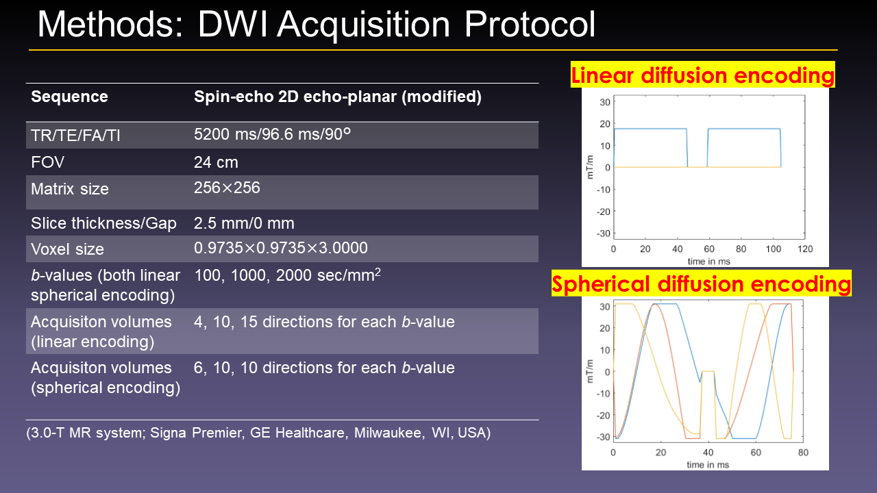
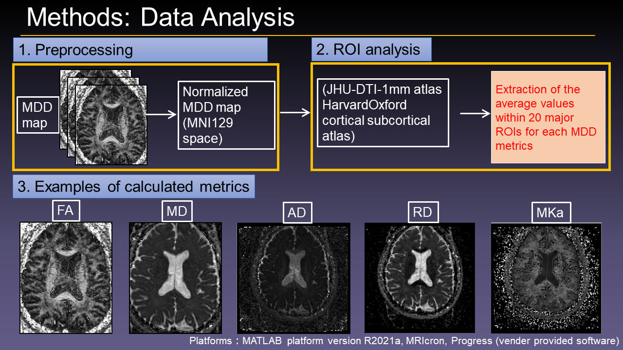
Materials and Methods: Ten healthy subjects (8 men; mean age 29 years, range 23-37 years) were included in this study. During March 2021 to June 2021, whole-brain diffusion-weighted imaging was performed using MDE twice with an interval of more than 1 week using a 3T scanner with a 48-channel head coil. The DIVIDE sequence is a modification of the spin-echo 2D echo-planar imaging (EPI). Imaging parameters were as follows: TR/TE = 5200/96.6 ms, FA = 90°, 60 slices without gaps, FOV = 256×256 mm2, spatial resolution = 2 × 2 × 2.5 mm2, bandwidth = 3906 Hz/pixel, parallel imaging factor = 2 (anterior-posterior), partial-Fourier factor = 0.75 . MDE encoding was performed with 29 linear and 26 spherical b-tensors in an interleaved fashion using optimized gradient waveforms for b = 100, 1000, 2000 s/mm2 with 4, 10, 15 directions, respectively, for linear encodings and with 6, 10, 10 directions, respectively, for spherical encodings, giving a total scan time about 5 min. Automated extraction of 20 major regions of interest (ROI) based on JHU DTI-based white-matter atlas was performed to obtain DIVIDE metrics including fractional anisotropy (FA), mean diffusivity (MD), axial diffusivity (AD), radial diffusivity (RD), anisotropic mean kurtosis (MKA), isotropic mean kurtosis (MKI), total mean kurtosis (MKT), and microscopic fractional anisotropy (μFA). The coefficient of variation (CV) and interclass correlation coefficient (ICC) were calculated to assess the intra-subject (scan-rescan) and inter-subject reproducibilities of the regional DIVIDE metrics.
Results: The intra-subject CV was less than 5% in 20/20 ROIs for FA, 20/20 for MD, 20/20 for AD, 19/20 for RD, 18/20 for MKA, 13/20 for MKI, 19/20 for MKT, 20/20 for μFA. The intra-subject CV was lower than that of inter-subject CV in all the metrics of all the regions. The ICC showed almost perfect agreements for all of the regional values of FA, MD, AD, RD, MKA, MKT, μFA. The ICC showed substantial to almost perfect agreement in 17/20 ROIs for MKI.
Conclusion: Scan-rescan repeatability was acceptable in all the metrics. DIVIDE may be reliably used for measuring diffusion metrics.
Acknowledgements
No acknowledgement found.References
1.Lampinen B, Szczepankiewicz F, Mårtensson J, et al. Neurite density imaging versus imaging of microscopic anisotropy in diffusion MRI: A model comparison using spherical tensor encoding. Neuroimage. 2017;147:517-531.
2.Westin CF, Knutsson H, Pasternak O, et al. Q-space trajectory imaging for multidimensional diffusion MRI of the human brain. Neuroimage. 2016;135:345-62.
3.Li S, Zheng Y, Sun W, et al. Glioma grading, molecular feature classification, and microstructural characterization using MR diffusional variance decomposition (DIVIDE) imaging. Eur Radiol. 2021;31:8197-8207.
4.Szczepankiewicz F, Lasič S, van Westen D, et al., Quantification of microscopic diffusion anisotropy disentangles effects of orientation dispersion from microstructure: applications in healthy volunteers and in brain tumors. Neuroimage. 2015;104:241-52.
5.Lawrenz M, Brassen S, Finsterbusch J. Microscopic diffusion anisotropy in the human brain: reproducibility, normal values, and comparison with the fractional anisotropy. Neuroimage. 2015;109:283-97.
Figures