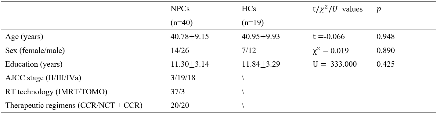
2864
Dynamic Changes of Cortical Microstructure in Nasopharyngeal Carcinoma Patients After Radiotherapy: A Multi-Shell Diffusion Imaging Study1School of Biomedical Engineering, Southern Medical University, Guangzhou, China, 2Departments of Nuclear Medicine, The First Affiliated Hospital of Sun Yat-Sen University, Guangzhou, China, 3Department of Medical Imaging, Sun Yat-sen University Cancer Center, Guangzhou, China
Synopsis
Keywords: DWI/DTI/DKI, Gray Matter, Nasopharyngeal carcinoma, radiation-induced brain injury, GM-based spatial statistics
Motivation: Radiotherapy for nasopharyngeal carcinoma induces brain structural abnormalities, altering cortical microstructure.
Goal(s): Longitudinally explore dynamic cortical microstructure changes over one year after Radiotherapy.
Approach: We performed GM-based spatial statistics analysis (GBSS) on DTI and NODDI data, utilizing nonparametric permutation inference to identify dynamic alterations in diffusion metrics. Cortical regions were located in gray matter clusters by referring to the Desikan-Killiany atlas.
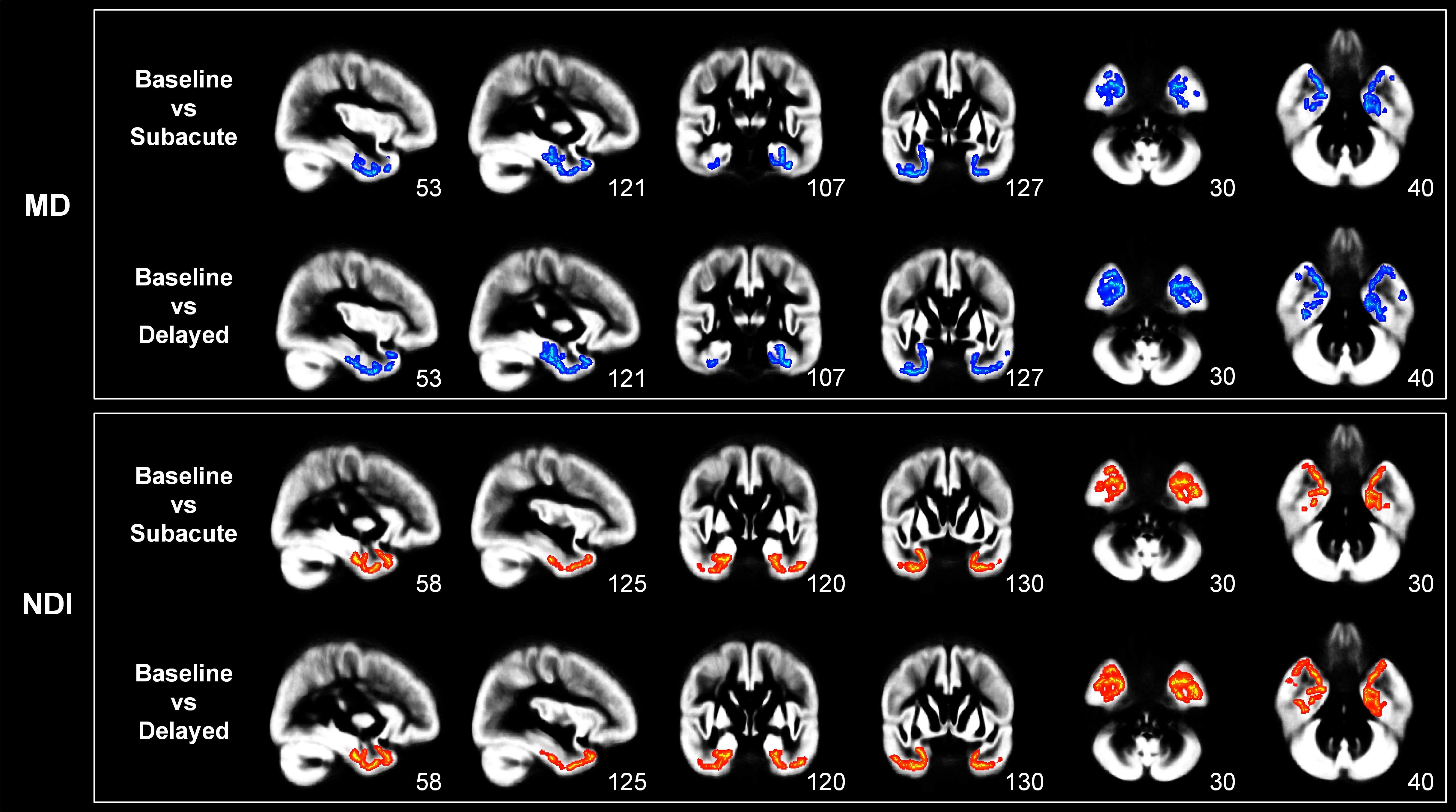
Results: Bilateral temporal lobe MD reduced significantly, and NDI increased notably at six months post-RT, and these changes remained with larger clusters at 12 months. The affected gray matter regions included the entorhinal cortex, temporal pole, inferior temporal gyrus, and fusiform gyrus.
Impact: The combination of DTI and NODDI allows for a more comprehensive understanding of the patterns of cortical microstructural changes induced by RT, offering the potential for early interventions in radiation-induced brain injury.
Introduction
Nasopharyngeal carcinoma (NPC) is prevalent in South China and Southeast Asia, commonly treated with radiotherapy (RT) [1]. Despite its tumor-targeted effectiveness, RT damages brain tissue, causing cognitive dysfunctions. Post-RT periods include acute (days–weeks); subacute (3–6 months); and delayed (>6 months) periods [2]. Diffusion-weighted imaging (DWI) non-invasively assesses nerve cell integrity and brain tissue microstructure using water molecule diffusion patterns [3]. Most studies adopted the diffusion tensor imaging (DTI) model to detect the changes of white matter microstructure in RT-induced brain injury research. However, DTI model assumes Gaussian diffusion, limiting cortical gray matter analysis due to signal contamination from adjacent cerebrospinal fluid (CSF) [4]. Recent advancements in diffusion models have enabled more precise in vivo examination of gray matter (GM) microstructure. We utilized neurite orientation dispersion and density imaging (NODDI) [5] to enhance MRI sensitivity, capturing detailed information about neurite density and direction. NODDI offers specific insights into cortical pathology compared to traditional DTI metrics. Our study longitudinally examined cortical changes in NPC patients at baseline, acute, subacute, and delayed phases [6][7], by performing the gray matter-based spatial statistics (GBSS) analysis on NODDI and DTI metrics.Methods
We studied 40 treatment-naïve NPC patients undergoing radiotherapy at four intervals: before radiotherapy (baseline phase), 0-3 months (acute phase), 6 months (subacute phase), and 12 months (delayed phase) post-radiotherapy. Additionally, 19 age- and gender-matched healthy volunteers were included (Table 1). MRI images were acquired using a GE Discovery MR 750 3.0T scanner. Multi-shell DWI data were acquired along 25 gradient directions for each of b-values (1000 and 2000 s/mm²) with two b=0 s/mm² scan. DTI metrics (MD and FA) were derived from single-shell diffusion-weighted images using MRtrix3's dwi2tensor tool. NODDI metrics (NDI, ODI, VISO) were obtained using the AMICO package [8]. DTI and NODDI metrics were assessed using the GBSS method, employing a tract-based spatial statistics framework. GM fraction maps were generated and transformed into 'pseudo T1-weighted' images. DTI and NODDI parameter maps were warped to a population-specific template. Diffusion metrics were projected onto the GM skeleton for analysis. Voxel-wise permutation analysis was conducted using FSL's palm and randomise tools. Mixed-design ANOVA was applied to test group-by-time interaction effects. Significant differences were determined with clusters exceeding 200 voxels, corrected for multiple comparisons using threshold-free cluster enhancement. Paired T-tests were performed to analyze simple effects across the four time points in both NPC and healthy control groups. Additionally, the Desikan-Killiany atlas was utilized to assess GM cluster distribution across various cortical regions.Results
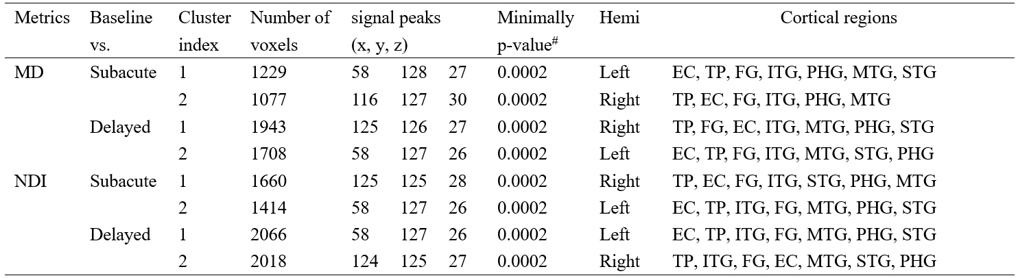
The FA, ODI, and VISO did not exhibit significant group-by-time interaction effects. Compared to the baseline, no significant differences were found for the MD and NDI at the acute phase. Their significant differences emerged in the subacute period, as illustrated in Figure 1. At the subacute and delayed phases, MD significantly decreased in both temporal lobes, while NDI significantly increased. Table 2 reveals that the significant differences in NDI covered a larger area than MD. Notably, both MD and NDI exhibited expanded significance in the delayed phase compared to the subacute phase. The affected GM regions were bilaterally located in the temporal lobes, including the entorhinal cortex, temporal pole, inferior temporal gyrus, fusiform gyrus, and others.Discussion
After radiotherapy, significant alterations of diffusion metrics occurred mainly in the temporal lobe, near the radiation target. Specifically, compared to baseline, NPC patients exhibited decreased MD and increased NDI in the temporal cortex, possibly due to processes such as neuronal regeneration, synaptic reconstruction, and axonal myelination. Neuronal regeneration and synaptic reconstruction led to reduced MD, indicating increased cellular compactness and decreased intercellular spacing. Concurrently, the formation of new synaptic connections and axonal myelination increased neurite terminal density and axonal density, leading to higher NDI values. Based on our results, these changes were predominantly localized in the basal temporal lobes, crucial for cognition and emotion, and were detected as early as six months post-radiotherapy in our study. The sensitivity of diffusion metrics to GM changes highlights the potential for targeted early interventions in brain regions affected by radiation-induced microstructural alterations. Additionally, integrating radiation dosage with these microstructural indices for personalized radiotherapy plans, especially safeguarding the temporal lobe region, appears to be a viable strategy in the future.Conclusions
Our research reveals dynamic cortical microstructural changes in NPC patients post-radiotherapy. These changes, starting in the subacute phase, involve reduced MD and increased NDI in the temporal cortex. The regions with significant differences dynamically expand over time.Acknowledgements
This work was supported by the National Natural Science Foundation of China (61971214) and Natural Science Foundation of Guangdong Province (2023A1515012093).References
[1] Chen, Y.-P., Chan, A.T., Le, Q.-T., Blanchard, P., Sun, Y., Ma, J. (2019) Nasopharyngeal carcinoma. The Lancet, 394:64-80.
[2] Lell, M.M. Therapy-induced changes in head and neck. (2015) Imaging of complications and toxicity following tumor therapy: Springer. p 95-111.
[3] Le Bihan, D. (2003) Looking into the functional architecture of the brain with diffusion MRI. Nature reviews neuroscience, 4:469-480.
[4] Henf, J., Grothe, M.J., Brueggen, K., Teipel, S., Dyrba, M. (2018) Mean diffusivity in cortical gray matter in Alzheimer's disease: The importance of partial volume correction. NeuroImage: Clinical, 17:579-586.
[5] Zhang, H., Schneider, T., Wheeler-Kingshott, C.A., Alexander, D.C. (2012) NODDI: practical in vivo neurite orientation dispersion and density imaging of the human brain. Neuroimage, 61:1000-1016.
[6] Nazeri, A., Chakravarty, M.M., Rotenberg, D.J., Rajji, T.K., Rathi, Y., Michailovich, O.V., Voineskos, A.N. (2015) Functional consequences of neurite orientation dispersion and density in humans across the adult lifespan. Journal of Neuroscience, 35:1753-1762.
[7] Nazeri, A., Mulsant, B.H., Rajji, T.K., Levesque, M.L., Pipitone, J., Stefanik, L., Shahab, S., Roostaei, T., Wheeler, A.L., Chavez, S. (2017) Gray matter neuritic microstructure deficits in schizophrenia and bipolar disorder. Biological psychiatry, 82:726-736.
[8] Daducci, A., Canales-Rodríguez, E.J., Zhang, H., Dyrby, T.B., Alexander, D.C., Thiran, J.-P. (2015) Accelerated microstructure imaging via convex optimization (AMICO) from diffusion MRI data. Neuroimage, 105:32-44.
Figures