2863
Resting-state fMRI network efficiency mediates the association between glymphatic system and cognition function in OSAHS: a DTI-ALPS study1Department of Radiology, Guizhou Provincial People’s Hospital, Guiyang, China, 2College of Computer Science and Technology, Guizhou University, Guiyang, China, 3GE HealthCare MR Research, Beijing, China
Synopsis
Keywords: DWI/DTI/DKI, Diffusion Tensor Imaging
Motivation: Obstructive Sleep Apnea Hypopnea Syndrome (OSAHS) is a serious sleep disorder linked to cognitive impairment. This study investigates the glymphatic system's role in OSAHS and its impact on cognition.
Goal(s): Explore glymphatic system changes in OSAHS and their influence on cognitive function. We aim to uncover the mechanisms connecting OSAHS and cognitive issues.
Approach: We use DTI-ALPS and rs-fMRI to assess the glymphatic system and brain network properties. Correlations and mediation analysis examine the link between glymphatic function, cognitive abilities, and OSAHS severity.
Results: OSAHS patients exhibit reduced glymphatic function, affecting cognitive performance.
Impact: This study may enhance comprehension of cognitive problems in OSAHS, suggesting dementia risk. It could inform early intervention, enhancing OSAHS patients' quality of life.
Introduction
Obstructive Sleep Apnea Hypopnea Syndrome (OSAHS) is characterized by upper airway collapse during sleep, causing hypoxia and organ damage1. Cognitive impairment is a severe consequence. Understanding its mechanisms is vital. The glymphatic system, responsible for waste clearance in the brain, is linked to neurological conditions. Diffusion Tensor Image Analysis Along the Perivascular Space (DTI-ALPS)2 provides a non-invasive method to assess glymphatic function. Graph theory analysis on resting-state fMRI data evaluates brain network efficiency and cognitive function. This study aims to investigate glymphatic system changes in OSAHS and their impact on brain connectivity.Methods
Our prospective study received approval from the institutional ethics committee. Prior to their participation in the study, all subjects provided written informed consent.Participants
This study included 25 OSAHS patients and 23 age- and gender-matched healthy controls (HC).
MRI Acquisition
All participants underwent magnetic resonance imaging (MRI) with diffusion tensor imaging (DTI) and resting-state fMRI scans on 3.0T MR scanner (Discovery MR 750w, GE Healthcare). The following key sequence parameters were used: DTI of the whole brain, single-shot EPI; TR/TE: 9700/74.2 ms, b = 0 and 1000 s/mm2, diffusion directions = 64, FOV: 256 mm, matrix: 128 × 128, 47 slice with 3 mm thickness without intersection gap, pixel size: 2×2 mm2. The rs-fMRI images were obtained using an echo-planar imaging sequence (TR = 2000 ms, TE = 30 ms, flip angle = 70°, FOV = 216 mm × 216 mm, matrix = 72 × 72, thickness = 3.5 mm, slice gap = 0.5 mm, slices = 34, voxel size = 3 mm × 3 mm × 3.5 mm, volume = 250).
Data Analysis
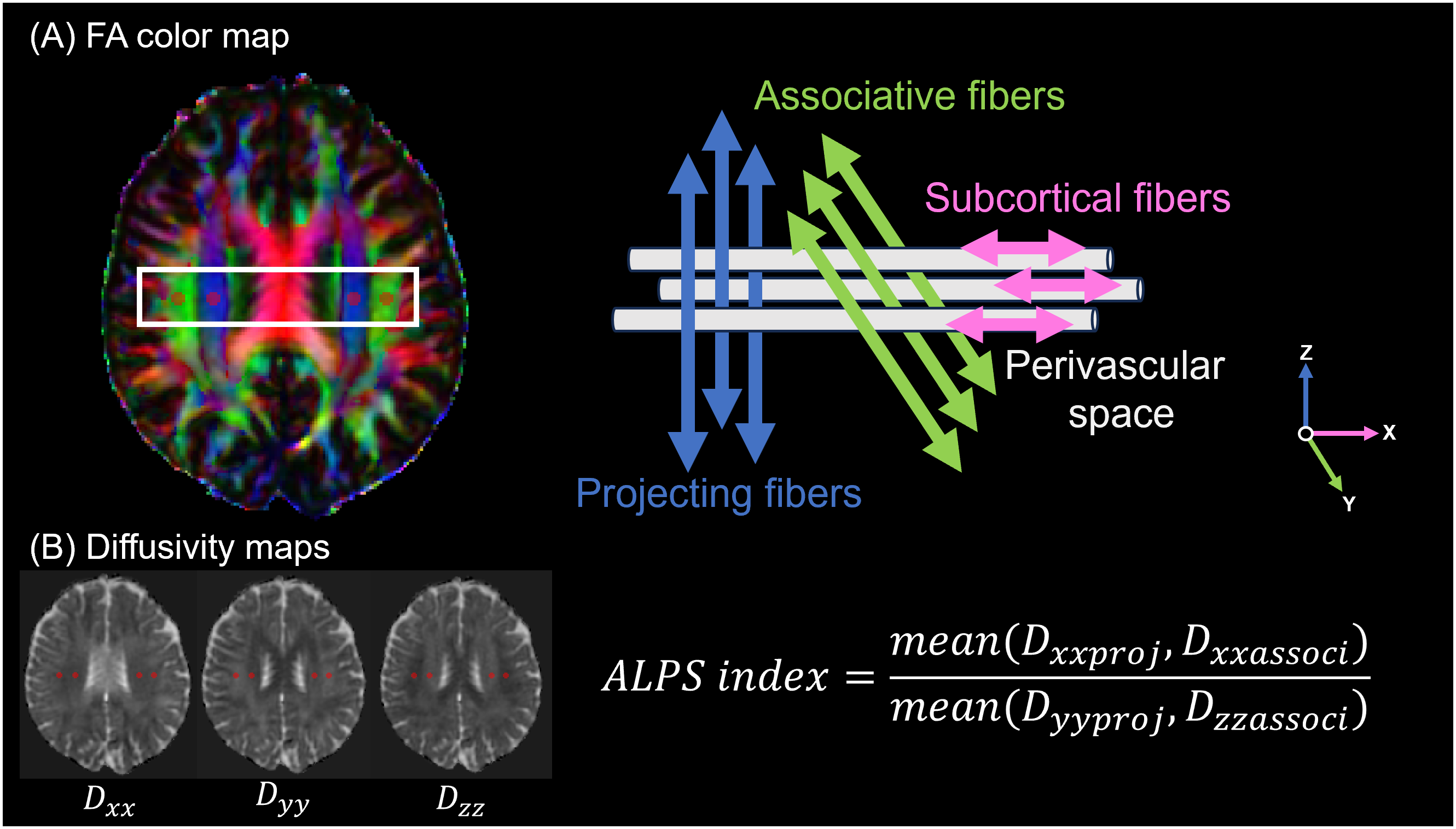
The DTI-ALPS index was derived from DTI data, as depicted in Figure 1, and the data post-processing was executed using FSL (https://fsl.fmrib.ox.ac.uk/fsl/). Additionally, brain network properties were evaluated through the analysis of resting-state fMRI data using the GRETNA toolbox3. The severity of OSAHS in patients is assessed through the Apnea Hypopnea Index (AHI), while the cognitive function level of OSAHS patients is evaluated using the Montreal Cognitive Assessment (MoCA). Statistical analysis was conducted using Python language (https://www.python.org/). Differences in DTI-ALPS index and brain network properties between OSAHS patients and healthy controls were evaluated. Additionally, correlations between the DTI-ALPS index and clinical characteristics were analyzed. Furthermore, the mediating role of brain network efficiency in the association between brain glymphatic system impairment and cognitive function in OSAHS patients was explored. P < 0.05 was considered significant.
Results
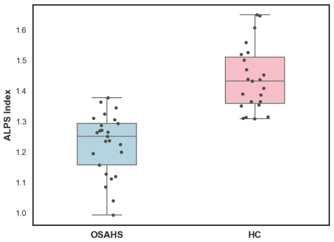
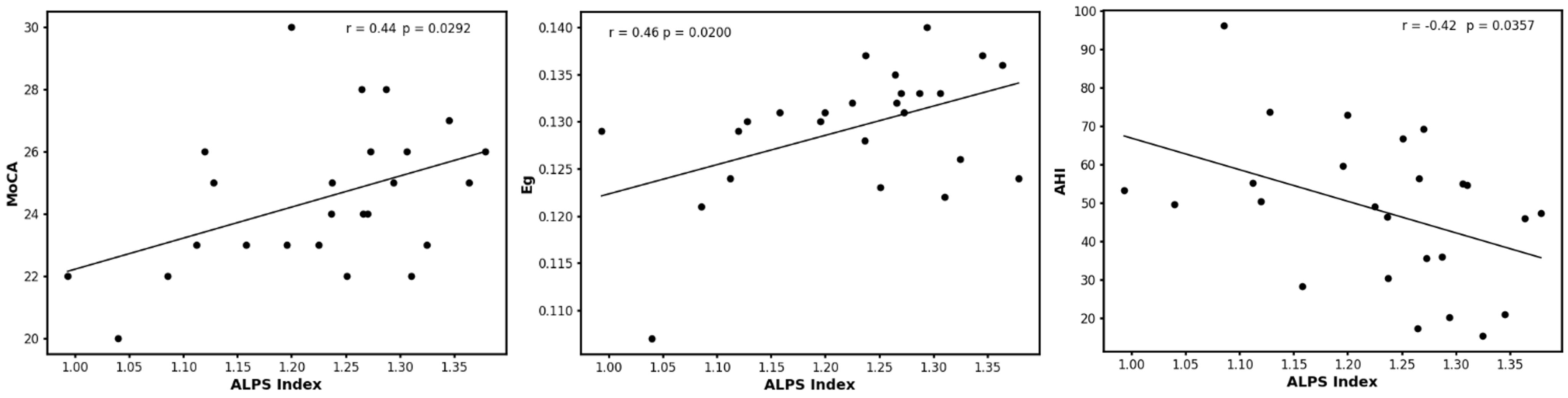
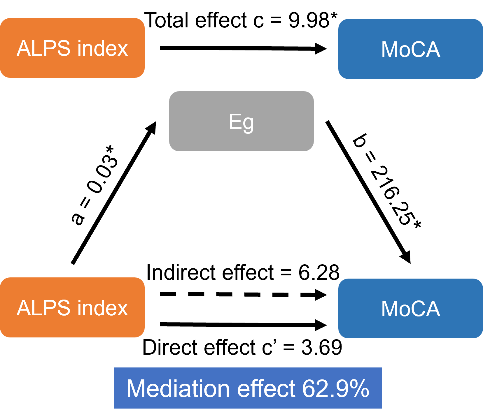
The DTI-ALPS index exhibited significant intergroup differences, with OSAHS patients displaying a notably lower DTI-ALPS index compared to the HC group (1.23 vs. 1.44, p < 0.001), as shown in Figure 2. Correlation analysis unveiled a negative correlation between the DTI-ALPS index and the Apnea Hypopnea Index (AHI) (r = −0.42, p < 0.05), as well as positive correlations with MoCA scores (r = 0.44, p < 0.05) among OSAHS patients, as illustrated in Figure 3. Additionally, global efficiency (Eg) of the brain network exhibited a positive correlation with the DTI-ALPS index in OSAHS patients (r = 0.46, p < 0.05), as depicted in Figure 3. Furthermore, mediation analysis revealed that Eg partially mediated the impact of brain glymphatic system dysfunction on cognitive impairment in OSAHS patients (indirect effect = 6.28, mediation effect 62.9%), as presented in Figure 4.Discussion and Conclusion
The study underscores the importance of investigating the glymphatic system in OSAHS. OSAHS patients had a significantly lower DTI-ALPS index, indicating glymphatic system impairment, consistent with previous research on OSAHS-related physiological changes4. Notably, the DTI-ALPS index independently influenced cognitive function, as evidenced by positive correlations with MoCA scores, aligning with studies linking glymphatic dysfunction to cognitive deficits and neurodegenerative conditions5. Furthermore, the negative correlation between the DTI-ALPS index and AHI suggests that more severe OSAHS may relate to greater glymphatic dysfunction, with hypoxia-reoxygenation cycles playing a role6. The positive correlation between the DTI-ALPS index and global brain network efficiency (Eg) adds complexity, as mediation analysis suggests Eg partially mediates glymphatic system effects on cognitive impairment.In conclusion, OSAHS patients exhibit decreased DTI-ALPS index, signifying compromised glymphatic function. This may disrupt brain network connectivity, increasing dementia risk, and revealing an underlying pathological mechanism in OSAHS. The DTI-ALPS index holds promise as an emerging OSAHS imaging marker.
Acknowledgements
This study has received funding from National Natural Science Foundation of China (82060314) and Guizhou Province science and technology plan project [2021]037.References
1. Kapur VK, Auckley DH, Chowdhuri S, Kuhlmann DC, Mehra R, Ramar K, Harrod CG. Clinical practice guideline for diagnostic testing for adult obstructive sleep apnea: an American Academy of Sleep Medicine clinical practice guideline. Journal of clinical sleep medicine. 2017;13:479–504
2. Taoka T, Masutani Y, Kawai H, Nakane T, Matsuoka K, Yasuno F, Kishimoto T, Naganawa S. Evaluation of glymphatic system activity with the diffusion MR technique: diffusion tensor image analysis along the perivascular space (DTI-ALPS) in Alzheimer’s disease cases. Japanese Journal of Radiology [Internet]. 2017;35:172–178. Available from: https://doi.org/10.1007/s11604-017-0617-z
3. Wang J, Wang X, Xia M, Liao X, Evans A, He Y. GRETNA: a graph theoretical network analysis toolbox for imaging connectomics. Front Hum Neurosci [Internet]. 2015;9:386. Available from: http://journal.frontiersin.org/Article/10.3389/fnhum.2015.00386/abstract
4. Roy B, Nunez A, Aysola RS, Kang DW, Vacas S, Kumar R. Impaired Glymphatic System Actions in Obstructive Sleep Apnea Adults. Frontiers in Neuroscience [Internet]. 2022;16. Available from: https://www.frontiersin.org/articles/10.3389/fnins.2022.884234
5. Ke Z, Mo Y, Li J, Yang D, Huang L, Yang Z, Qin R, Mao C, Lv W, Huang Y, Hu Z, Zhang B, Xu Y. Glymphatic Dysfunction Mediates the Influence of White Matter Hyperintensities on Episodic Memory in Cerebral Small Vessel Disease. Brain Sciences [Internet]. 2022;12. Available from: https://www.mdpi.com/2076-3425/12/12/1611
6. Lee H-J, Lee DA, Shin KJ, Park KM. Glymphatic system dysfunction in obstructive sleep apnea evidenced by DTI-ALPS. Sleep Medicine [Internet]. 2022;89:176–181. Available from: https://www.sciencedirect.com/science/article/pii/S1389945721005918
Figures