2862
Glymphatic system impairment in Type 2 diabetes mellitus associated with cognitive function1Department of MRI, Shaanxi Provincial People’s Hospital, Xi’an, China, 2Philips Healthcare, Xi’an, China, 3Philips Healthcare, Shanghai, China
Synopsis
Keywords: DWI/DTI/DKI, Diabetes
Motivation: T2DM has emerged as an important risk factor for cognitive impairment. However, the neuropathological basis of the cognitive impairment associated with T2DM is still unclear.
Goal(s): To investigate whether the impaired glymphatic function is one of the mechanisms in T2DM-associated cognitive dysfunction.
Approach: We compared the glymphatic function by calculating the diffusion along the perivascular space based on a diffusion tensor image, namely the DTI-ALPS index between HCs and patients with T2DM and investigated the relationship between DTI-ALPS index and cognitive functions.
Results: The lower DTI-ALPS index was found in patients with T2DM, and DTI-ALPS index was correlated with worse cognitive performance.
Impact: DTI-ALPS index may be applied as a useful indicator to evaluate the glymphatic system function. The impaired glymphatic system in patients with T2DM may provide a new perspective for understanding the pathophysiology of T2DM related cognitive impairment.
Introduction
Type 2 diabetes mellitus (T2DM) has emerged as an important risk factor for cognitive impairment and dementia1-2. However, the neuropathological basis of the cognitive impairment associated with T2DM is still unclear. The glymphatic system is a highly organized cerebrospinal-fluid-interstitial transport system that clears abnormal proteins and metabolites to maintain brain homeostasis. Glymphatic disruption is considered the final common pathway for dementia3, and degradation of the glymphatic system has been demonstrated to be involved in T2DM-associated cognitive dysfunction4-5. Taoka et al6 proposed a noninvasive alternative to evaluate the glymphatic function by calculating the diffusion along the perivascular space based on a diffusion tensor image, namely the DTI-ALPS index. Thus, this study aims to investigate glymphatic function in T2DM using the DTI-ALPS method and to explore the relationship between DTI-ALPS index and cognitive functions.Methods
This study was approved by the local institutional review board. Seventeen patients with T2DM and 20 age- and sex-matched health controls (HCs) were included in this study. All participants underwent neurological MRI and cognitive assessments. A battery of neuropsychological tests including Montreal Cognitive Assessment (MoCA), Mini-Mental State Examination (MMSE), Trail-Making Test A (TMT-A), Trail-Making Test B (TMT-B), Auditory Verbal Learning Test (AVLT) and Clock Drawing Task (CDT) were performed. Conventional MRI and DTI were performed on a 3.0T scanner (Ingenia, Philips Healthcare, The Netherlands) with 32-channel head coil. DTI protocols were: 32 directions, b value = 0, 1000 s/mm2, TR/TE=5000/150 ms, slice thickness= 2 mm, field of view = 256 mm × 256 mm, matrix = 128 × 128, spatial resolution = 2 × 2 × 2 mm3. Bilateral DTI-ALPS indices were calculated using an in-house software which developed by Philips. DTI-ALPS index, clinical and cognitive measurements were compared between the two groups. The correlations between the DTI-ALPS index and cognitive measurements were analyzed by using multiple linear regression analyses, with age, sex, and years of education as the covariates. The Bonferroni correction was applied to correct for multiple comparisons. All tests were taken to be significant at P<0.05.Results
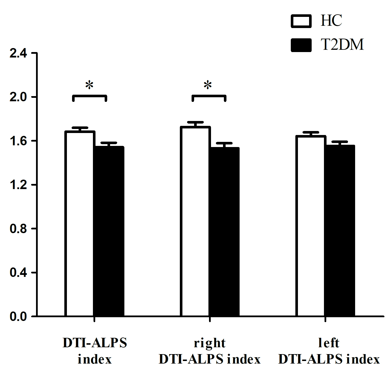
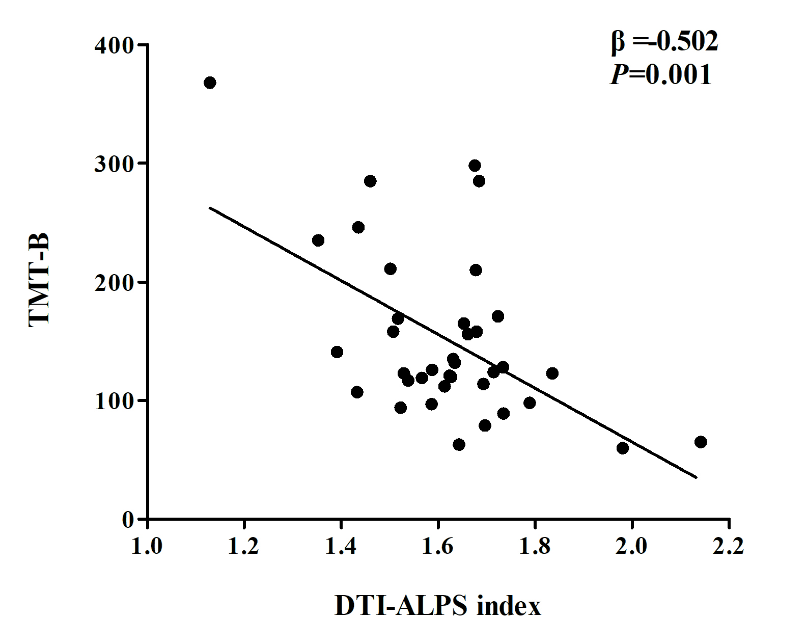
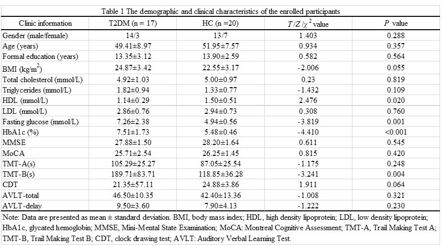
Table 1 showed the demographic and clinical characteristics of the enrolled participants. There were no significant differences in age, sex, years of education, general cognitive function (MoCA and MMSE), TMT-A, AVLT or CDT between HCs and patients with T2DM. Compared with HCs, patients with T2DM showed a significantly worse cognitive domain in executive function (TMT-B, P=0.004), as well as a significantly lower DTI-ALPS index in the whole brain (1.68±0.15 vs. 1.54±0.17, P=0.011) and in the right hemisphere (1.73±0.19 vs. 1.53±0.20, P=0.005), as shown in Fig.1. Furthermore, lower DTI-ALPS index was significantly correlated with worse performance in the TMT-B (β=-0.502, P=0.001), as shown in Fig.2.Discussion
In this study, we investigated in vivo glymphatic system function in T2DM patients using the DTI-ALPS index, a non-invasive diffusion-derived MRI index of diffusivity along the perivascular space. Our results demonstrated impaired glymphatic system function in T2DM patients, and it is related with worse performance in executive functions (TMT-B). The glymphatic system is a well-organized cerebrospinal-fluid-interstitial transport system that responsible for clearing abnormal proteins and metabolites to maintain brain homeostasis. Its function is impaired with aging and in pathological conditions7-9. From evidence in a T2D rat model10, MRI analysis revealed that clearance of cerebrospinal fluid contrast agent Gd-DTPA from the interstitial space was slowed in the hippocampus of T2DM rats and cognitive deficits detected by behavioral tests were highly and inversely correlated to the retention of Gd-DTPA contrast and fluorescent tracer in the hippocampus of T2DM rats. Thus, we speculate that impairment of the glymphatic system might be one of the mechanisms of T2DM-associated cognitive impairment, and that the DTI-ALPS index might be a prospective biomarker of domain-specific cognitive decline in patients with T2DM. Furthermore, our results indicated asymmetric impairment of the glymphatic system in T2DM (more obvious in the right hemisphere rather than left hemisphere), which suggested that brain lateralization or selective vulnerability also existed in glymphatic system function.Conclusions
This study demonstrated impaired glymphatic system function in T2DM patients and it is associated with worse performance in executive functions. DTI-ALPS index may be applied as a useful indicator to evaluate the glymphatic system function and the impaired glymphatic system in patients with T2DM may provide a new perspective for understanding the pathophysiology of T2DM-associated cognitive impairment.Acknowledgements
The authors thank all participants who volunteered for this study as well as the researchers who participated in this project.References
1. Moheet A, Mangia S, Seaquist ER. Impact of diabetes on cognitive function and brain structure. Ann N Y Acad Sci 2015; 1353:60-71.
2. Damanik J, Yunir E. Type 2 Diabetes Mellitus and Cognitive Impairment. Acta Med Indones 2021; 53(2):213-220.
3. Nedergaard M, Goldman SA. Glymphatic failure as a final common pathway to dementia. Science. 2020;370(6512):50-56.
4. Jiang Q, Zhang L, Ding G, et al. Impairment of the glymphatic system after diabetes. J Cereb Blood Flow Metab. 2017;37(4):1326-1337.
5. Kim YK, Nam KI, Song J. The Glymphatic System in Diabetes-Induced Dementia. Front Neurol. 2018;9:867.
6. Taoka T, Masutani Y, Kawai H, et al. Evaluation of glymphatic system activity with the diffusion MR technique: diffusion tensor image analysis along the perivascular space (DTI-ALPS) in Alzheimer's disease cases. Jpn J Radiol. 2017;35(4):172-178.
7. Kamagata K, Andica C, Takabayashi K, et al. Association of MRI Indices of Glymphatic System With Amyloid Deposition and Cognition in Mild Cognitive Impairment and Alzheimer Disease [published online ahead of print, 2022 Sep 19]. Neurology. 2022;99(24):e2648-e2660.
8. Rasmussen MK, Mestre H, Nedergaard M. The glymphatic pathway in neurological disorders. Lancet Neurol. 2018;17(11):1016-1024.
9. Reeves BC, Karimy JK, Kundishora AJ, et al. Glymphatic System Impairment in Alzheimer's Disease and Idiopathic Normal Pressure Hydrocephalus. Trends Mol Med. 2020;26(3):285-295.
10. Jiang Q, Zhang L, Ding G, et al. Impairment of the glymphatic system after diabetes. J Cereb Blood Flow Metab. 2017;37(4):1326-1337.
Figures