2861
Relationship Between Apparent Fiber Density in Corpus Callosum and Cognitive Function.1Department of Radiology, Tokyo Medical University Hospital, Tokyo, Japan, 2Department of Radiological Sciences, Graduate School of Health Sciences, Tokyo Metropolitan University, Tokyo, Japan, 3Department of Radiology, Tokyo Medical University, Tokyo, Japan, 4Division of Regenerative Medicine, Jikei University School of Medicine, Tokyo, Japan, 5Department of Radiological Technonlogy, Faculty of Health Science, Juntendo University, Tokyo, Japan, 6Department of Geriatric Medicine, Tokyo Medical University, Tokyo, Japan
Synopsis
Keywords: DWI/DTI/DKI, Diffusion Tensor Imaging
Motivation: Evaluation of fiber density in the corpus callosum (CC) is expected to show a higher sensitivity in relation to cognitive function than changes in volume.
Goal(s): The goal is to clarify the association between the volume and AFD of the CC5 region and neuropsychological test scores.
Approach: We examined the relationship between neuropsychological test scores, and calculated AFD and CC volume for 180 patients with suspected dementia.
Results: CC volume showed no significant association with neuropsychological test scores. On the other hand, AFD showed associations with several neuropsychological tests.
Impact: This study demonstrated that AFD correlated better with cognitive function than the previously reported CC volume. It was indicated the superior sensitivity of AFD in assessing cognitive function in dementia diagnosis.
INTRODUCTION
In diagnosing dementia, it is important to assess both functional aspects including neuropsychological tests, and structural aspects such as MRI. While cortical areas with neuronal cells generally garner focus, the corpus callosum (CC) responsible for left-right neural connections has been shown to play a role in cognitive functions. Decreases in CC volume have been reported with cognitive decline1,2. However, volume assessments of the CC do not equate to evaluations of actual neural connections and are also influenced by the overall brain volume. Therefore, we hypothesized that neural density might provide a more sensitive correlation with cognitive functions than volume changes. We have concentrated on the detailed neural density distribution within the CC using Apparent Fiber Density (AFD)3, a technique derived from Diffusion Tensor Imaging (DTI). The purpose of this study is to analyze the volume and AFD across various regions of the CC and to determine their association with multiple neuropsychological test scores.METHODS
The study population consisted of all consecutive patients with suspected dementia (n=180, female=100, age=76.8±8.8 years) who had undergone neuropsychological assessments (MMSE, MOCA-J, ADAS, FAB, TMT). All MR studies were performed on a 3 T system (MAGNETOM Vida; Siemens). The patients were imaged with 3D T1 weighted Image and DTI (SE-EPI, b-value = 0, 2000 s/mm², the number of MPG-directions = 30.) The volume of the CC was analyzed using FreeSurfer 7.3.2, dividing it into five regions: CC_Posterior, CC_Mid_Posterior, CC_Central, CC_Mid_Anterior, and CC_Anterior. For DTI analysis, FSL and MRtrix3 were utilized to preprocess for noise reduction and correct for B0 field inhomogeneity and eddy currents before calculating AFD. The AFD maps obtained were registered to each individual's atlas derived from FreeSurfer using ANTs to measure the AFD of each CC region. Statistical analysis involved multiple regression with age and sex as covariates, using neuropsychological test scores as explanatory variables for volume and AFD in each CC region. P-values were corrected using FDR, and the statistical significance level was set at 0.05. These statistical analyses were conducted using R and JMP.RESULTS
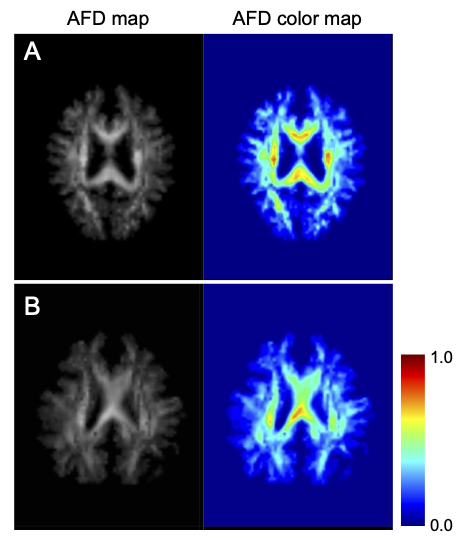
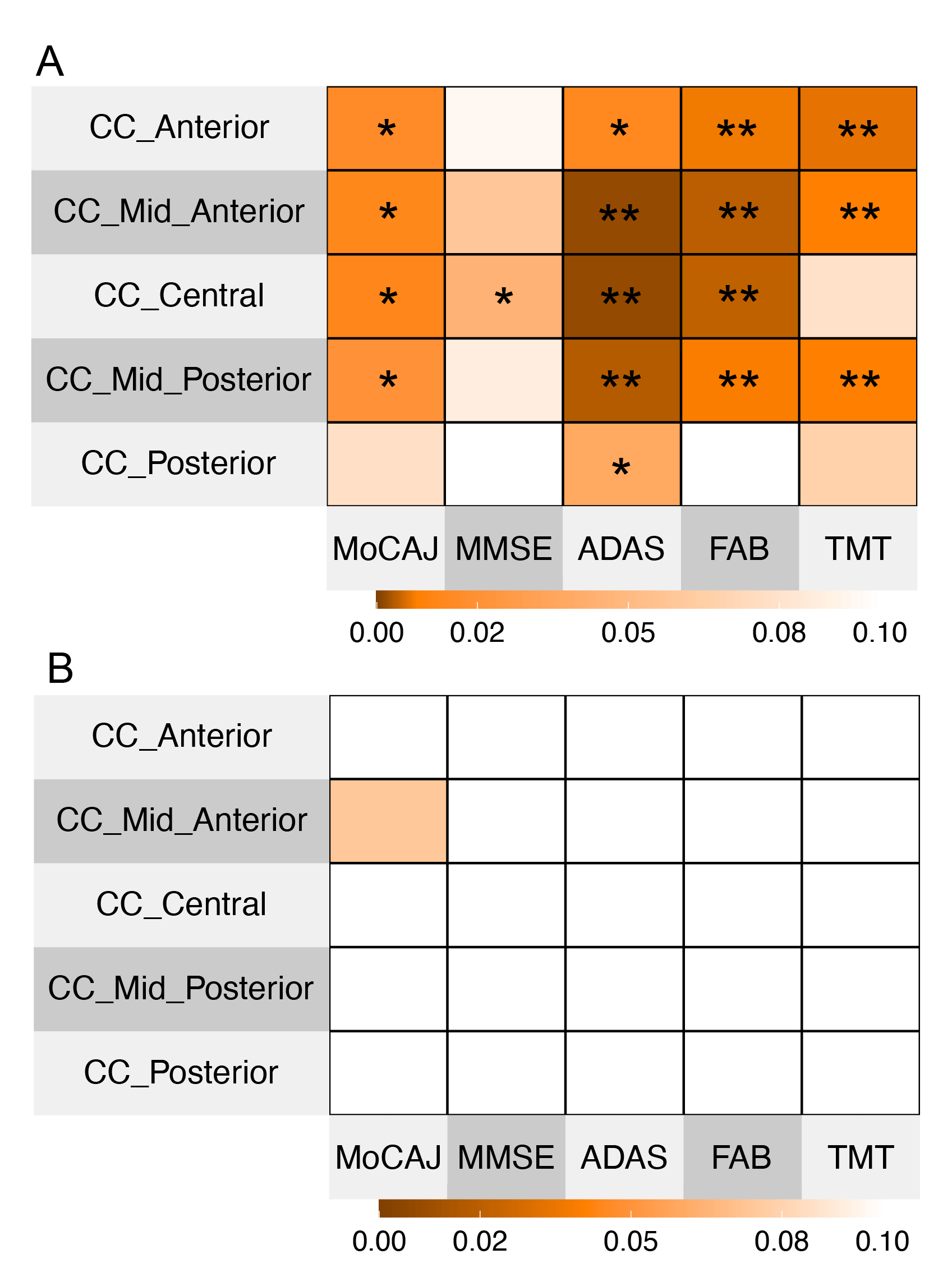
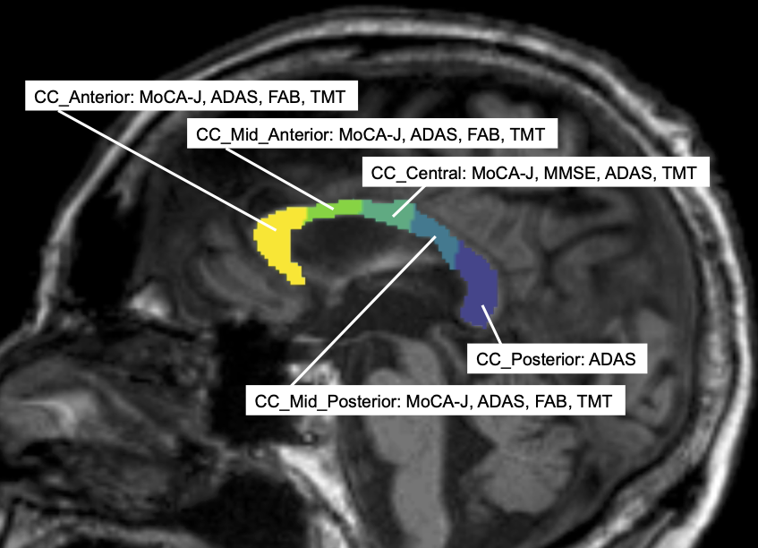
Figure 1 displays the AFD maps for patients with low and high neuropsychological test scores. Multiple regression analyses with CC volume as the dependent variable showed no significant differences with neuropsychological test scores (FDR corrected p > 0.05). Conversely, multiple regions of the CC showed significant associations with various neuropsychological test scores when AFD was the dependent variable: MoCA-J (for all = FDR corrected p < 0.05, except for CC_Posterior), MMSE (CC_Central = FDR corrected p < 0.05), ADAS (CC_Anterior; CC_Posterior = FDR corrected p < 0.05, CC_Central; CC_Mid_Central = FDR corrected p < 0.01), FAB (for all = FDR corrected p < 0.01, except for CC_Posterior), TMT (CC_Anterior; CC_Mid_Central; CC_Mid_Posterior = FDR corrected p < 0.01, Figure 2). The functional relevance of each CC region is presented in Figure 3.DISCUSSION
No significant differences were observed between cognitive function and CC volume. However, significant differences in the AFD of CC suggest a strong association with cognitive functions. Regions associated with the AFD of CC were more numerous for ADAS, TMT, and FAB than for MoCA-J and MMSE. This may be due to the fact that ADAS, TMT, and FAB are test-form assessments, as opposed to the conversational style of MoCA-J and MMSE4-8, with the former potentially engaging areas not primarily associated with conversational tasks typically involving the temporal lobe. The significant findings for ADAS in the Posterior CC could reflect abnormalities reported in the posterior part of the CC in Alzheimer's disease9. Furthermore, since TMT and FAB are specialized for frontal lobe functions7,8, their significance in the anterior CC regions is notable. Future work will expand on the relationship between the functions governed by each CC region, AFD, and neuropsychological test scores.CONCLUSION
AFD in the CC is suggested to have a stronger association with cognitive functions than CC volume.Acknowledgements
No acknowledgement found.References
[1] Hensel A, Wolf H, Kruggel F, Riedel-Heller SG, Nikolaus C, Arendt T, Gertz HJ. Morphometry of the corpus callosum in patients with questionable and mild dementia. J Neurol Neurosurg Psychiatry. 2002 Jul;73(1):59-61.
[2] Huang X, Du X, Song H, Zhang Q, Jia J, Xiao T, Wu J. Cognitive impairments associated with corpus callosum infarction: a ten cases study. Int J Clin Exp Med. 2015 Nov 15;8(11):21991-8.
[3] Raffelt D, Tournier JD, Rose S, et al. Apparent Fibre Density: a novel measure for the analysis of diffusion-weighted magnetic resonance images. Neuroimage. 2012;59(4):3976-3994.
[4] Elkana O, Nimni Y, Ablin JN, Shorer R, Aloush V. The Montreal Cognitive Assessment Test (MoCA) as a screening tool for cognitive dysfunction in fibromyalgia. Clin Exp Rheumatol. 2022 Jun;40(6):1136-1142.
[5] Tariq SH, Tumosa N, Chibnall JT, Perry MH 3rd, Morley JE. Comparison of the Saint Louis University mental status examination and the mini-mental state examination for detecting dementia and mild neurocognitive disorder--a pilot study. Am J Geriatr Psychiatry. 2006 Nov;14(11):900-10.
[6] Kueper JK, Speechley M, Montero-Odasso M. The Alzheimer's Disease Assessment Scale-Cognitive Subscale (ADAS-Cog): Modifications and Responsiveness in Pre-Dementia Populations. A Narrative Review. J Alzheimers Dis. 2018;63(2):423-444.
[7] Llinàs-Reglà J, Vilalta-Franch J, López-Pousa S, Calvó-Perxas L, Torrents Rodas D, Garre-Olmo J. The Trail Making Test. Assessment. 2017 Mar;24(2):183-196.
[8] Dubois B, Slachevsky A, Litvan I, Pillon B. The FAB: a Frontal Assessment Battery at bedside. Neurology. 2000 Dec 12;55(11):1621-6.
[9] Khasawneh RR, Abu-El-Rub E, Alzu'bi A, Abdelhady GT, Al-Soudi HS. Corpus callosum anatomical changes in Alzheimer patients and the effect of acetylcholinesterase inhibitors on corpus callosum morphometry. PLoS One. 2022 Jul 27;17(7):e0269082.
Figures