2858
Denoising diffusion MRI with an improved non-local principal component analysis approach1Wellcome Centre for Integrative Neuroimaging, FMRIB, Nuffield Department of Clinical Neurosciences, Univeristy of Oxford, Oxford, United Kingdom, 2Department of Radiology and Imaging Sciences, University of Utah, Salt Lake City, UT, United States, 3United Imaging, Shanghai, China, 4Tiantan Neuroimaging Center of Excellence, Beijing Tiantan Hospital, Capital Medical University, Beijing, China, 5Center for Magnetic Resonance Research, Radiology, Medical School, University of Minnesota, Minneapolis, MN, United States, 6Tsinghua University, Beijing, China
Synopsis
Keywords: DWI/DTI/DKI, Diffusion/other diffusion imaging techniques
Motivation: Previously, we proposed an improved 2-step non-local principal component analysis (PCA) approach and demonstrated its utility for denoising diffusion MRI with many diffusion directions.
Goal(s): Our goal here was to investigate how our approach would benefit diffusion tensor MRI (DTI) with a few diffusion directions.
Approach: we evaluated our approach’s denoising performances using both simulation and human-data experiments, and compared the results to those obtained with existing local-PCA-based methods.
Results: Our approach substantially enhanced image quality relative to the noisy counterpart, yielding improved performances for estimation of relevant DTI metrics. It also outperformed existing local-PCA-based methods in reducing noise while preserving anatomic details.
Impact: Capable of improving image quality for DTI with reduced diffusion directions, our improved non-local PCA denoising approach is believed to have utility for many applications, especially those targeting quality DTI or parametric mapping or both within a clinically relevant timeframe.
Introduction
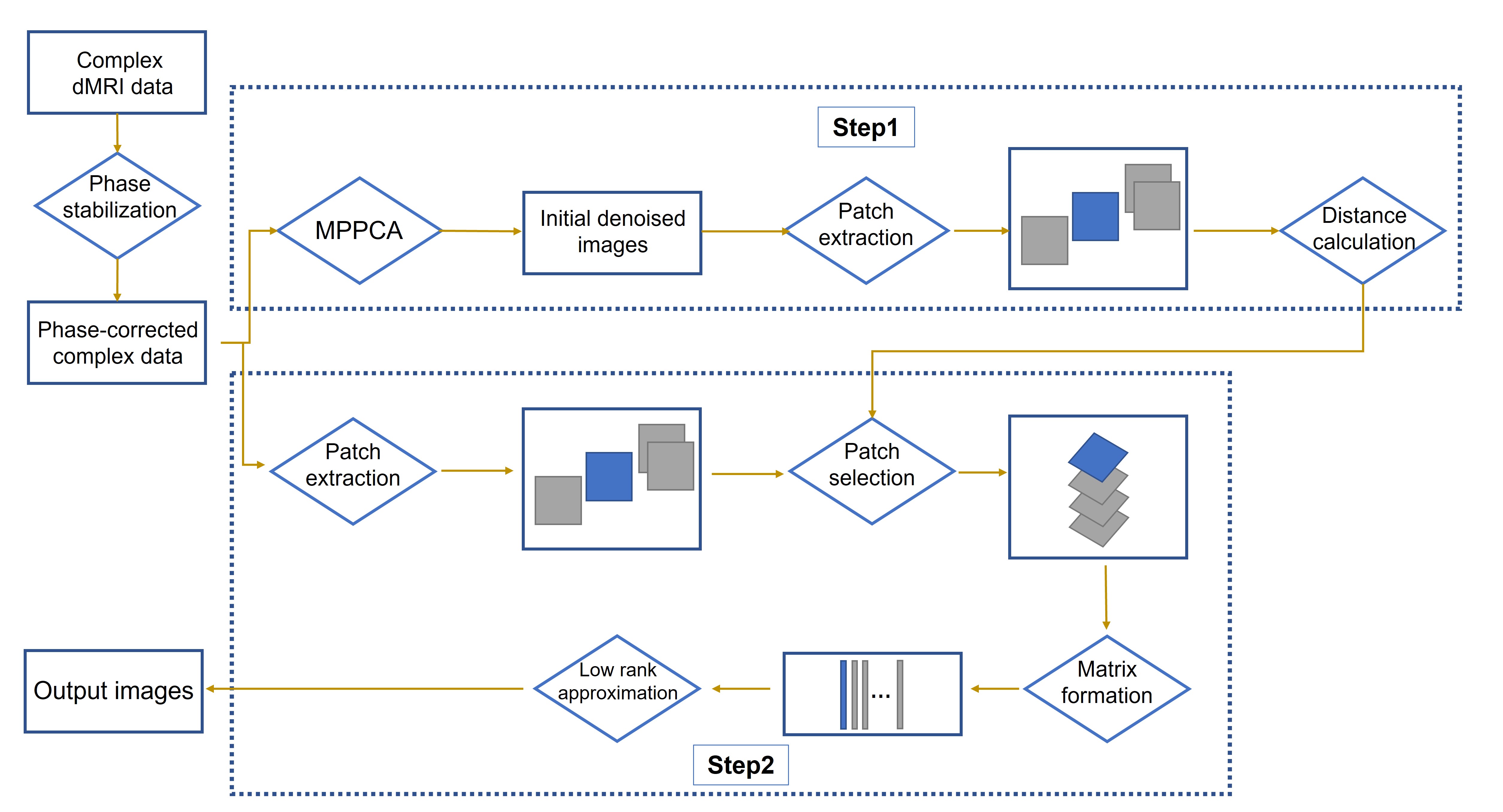
Previously, we introduced a 2-step non-local principal component analysis (PCA) approach and demonstrated its utility for denoising complex-valued diffusion with many diffusion directions1. Here we studied how our approach would help improve high-resolution diffusion tensor imaging (DTI) with reduced diffusion directions, in comparison to existing local-PCA-based methods2-4.Method
2-step non-local PCA methodBriefly, each noisy patch was grouped with 140 similar non-local patches (selected based on Euclidian distance calculated from the initially denoised images in step1) to form the Casorati matrix, of which the low-rank components were estimated using optimal singular value shrinkage5. The flowchart is shown in Fig.1.
Simulation data
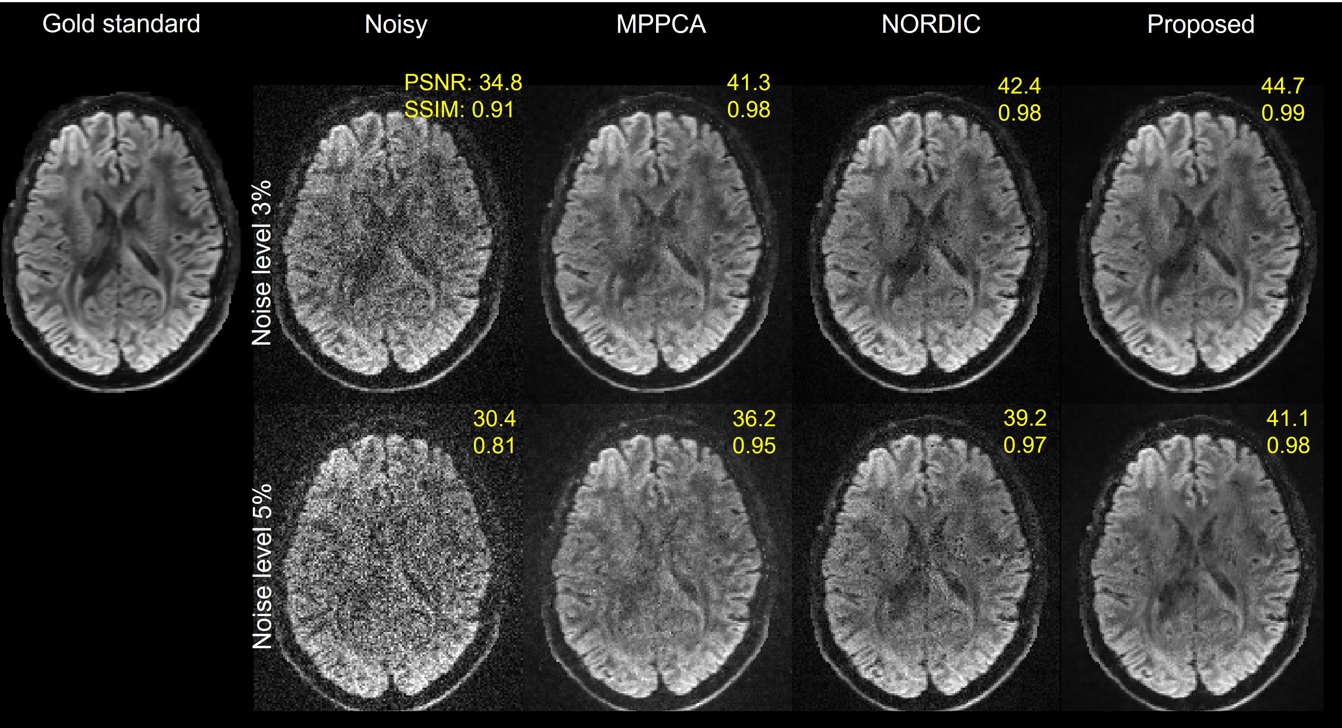
To demonstrate the utility of our approach for DTI, we conducted a simulation experiment. Synthetic data were generated as follows. Noise-free complex-valued data (serving as a gold standard) were synthesized based on part of a single subject’s 3T Human-Connectome-Project (HCP) diffusion data6.
A total of 108 images (including 18 b=0 and 90 b=1000 s/mm2 images) obtained at 1.25-mm resolutions was used to fit a tensor model7 in fsl8, which in turn was used to create noise-free magnitude data comprising a total of 15 images (including one b=0 and 14 b=1000 s/mm2 images). Second-order smooth phase variations were imposed to synthesize the noise-free complex-valued data. Noisy data were created by corrupting the noise-free complex-valued data with 3D spatially-varying Gaussian noise.
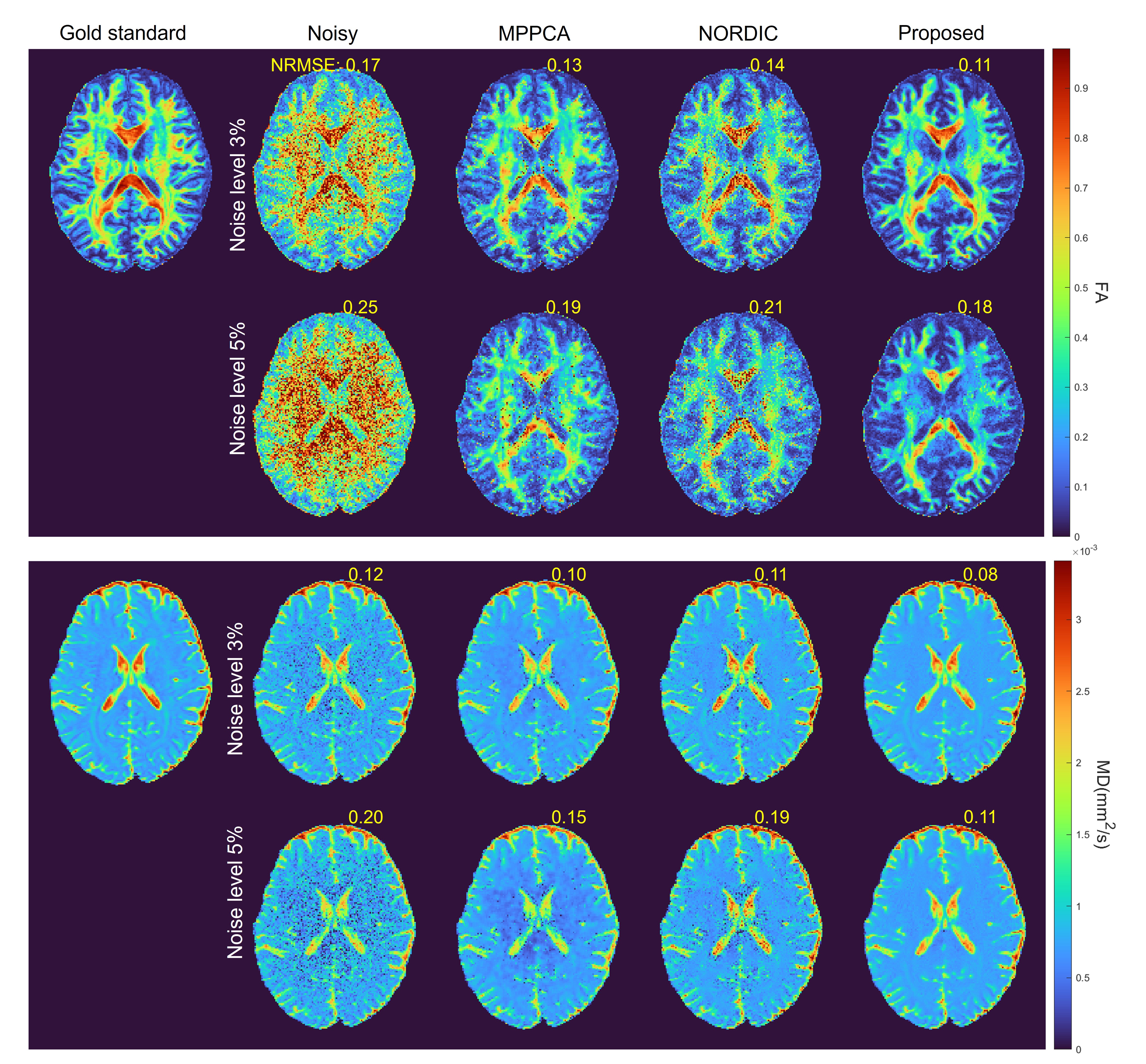
The denoising performances were evaluated in the image domain by calculating peak signal-to-noise ratio (PSNR) and structural similarity index measure (SSIM), and in the DTI metrics domain by calculating normalized root-mean-squared error (NRMSE), all in reference to the gold standard.
Human data acquisition
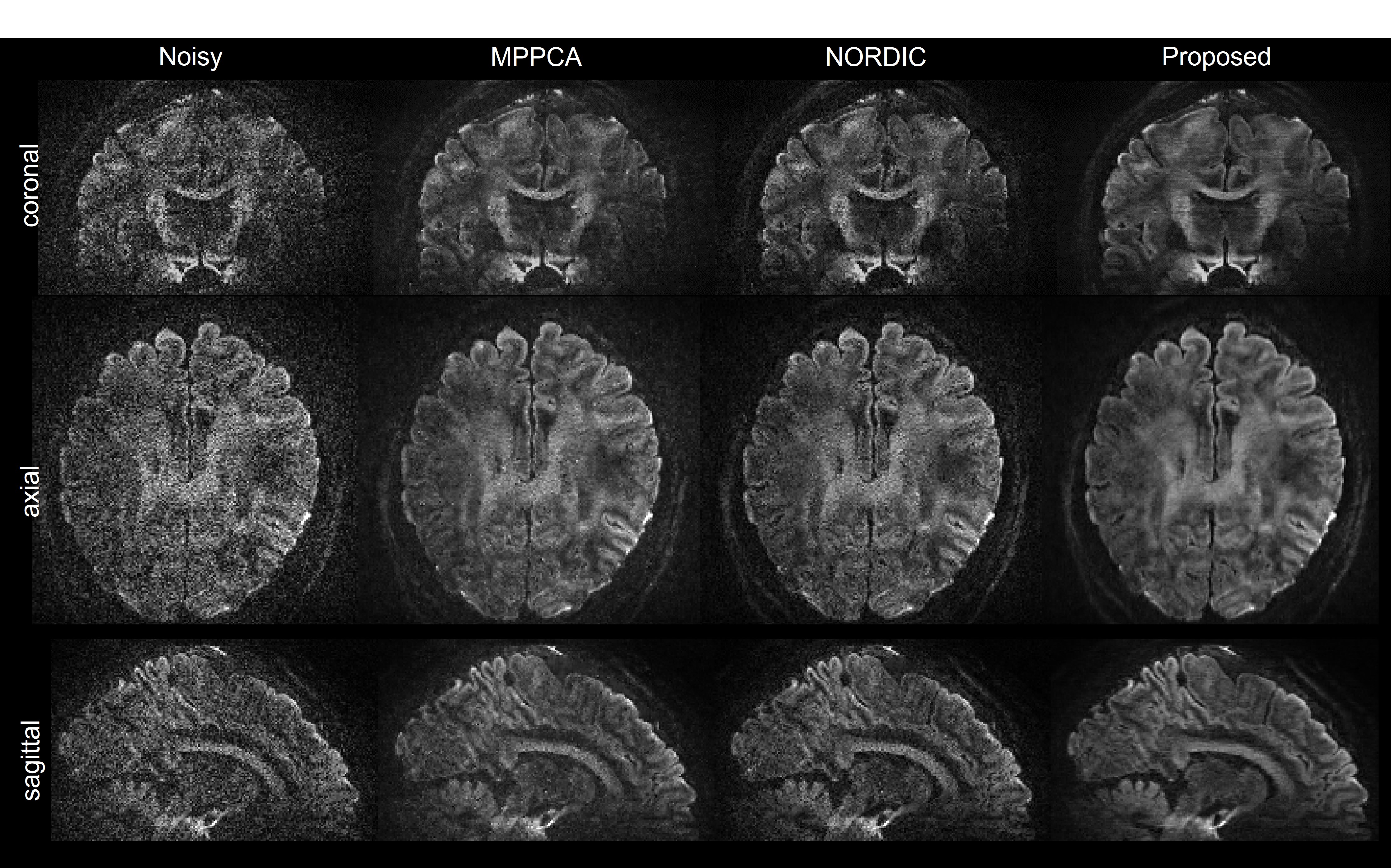
We also performed a human-data experiment to demonstrate the utility of our approach. Images were collected at higher resolutions on a 7T Siemens Terra scanner (Siemens, Erlangen, Germany) equipped with a body gradient (80 mT/m Gmax and 200 T/m/s slew rate). One healthy adult who signed a consent form approved by the local Institutional Review Board was scanned using the commercial Nova 32-channel receive coil. Slice-accelerated whole-brain DTI data were acquired at 0.9-mm isotropic resolutions using single-shell q-space sampling (b=1500 s/mm2) and the multiband sequence as in the 7T HCP9. Other imaging parameters were: 2-fold slice acceleration, 3-fold in-plane acceleration, and TR/TE=7000/70 ms. The dataset comprised 20 averages, each having nine images (corresponding to one b0 and eight diffusion directions). A single average was selected for denoising.
Multichannel images were reconstructed using a custom 3D GRAPPA algorithm (involving a new 2-stage N/2 ghost correction and the GRE single-band reference for improved reconstruction)10, and were combined via adaptive combination11.
In both simulation and human-data experiments, the results were compared to those obtained using MPPCA2 and NORDIC3.
Results
In simulation, our proposed method substantially improved the image quality relative to the noisy counterparts (Fig. 2), increasing PSNR by as much as ~38% at both 3% and 5% noise levels. It also appeared to outperform existing approaches, increasing PSNR by up to ~17% (vs. MPPCA) and up to ~5% (vs. NORDIC) while leading to greatest SSIM values at both noise levels.The improvement in image quality (relative to the noisy images) translated into increased performances for estimation of DTI metrics (Fig. 3), bringing both FA and MD maps close to the gold standard, with fine brain structures starting to be visualized. The FA and MD maps also presented less noise levels than those obtained with MPPCA and NORDIC especially around the center of the brain, leading to least NRMSE values at both noise levels.
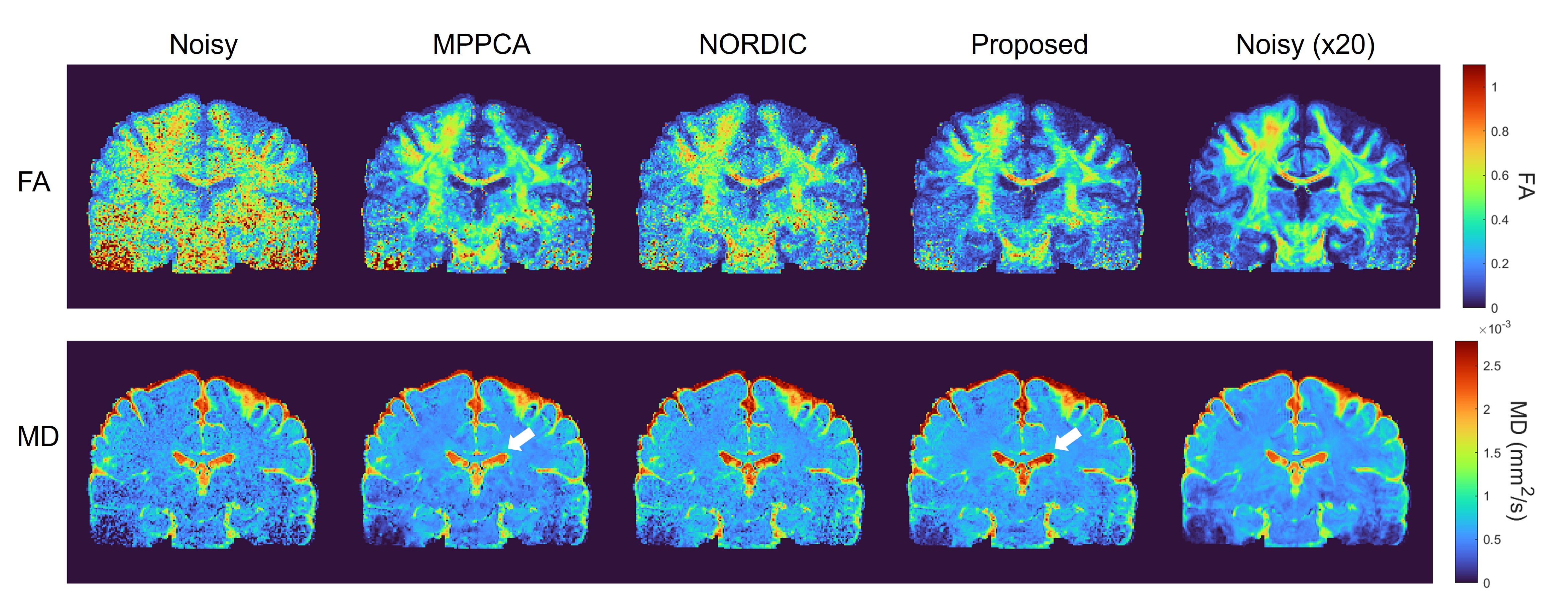
Likewise, our proposed method largely enhanced the image quality for the 0.9-mm human dMRI (Fig. 4), enabling fine brain structures to be visualized across the whole brain when compared to the noisy counterpart. Visually, it also outperformed both MPPCA and NORDIC, improving noise reduction in many brain regions. Similar results to simulation experiments were observed when comparing DTI metrics (Fig. 5).
Discussion
We have demonstrated the utility of our 2-step non-local PCA method for denoising complex-valued DTI data with a small number of image volumes. Our results for both simulation and human-data experiments show that our proposed method can largely improve image quality and estimation performances for DTI metrics (including FA and MD), when compared to the noisy case.Our results also show that our method can reduce noise more effectively than existing local PCA approaches, thanks to its ability to promote low rankness by integrating non-local similar patches. We believe that our method will benefit many applications especially those aiming to achieve quality parametric mapping using only a few image volumes.
Acknowledgements
The authors thank Steen Moeller for discussion on NORDIC. EA, KU, XW and all work conducted at the University of Minnesota were supported in part by USA NIH grants (NIBIB P41 EB027061, U01 EB025144, and S10 OD025256).References
1. Ye X, Ma X, Pan Z, et al. Non-local low rank denoising method for complex-valued DWI. In Proceedings of ISMRM 2022, 2022. p3041.
2.Veraart J, Novikov DS, Christiaens D, Ades-Aron B, Sijbers J, Fieremans E. Denoising of diffusion MRI using random matrix theory. Neuroimage 2016;142:384-396.
3. Moeller S, Pisharady PK, Ramanna S, et al. NOise reduction with DIstribution Corrected (NORDIC) PCA in dMRI with complex-valued parameter-free locally low-rank processing. Neuroimage 2021;
4. Ma XD, Ugurbil K, Wu XP. Denoise magnitude diffusion magnetic resonance images via variance -stabilizing transformation and optimal singular -value manipulation. Neuroimage 2020;215.
5. Gavish M, Donoho DL. Optimal Shrinkage of Singular Values. Ieee T Inform Theory. 2017;63(4):2137-2152. 6. Sotiropoulos SN, Jbabdi S, Xu JQ, et al. Advances in diffusion MRI acquisition and processing in the Human Connectome Project. Neuroimage 2013;80:125-143.
7. Tian QY, Bilgic B, Fan QY, et al. DeepDTI: High-fidelity six-direction diffusion tensor deep learning. Neuroimage 2020;219.
8. Jenkinson M, Beckmann CF, Behrens TE, Woolrich MW, Smith SM. Fsl. Neuroimage 2012;62(2):782-790.
9. Vu AT, Auerbach E, Lenglet C, et al. High resolution whole brain diffusion imaging at 7 T for the Human Connectome Project. Neuroimage. 2015;122:318-331.
10. Pan Z, Ma X, Dai E, Auerbach EJ, Guo H, Ugurbil K, Wu X. Reconstruction for 7T high-resolution whole-brain diffusion MRI using two-stage N/2 ghost correction and L1-SPIRiT without single-band reference. Magn Reson Med 2023;89(5):1915-1930.
11. Sotiropoulos SN, Moeller S, Jbabdi S, Xu J, Andersson JL, Auerbach EJ, Yacoub E, Feinberg D, Setsompop K, Wald LL, Behrens TEJ, Ugurbil K, Lenglet C. Effects of image reconstruction on fiber orientation mapping from multichannel diffusion MRI: Reducing the noise floor using SENSE. Magnetic Resonance in Medicine 2013;70(6):1682-1689.
Figures