2856
Accelerating Prostate DWI Scans: A Combination of Local SVD and Deep Learning for Enhanced Denoising1Pattern Recognition Lab, Friedrich-Alexander-University Erlangen-Nuremberg, Erlangen, Germany, 2Siemens Healthineers AG, Erlangen, Germany
Synopsis
Keywords: DWI/DTI/DKI, Prostate, Diffusion Denoising, Sparse and Low-Rank Models
Motivation: Diffusion-weighted MR images often suffer from low signal-to-noise ratio, particularly at high b-values, diminishing their diagnostic value. To counter this, multiple repetitions per diffusion direction are typically acquired and averaged, which is time-consuming and prone to motion artifacts.
Goal(s): Present a method that reduces the required number of repetitions in DWI, thus shortening scan times, while preserving diagnostic value.
Approach: The repetitions in DWI are jointly denoised through a combination of local Singular Value Decomposition and deep-learning-based denoising.
Results: Our evaluations indicate that this approach outperforms competing methods, offering a potential solution to the problem of prolonged acquisition times in DWI.
Impact: By combining local singular value decomposition with deep-learning-based denoising techniques, the necessary number of repetitions for the acquisition of diffusion-weighted MR images is substantially decreased and thus the acquisition is accelerated, while retaining comparable image quality.
Introduction
In magnetic resonance imaging (MRI), obtaining a favorable signal-to-noise ratio (SNR) within limited acquisition time is a persistent challenge. This is particularly pronounced in diffusion-weighted imaging (DWI), especially at high b-values, where additional gradient pulses for diffusion weighting are required. In certain anatomical regions like the prostate, this results in images with low diagnostic value1. The prevailing approach to address SNR limitations involves acquiring multiple repetitions and computing their average, this is time-consuming and susceptible to motion artifacts. Consequently, the goal of this work is to significantly reduce the number of repetitions in prostate DWI scans through a combination of local singular value decomposition (SVD) and deep-learning-based denoising, while retaining comparable image quality.Methods
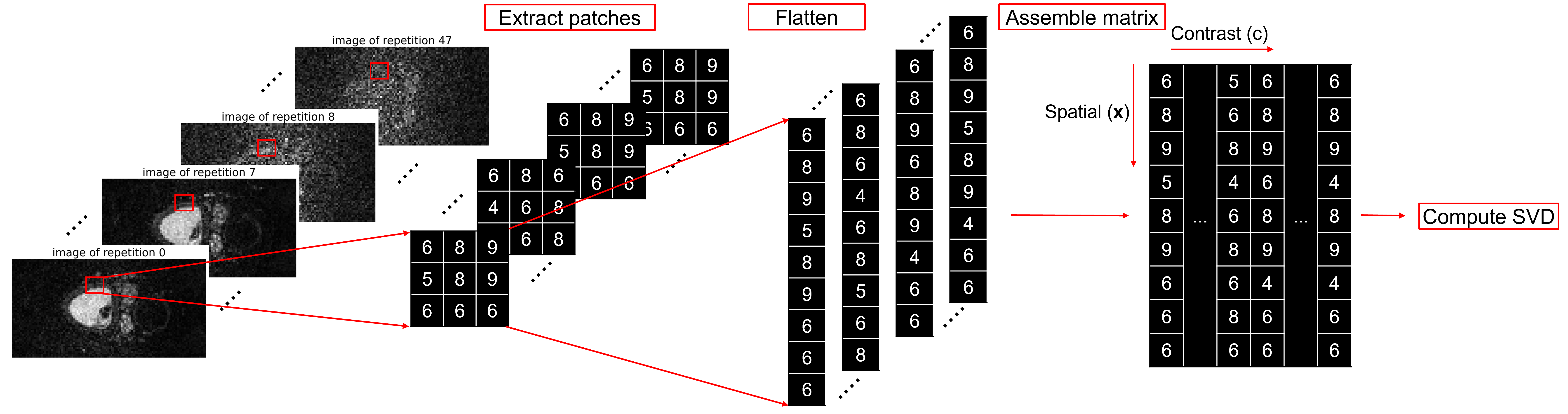
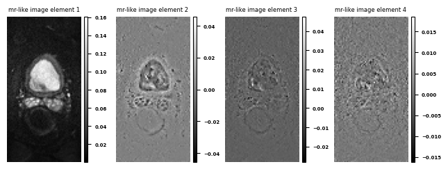
We use a patch-based processing of image stacks of the form $$$\mathbf{A}_{c,\mathbf{x}}$$$, where c is an index for different contrasts, i.e., b-values and diffusion directions for the DWI case, and $$$\mathbf{x}$$$ are spatial coordinates. The processing per patch ($$$p$$$), shown in Figure 1, is then based on SVD along the contrast and spatial dimensions of all acquired repetitions in a scan: $$$\mathbf{A}_{c,\mathbf{x}}=\sum_p\mathbf{U}_{c,p}\mathbf{S}_p\mathbf{V}^*_{\mathbf{x},p}$$$.An established denoising method, after computing the SVD, reduces to soft thresholding the singular values2,3. In our approach the singular values get cropped to a larger and constant number of non-zero singular components which, compared to the conventional case, provides a minor denoising effect by itself. Subsequently supervised deep-learning-based denoising is applied to the product $$$\mathbf{M}_{p}(\mathbf{x})=\mathbf{S}_p\mathbf{V}^*_{\mathbf{x},p}$$$, which represents the spatial distribution of the signal throughout the different contrasts and therefore an ‘MR-like image’, shown in Figure 2.
Having those MR-like images $$$\mathbf{M}_{p}(\mathbf{x})$$$ we train a deep neural network (DNN) on MR-like images $$$\mathbf{M}_{p}(\mathbf{x})$$$ to denoise them. A natural choice is to take a fixed number of singular values to use them in channel dimension and therefore take the correlation between the MR-like images into account. Additionally, the number of different contrasts, repetitions and directions can be variable and is not fixed to a specific number as the channels of the DNN are. Conventionally this variability is only possible when processing the different contrasts in the batch dimension, which neglects the correlation between them.
After applying deep-learning-based denoising, the denoised MR images are recovered by applying $$$\mathbf{A}_{c,\mathbf{x}}=\sum_p\mathbf{U}_{c,p}\mathbf{M}_{p}(\mathbf{x})$$$, again patch-based.
271 prostate image volumes were obtained from volunteers using 1.5T and 3T scanners (MAGNETOM scanners, Siemens Healthineers, Erlangen, Germany) and standard DWI acquisition protocols. The dataset includes images at b-values of 50 s/mm² and 800 s/mm², with four diffusion directions. For high b-values, ten repetitions were acquired per diffusion direction, while for low b-values, two repetitions were obtained per diffusion direction, resulting in a combined total of 48 repetitions, which served as ground truth. The respective half repetition count (24) served as input to our proposed denoising pipeline.
We chose a U-Net4 based architecture, implemented in PyTorch. The number of singular values was cropped to four, therefore we employed four input and output channels. We trained on 4065 slices using mean-squared-error (MSE) as supervised loss function.
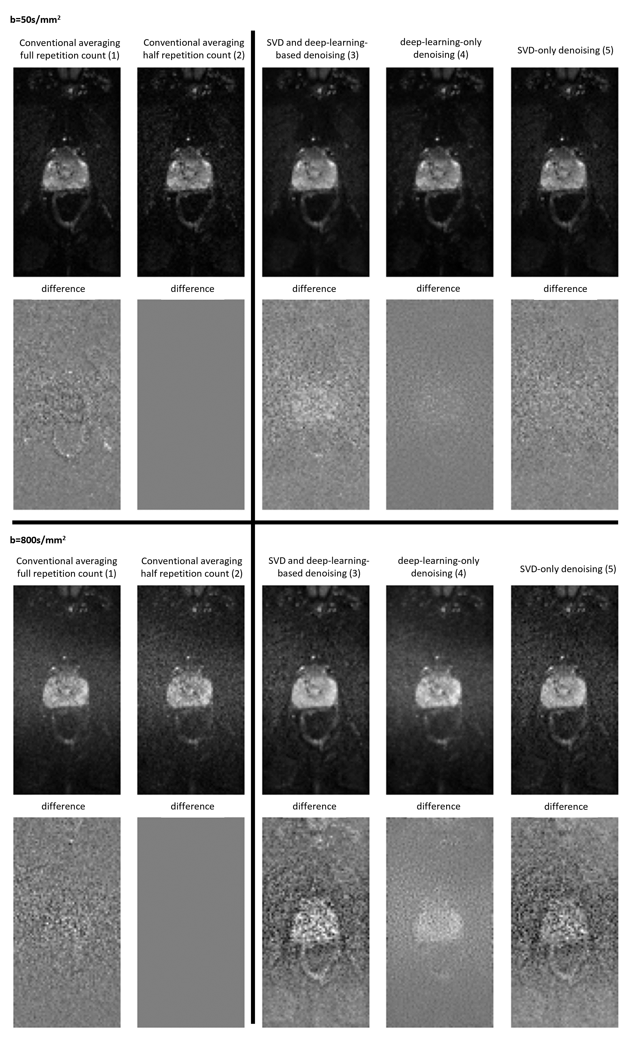
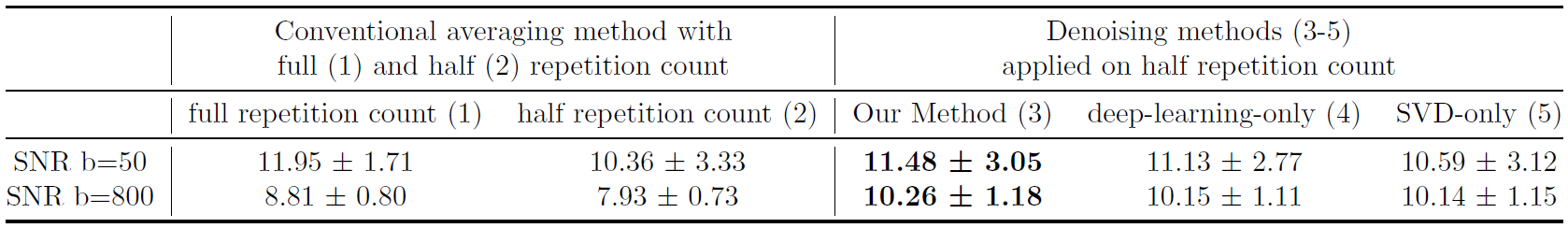
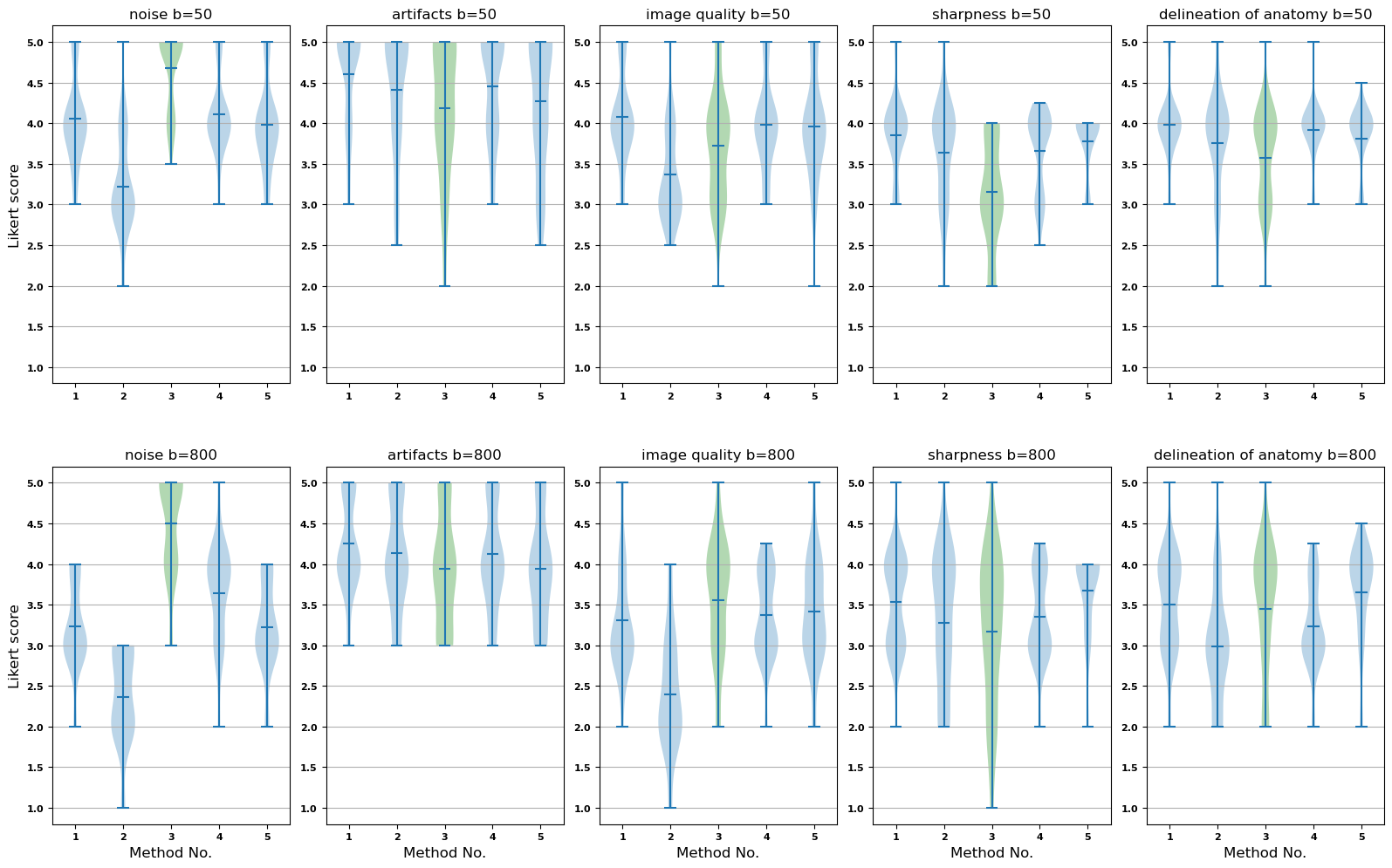
For qualitative evaluation, four MR DWI specialists rated the results for five different methods (1-5): two conventional methods by averaging the full repetition count (1) and half repetition count (2), which also served as input to the three denoising methods: our proposed combination of SVD and deep learning (3), deep learning-only denoising (4) and SVD-only denoising (5). To quantitatively evaluate the results, we calculated SNR for each of the methods.
Results and Discussion
Representative results are illustrated in Figure 3. The results of the quantitative evaluation are shown in Table 1. It becomes clear that the denoising characteristics of the presented method (3) results in the best SNR compared to the two other denoising methods (4, 5) and for the high b-value it even outperforms the two conventional methods (1, 2) clearly. The tradeoff between good denoising performance and loosing perceived sharpness and delineation of anatomy becomes visible in the results of the reader study (Figure 4) and is a fundamental aspect of performing denoising.Conclusion
We demonstrated a combined method employing local SVD and deep-learning-based denoising in prostate DWI, leading to improved image quality and higher SNR even with a 50% reduction in repetitions and thus scan time, especially for high b-values. Importantly, this approach is not limited to prostate DWI, finding application in scenarios involving multiple repetitions with varying contrasts, such as brain diffusion tensor imaging. Furthermore, this method offers flexibility with respect to the repetition count and the number of acquired directions, and the possibility to further enhance images acquired with the full repetition count.Acknowledgements
No acknowledgement found.References
1. Kazuhiro Kitajima, Yasushi Kaji, Kagayaki Kuroda, Kazuro Sugimura, "High b-value diffusion-weighted imaging in normal and malignant peripheral zone tissue of the prostate: effect of signal-to-noise ratio," Magn Reson Med Sci, pp. 7(2):93-9, 2008.2. Jelle Veraart, Dimitry S. Novikov, Daan Christiaens, Benjamin Ades-aron, Jan Sijbers, Elsa Fieremans, "Denoising of diffusion MRI using random matrix theory," NeurImage, vol. 142, pp. 394-406, 2016.
3. Joshua D. Trzasko, Armando Manduca, "Calibrationless Parallel MRI Using CLEAR," in Asilomar Conference on Signals, Systems and Computers, Pacific Grove, CA, USA, 2011.
4. Olaf Ronneberger, Philipp Fischer, Thomas Brox, "U-Net: Convolutional Networks for Biomedical Image Segmentation," 2015.
Figures