2851
Trajectory optimization of field of view within a Nonlinear Magnetic Field by a Single-sided Magnet for spatial encoding of portable MRI1Department of Medical Engineering, Chiba University, Chiba, Japan, 2Engineering Product Development Department, Singapore University of Technology and Design, Singapore, Singapore, 3Department of Surgery, National University of Singapore, Singapore, Singapore, 4Center for Frontier Medical Engineering, Chiba University, Chiba, Japan
Synopsis
Keywords: Low-Field MRI, Low-Field MRI
Motivation: Single-sided MRI offers flexibility for the movements of the field-of-view (FoV) with respect to the magnet. This adds a degree-of-freedom for signal encoding especially when the gradients are non-linear.
Goal(s): We aim to optimize the trajectory of the FoV within a non-linear magnet field, generated by a single-sided magnet array for good signal encoding.
Approach: Genetic algorithm was used for the optimization. The fitness function includes the filling area of local k-spaces, an indicator of the encoding capability of non-linear gradient field.
Results: The optimized trajectories result in improved signal encoding and thus improved image quality.
Impact: This work provides an additional degree of freedom to encode signals using single-sided magnet/arrays besides the gradients of field patterns, to improve image quality. It extends the possibility to use a moving single-sided magnet/arrays for imaging.
Introduction
Traditional MRI is constrained by the cost, size, siting requirements, etc. and is not accessible to the resource-scarce regions nor supporting regular monitoring of a patient1. Portable MRI system as an alternative may increase the accessibility. The systems using enclosed permanent magnet arrays are proposed dedicated to head/extremity imaging with small field-of-view (FoV) (< 20 cm diameter of spherical volume)2,3. For organs in the torso, the size of an enclosed magnet increases significantly, which makes the system less portable4. On the other hand, single-sided magnet5,6 allows the FoV to be outside the magnet and more flexible movements of the FoV with respect to the magnet, which may offer opportunities to image an organ in the torso without a big magnet.In this study, we optimize the trajectory of the movement of the FoV with respect to a single-sided magnet within a magnetic field with non-linear gradients. GA (Genetic Algorithm) was used as an optimization tool and fitness function contains the filling areas of local k-spaces that are used to evaluate the encoding of different trajectory. This offers one more degree of freedom for encoding to improve the image quality. The effectiveness of the optimization is demonstrated using simulation results.
Methods
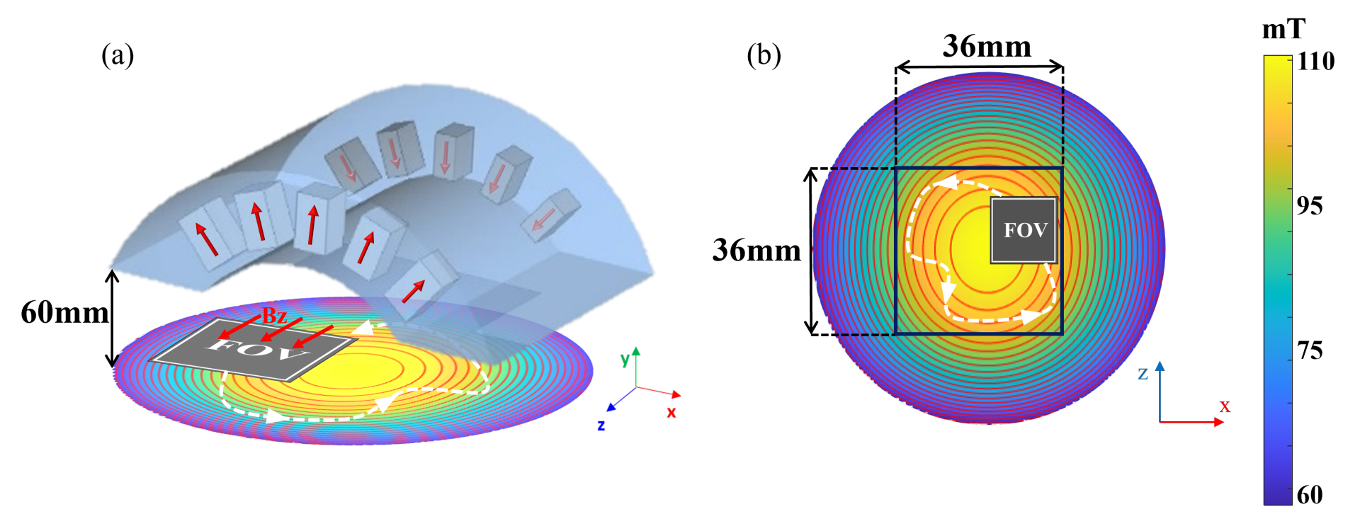
Fig 1(a) shows the single-sided magnet array used for this study. It is a partial inward-outward ring array4 consisting of 20mm$$$\times$$$40mm$$$\times$$$20mm N52 NdFeB magnet blocks arranged along two arcs. The FoV moves on a plane 60 mm away. The pattern and the direction of the magnetic field on this plane are shown. Fig 1(b) shows a top view of this plane, where the dashed line indicates the trajectory of the center of the FoV. For this study, only translational movements are included.The signal equation of this encoding is shown as follows,
$$s(t, n)=\int_V m(\vec{r}) c(\vec{r}) e^{-i \gamma \int_0^t B_{S E M}^\mu(\vec{r}, n) d \tau} d \vec{r}\tag1$$
where $$$m$$$ represents scanned object, $$$\vec{r}$$$ is the position factor, $$$c$$$ is the sensitivity map of receive coil, $$$\gamma$$$ is gyromagnetic ratio, $$$B_{S E M}^\mu(\vec{r}, n)$$$ is the SEM at the nth location of the FoV. Eq (1) is discretized in space and time to have $$$\bar{\bar{E}} \cdot \bar{m}=\bar{s}$$$, for imaging9.
For the acquisitions taken at the nth SEMs with $$$n_t$$$ time steps and a step size of $$$\Delta t$$$, the local k-space during the time $$$t=n_t \cdot \Delta t$$$ were generated using the following expression.
$$\vec{k}(\vec{r}, t, n)=\gamma \sum_0^{n_t} \nabla \vec{B}_{S E M}(\vec{r}, n) \cdot \Delta t\tag2$$
The FoV is split into 5$$$\times$$$5 sub-regions, the local k-spaces were plotted based on the gradients of the center point of each sub-region. As shown in the following three equations, the values of ratio of the actual sampled area $$$S_{actual}$$$ to the fully sampled area $$$S_{full}$$$ of individual sub-regions $$$k_L$$$ were calculated and averaged as an indicator $$$k$$$, and the variance of all $$$k_L$$$ was calculated as $$$k_{var}$$$, to evaluate the encoding capability of a trajectory.
$$k_L=\frac{S_{\text {actual }}}{S_{\text {full }}}\tag3$$
$$k=\frac{\sum_L k_L}{L}\tag4$$
$$k_{v a r}=\frac{\sum_{L=1}^N\left(k_L-k\right)^2}{N}\tag5$$
In the experiment, the region for the FoV to move was set to be 36mm$$$\times$$$36mm. The number of stopping points in one trajectory was set to be 36.
The fitness function of GA was defined as below,
$$Fitness function=k-w \cdot k_{\text {var }}\tag6$$
Herein, $$$w$$$ represents the weight, subject to further exploration. The population size of GA is 70000. A Shepp-logan phantom was used to test the imaging quality in the simulation.
Results
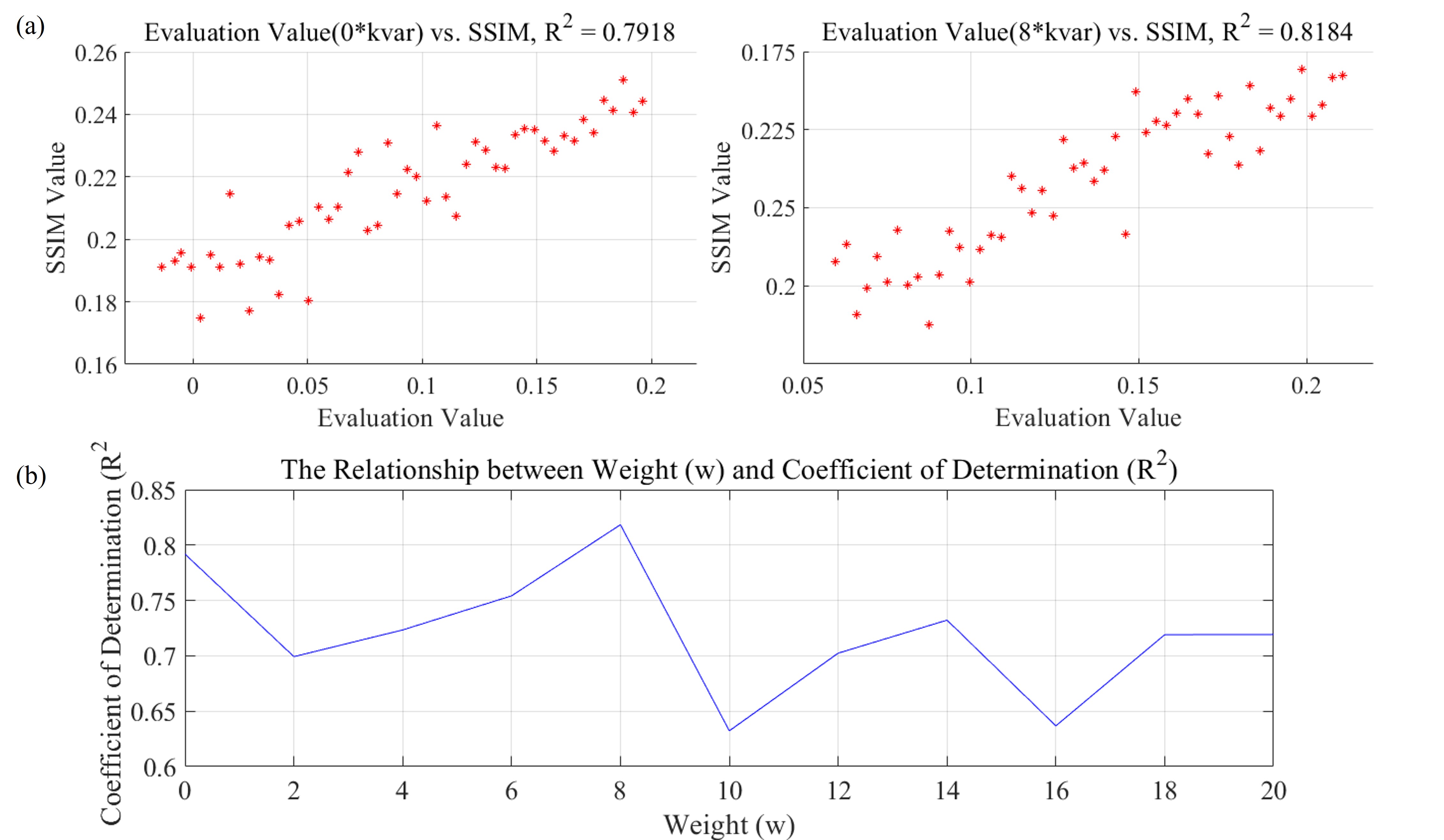
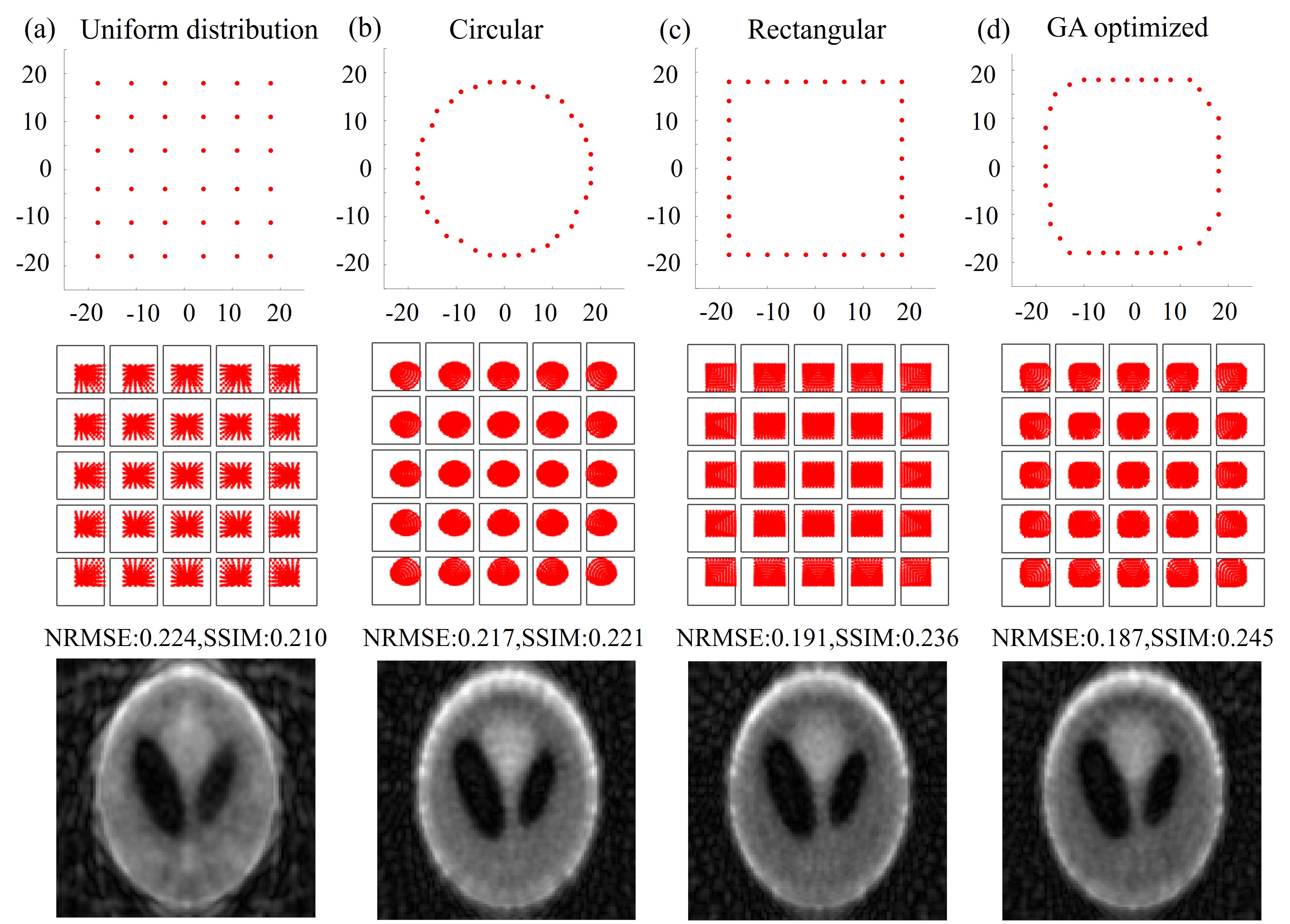
The results of exploration of $$$w$$$ are shown in Fig 2. When $$$w=8$$$, the fitness function has the highest correlation to the SSIM of reconstructed images.The uniform distribution, circular, rectangular and the optimized trajectory $$$(w=8)$$$ of stopping point are shown in Fig 3(a), (b), (c), (d), respectively. The corresponding local k-spaces and reconstructed images are shown in the second and the third row. As shown, the trajectory obtained through optimization yields better reconstruction compared to other commonly used trajectories. It results in large local k-space filling area and uniform filling levels across different sub-regions.
Discussion and Conclusion
Our research utilized GA to optimize FoV trajectories for single-sided magnet, resulting in improved signal encoding and reconstruction quality compared to common trajectories. The parameter $$$w$$$ of fitness function was explored to acquire a high correlation between the fitness function and the quality of reconstructed images.It was an initial study where only translational movements are included, and the phantom is not a realistic one for the targeted scenario. This study can be extended to encoding using more flexible movements for single-sided magnets, which can make an MRI system further portable.
Acknowledgements
No acknowledgement found.References
- Huang SY, Ren ZH, Obruchkov S, Gong J, Dykstra R, Yu W. Portable low-cost MRI system based on permanent magnets/magnet arrays. arXiv preprint arXiv:1812.10474. 2018.
- Cooley CZ, Stockmann JP, Armstrong BD, Sarracanie M, Lev MH, Rosen MS, Wald LL. Two-dimensional imaging in a lightweight portable MRI scanner without gradient coils. Magn Reson Med. 2015;73(2):872-883. doi:10.1002/mrm.25135
- Ren ZH, Mu WC, Huang SY. Design and optimization of a ring-pair permanent magnet array for head imaging in a low-field portable MRI system. IEEE Trans Magn. 2018;55(1):1-8. doi:10.1109/TMAG.2018.2866572
- Liang TO, Li E, Yu W, Huang SY. Compact and lightweight single-sided inward-outward (IO)-ring permanent magnet array for back imaging. Presented at: ISMRM; 2023.
- McDaniel PC, Cooley CZ, Stockmann JP, Wald LL. The MR cap: a single-sided MRI system designed for potential point-of-care limited field-of-view brain imaging. Magn Reson Med. 2019;82(5):1946-1960. doi:10.1002/mrm.27869
- Greer M, Chen C, Mandal S. An easily reproducible, hand-held, single-sided, MRI sensor. J Magn Reson. 2019;308:106591. doi:10.1016/j.jmr.2019.106591
- Cooley CZ, Stockmann JP, Armstrong BD, et al. Two-dimensional imaging in a lightweight portable MRI scanner without gradient coils. Magn Reson Med. 2015;73(2):872-883. doi:10.1002/mrm.25135
- Ren ZH, Obruchkov S, Lu DW, Dykstra R, Huang SY. A low-field portable magnetic resonance imaging system for head imaging. In: 2017 Progress in Electromagnetics Research Symposium-Fall (PIERS-FALL). 2017:3042-3044
- Gong J, Huang SY, Ren ZH, Yu W. Effects of encoding fields of permanent magnet arrays on image quality in low-field portable MRI systems. IEEE Access. 2019;7:80310-80327 doi:10.1109/ACCESS.2019.2922916
Figures