2849
Short T2* imaging in a portable and low-field MRI scanner1i3M, CSIC, Valencia, Spain, 2Tesoro Imaging SL, Valencia, Spain
Synopsis
Keywords: Low-Field MRI, Low-Field MRI, Short T2, hard tissue, extremity imaging
Motivation: We have previously demonstrated the versatility of a portable 72mT extremity MRI scanner. Hard tissue imaging would enhance the system’s potential, but this remains to be demonstrated in low-field systems (<0.1T)
Goal(s): To explore the possibility of imaging samples with T2*<1ms, comparable to those bone or ligament.
Approach: We programmed a PETRA sequence into the MaRCoS opens-source console, and we compared images of short and long T2* samples resulting from PETRA and Spin Echo.
Results: Image reconstructions show that samples with T2* as low as 800us can be successfully imaged with PETRA in conditions where Spin Echo outputs mostly noise.
Impact: By successfully capturing signals from short T2* tissues, our research enhances our 72mT portable MRI scanner utility designed for extremity imaging.
Introduction
The MRI community's heightened interest in low-field scanners (<100 mT) has led to the development of cost-effective, lightweight, and portable systems. Our 72 mT portable MRI scanner, based on a Halbach magnet, specifically targets musculoskeletal applications [1].In extremity imaging, details from hard tissues like bones (with T2* < 1 ms) can be of relevance. Conventional MRI sequences lack this capability, but special-purpose sequences like PETRA and other ZTE variations are used clinically at high field strengths [2]. In this work, we explore PETRA imaging for samples with T2* down to 800 us in our portable system [3, 4].
Methods
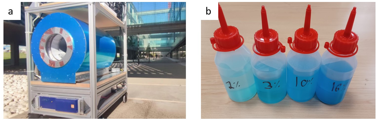
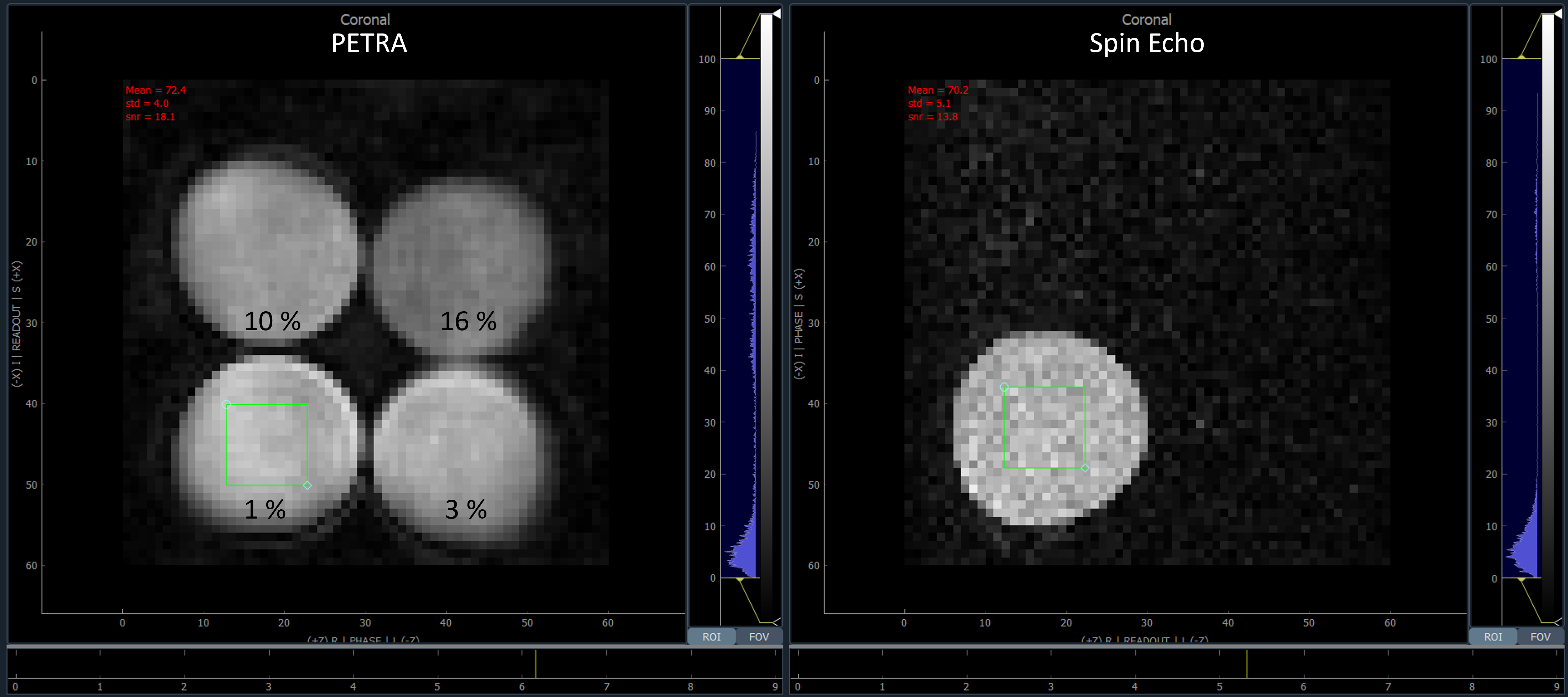
Experiments were performed in our portable MRI scanner of 72 mT (Figure 1.a). This extremity imaging system uses a discrete Halbach magnet and has been designed with the target to be portable, small foot-print, and low-cost. The magnetic field homogeneity can be shimmed down to 120 ppm over a spherical field of view of diameter 10 cm, the gradients can be ramped up to 25 mT/m, and the radiofrequency TxRx coil is a solenoid with 15 cm diameter and 15 cm length with 25 turns. The system is controlled by MaRCoS [5, 6], an open-source console based on a Red Pitaya board (SDRLab). In our imaging experiment, we utilized four water bottles with copper sulfate at concentrations of 1 %, 3 %, 10 % and 16 % (Figure 1.b), corresponding to T2* (T2) values of 7.5 (17) ms and 2.5 (3.1) ms, 1.2 (2.0) ms and 800 (950) us respectively, as measured with a FID (CPMG) sequence. With the sample in the scanner, we calibrated as usual: RF coil impedance matching to the Larmor frequency, B1 efficiency calibration, and B0 shimming.Subsequently, we acquired two images: one using the PETRA sequence and another with Spin Echo. Both images were obtained with a matrix size of 60x60x10 and a field of view of 12x12x10 cm³. The PETRA image was acquired with a flip angle of 90º, a repetition time (TR) of 50 ms. The Spin Echo image was acquired with TR = 50 ms and an echo time (TE) of 10 ms, allowing for a comparative analysis of the imaging outcomes. PETRA images are generated using Algebraic Reconstruction Techniques [7], while Spin Echo reconstruction is performed by conventional Inverse Fast Fourier Transform.
Results
Figure 2 displays the PETRA (left) and Spin Echo (right) images. The latter exclusively captures the long T2 sample, while PETRA distinctly reveals all four samples. PETRA images demonstrate a superior signal-to-noise ratio, possibly attributed to both the ART regularization process and the ability to capture signals hidden in Spin Echo due to short T2 samples. However, PETRA images exhibit degraded borders compared to Spin Echo, possibly due to the T2* effect.Discussion
We have shown the capability of PETRA to capture the signal from short T2* samples, exemplified by the simultaneous imaging of samples with copper sulfate concentrations ranging from 1 % to 16 %. Our study demonstrates the feasibility of employing ZTE sequences for in vivo hard tissue imaging at field strengths under 0.1 T. The ability to differentiate between tissues of varying T2* values is of potentially high clinical significance for e.g. extremity imaging. Additionally, the noise reduction observed in PETRA images increases the overall image quality, further enhancing its suitability for clinical applications.Conclusion
In conclusion, our study shows PETRA imaging is possible even in low-field and portable MRI scanners. By providing images of short T2 tissues, this advancement holds promise for improved diagnostic accuracy. Our portable system is hence being upgraded for an in vivo demonstration.Acknowledgements
Project funded by: the EU (EIC Transition, 101136407), Spanish MICINN (PID2022-142719OB-C22), the Valencian Government (CIPROM/2021/003) and the Valencian Innovation Agency (INNVA1/2022/4, INNVA1/2023/30).References
[1] Guallart-Naval, T., Algarín, J. M., Pellicer-Guridi, R., Galve, F., Vives-Gilabert, Y., Bosch, R., Pallás, E., González, J. M., Rigla, J. P., Martínez, P., Lloris, F. J., Borreguero, J., Marcos-Perucho, A., Negnevitsky, V., Martí-Bonmatí, L., Ríos, A., Benlloch, J. M., & Alonso, J. (2022). Portable magnetic resonance imaging of patients indoors, outdoors and at home. Scientific Reports, 12. https://doi.org/10.1038/s41598-022-17472-w
[2] Grodzki, D. M., Jakob, P. M., & Heismann, B. (2012). Ultrashort echo time imaging using pointwise encoding time reduction with radial acquisition (PETRA). Magnetic Resonance in Medicine, 67(2), 510–518. https://doi.org/10.1002/mrm.23017
[3] Grodzki, D. M., Jakob, P. M., & Heismann, B. (2012). Ultrashort echo time imaging using pointwise encoding time reduction with radial acquisition (PETRA). Magnetic Resonance in Medicine, 67(2), 510–518. https://doi.org/10.1002/mrm.23017
[4] Algarín, J. M., Díaz-Caballero, E., Borreguero, J., Galve, F., Grau-Ruiz, D., Rigla, J. P., Bosch, R., González, J. M., Pallás, E., Corberán, M., Gramage, C., Aja-Fernández, S., Ríos, A., Benlloch, J. M., & Alonso, J. (2020). Simultaneous imaging of hard and soft biological tissues in a low-field dental MRI scanner. Scientific Reports, 10, 21470. https://doi.org/https://doi.org/10.1038/s41598-020-78456-2
5] Negnevitsky, V., Vives-Gilabert, Y., Algarín, J. M., Craven-, L., Pellicer-Guridi, R., Reilly, T. O., Stockmann, J. P., Webb, A., Alonso, J., & Menküc, B. (2023). MaRCoS, an open-source electronic control system for low-field MRI. Journal of Magnetic Resonance, 350, 107424. https://doi.org/10.1016/j.jmr.2023.107424
[6] Guallart‐Naval, T., O’Reilly, T., Algarín, J. M., Pellicer‐Guridi, R., Vives‐Gilabert, Y., Craven‐Brightman, L., Negnevitsky, V., Menküc, B., Galve, F., Stockmann, J. P., Webb, A., & Alonso, J. (2022). Benchmarking the performance of a low‐cost magnetic resonance control system at multiple sites in the open MaRCoS community. NMR in Biomedicine, 36(1), 1–13. https://doi.org/10.1002/nbm.4825
[7] Gordon, R., Bender, R., & Herman, G. T. (1970). Algebraic Reconstruction Techniques (ART) for three-dimensional electron microscopy and X-ray photography. Journal of Theoretical Biology, 29(3), 471–481. https://doi.org/10.1016/0022-5193(70)90109-8
Figures