2847
Scan-specific deep learning-based denoising method for low-field MR images1Center for Adaptable MRI Technology (AMT Center), Institute of Medical Sciences, School of Medicine, Medical Sciences & Nutrition, University of Aberdeen, Aberdeen, United Kingdom
Synopsis
Keywords: Low-Field MRI, Low-Field MRI, denoising, self-supervised learning, zero-shot learning, low SNR
Motivation: Low SNR per unit-time in low-field MRI results in noisy images when targeting both clinically acceptable resolution and acquisition times which may limit their diagnostic effectiveness.
Goal(s): We seek to improve low-field MRI SNR by means of deep-learning while overcoming the limitations of traditional supervised learning and without compromising denoising performance.
Approach: We build on:1)self-supervised method enabling training without having to collect ‘noise-free’ data and 2)zero-shot concept to achieve dataset-free and scan-specific denoising.Additionally,we adopted simplified architecture for fast training times.
Results: Our method showed high denoising performance for different SNR levels and contrasts within few seconds of processing time competing with well-established BM4D.
Impact: Our proposed denoising method, based on self-supervised zero-shot deep-learning, enables high-performance denoising within short processing times. This approach shows promise for speedy acquisitions and enhanced imaged quality in low-field, point-of-care settings.
Introduction
Low magnetic field strength (LF) MRI is gaining interest worldwide, aspiring to develop complementary offers (i.e., more accessible, point-of-care, purpose-built) to conventional high-field (1.5T, 3T) clinical MRI scanners. Yet, the inherently impeded sensitivity in LF settings, mainly stemming from lower spin polarization, generally requires strategies to enhance signal-to-noise ratio (SNR) per unit-time for better imaging performance.With limited signal, denoising can be crucial to improve SNR. Recently, deep learning (DL)-based denoising methods have been proposed, showing promising performance for MRI at both low- and high-field1,2. Supervised learning is a commonly used DL approach where a network learns image and noise priors on extensive datasets consisting of pairs of noisy and “noise-free” images. Yet, this method raises questions about generalization which translates into performance drop when testing on data that do not belong to the same distribution as the training set (i.e., SNR, contrast, and anatomy mismatch)3.
In this work, we propose a scan-specific DL denoising method building upon a recently introduced self-supervised denoising technique, ‘Noisier2noise’4, and take it a step further by enabling learning without training datasets, a concept referred to as a zero-shot self-supervised learning (zs-ssl)5. The performance of this denoising approach was compared to BM4D6, a high-performance denoising algorithm often found in LF literature. Performance was assessed on MR images acquired at 0.1 T on a low-field extremity scanner7 for different SNR levels.
Methods
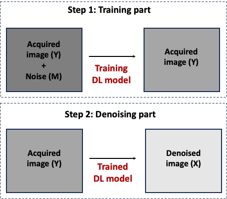
Let Y=X+N be a noisy acquired complex MR image, where X is the noise-free image and N is the random noise. In MRI, noise is well modelled by a Gaussian distribution with zero mean and variance in the real and imaginary parts of the complex data. The variance was estimated by computing the standard deviation of the real and imaginary parts of the MR complex images background. A noisier image Z=Y+M was then created by adding noise M drawn from the same noise distribution as N. Subsequently, a DL network was trained to predict Y given Z as an input (step1). Once trained, the model is used to denoise the acquired noisy image Y (step2) (figure1).A light version of DnCNN8 was adopted as a model architecture with only one single layer to increase training speed. Additionally, the complex nature of MR images was preserved by splitting real and imaginary data into 2 channels. Training was done using the ADAM optimizer and mean squared error (MSE) as a loss function. 500 epochs were sufficient for the model to converge with no sign of overfitting. Computation was carried out on a GPU GeForce RTX 4060.
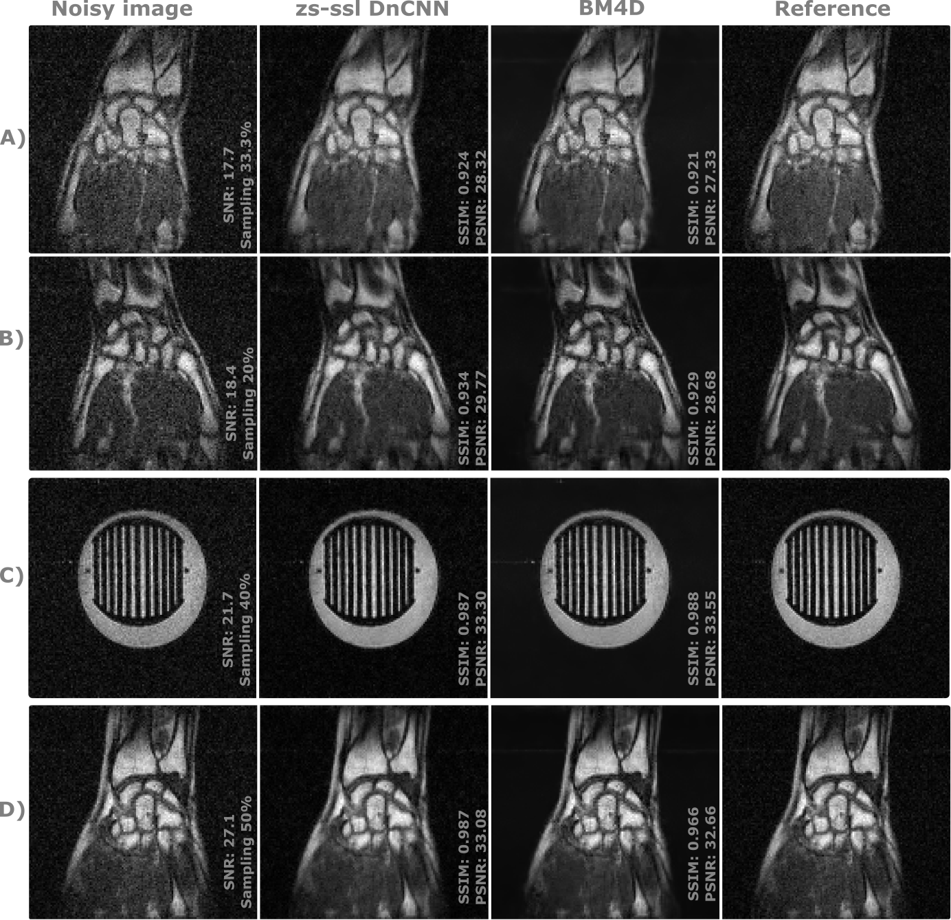
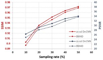
The performance of this approach was assessed on a total of 22 sets of 3D MR phantom and in-vivo human wrist data acquired on a 0.1 T scanner using heterogenous sequence parameters. During the acquisition, each average was individually stored in a fourth dimension, allowing retrospective manipulation of k-spaces to generate different SNR images. Accordingly, each set was degraded to simulate multiple reduced sampling rate. Denoising approaches were tested over SNR images ranging between 10 and 40. The performance of the proposed approach was compared to BM4D. SSIM9 and Peak SNR (PSNR) were chosen as evaluation metrics. Ultimately, the total processing time required by our proposed approach was reported.
Results
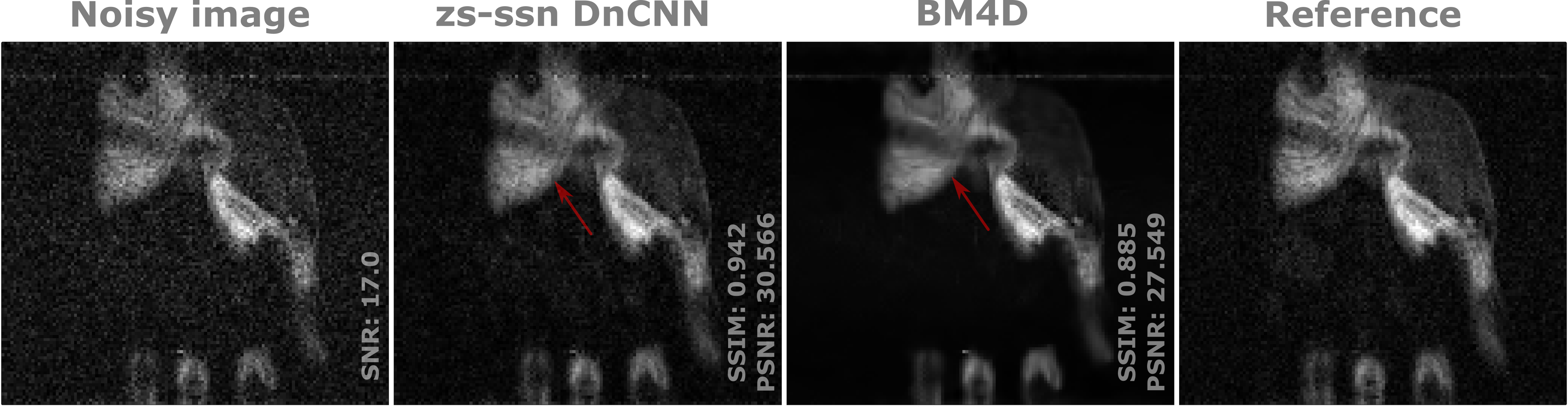
Figure 2 compares our zs-ssl DnCNN to BM4D for different SNR levels, contrasts and objects. It shows that our proposed approach is capable of denoising while preserving the overall texture, contrast and details. Qualitatively, the performance of both methods is very comparable. Nevertheless, our approach is immune to artifact in contrast to BM4D that occasionally shows artifacts when SNR is low (figure3). Quantitatively, our approach demonstrates slightly better performance when compared to BM4D (figure4). The training duration for DnCNN (step1) shows a dependency with matrix size. Here, it required 3 s to train over a [128x128x15]matrix and the denoising phase (step2) necessitated ~20 ms for the whole matrix.Discussion and Conclusion
We propose a scan-specific DL-denoising model that does not require any training database. The small DL network used allowed training and denoising steps to be achieved in a few seconds without compromising denoising performance. Network performance was validated on MR images with different contrasts, SNRs, and content (phantoms, in vivo hand/wrist images). Results showed comparable performance to BM4D while occasionally outperforming the latter at very low SNR. However, since the reference images are not ‘noise-free’, it is important to note that quantitative results may be impacted by the choice of evaluation metrics. With room for further improvement (tweaking the loss function or network hyperparameters) this approach holds significant promise for LF images denoising.Acknowledgements
No acknowledgement found.References
[1] Ran et al., Denoising of 3D magnetic resonance images using a residual encoder-decoder Wasserstein generative adversarial network, Medical Image Analysis (2019) DOI: 10.1016/j.media.2019.05.001
[2] Koonjoo et al. Boosting the signal-to-noise of low-field MRI with deep learning image reconstruction. Scientific Reports(2021). https://doi.org/10.1038/s41598-021-87482-7
[3] knoll. et al., Assessment of the generalization of learned image reconstruction and the potential for transfer learning, Magnetic Resonance in Medicine (2018), https://doi.org/10.1002/mrm.27355
[4] Moran et al., Noisier2noise: learning to denoise from unpaired noisy data. Proceedings of the IEEE/CVF Conference on Computer Vision and Pattern Recognition (2020). https://doi.org/10.48550/arXiv.1910.11908
[5] Yaman et al., Zero-shot self-supervised learning for MRI reconstruction. Conference paper at ICLR (2022).https://doi.org/10.48550/arXiv.2102.07737
[6] Dabov et al., Image denoising by sparse 3D transform-domain. IEEE transactions on image processing (2007). Doi:10.1109/TIP.2007.901238
[7] Constantinesco et al. Low-field dedicated and desktop magnetic resonance imaging systems for agricultural and food applications. Magn. Reson. Chem. (1997). https://doi.org/10.1002/(SICI)1097-458X(199712)35:13<S69::AID-OMR198>3.0.CO;2-5
[8] Zhang et al., Beyond a Gaussian denoiser: Residual learning of deep CNN for image denoising. IEEE Transactions on Image Processing (2017), 26(7), 3142–3155. https://doi.org/10.1109/TIP.2017.2662206
[9] Wang et al., Image Quality Assessment: From Error Visibility to Structural Similarity, IEEE Transaction Image Processing (2004). Doi: 10.1109/TIP.2003.819861
Figures