2846
Is low field always better for imaging around passive implants?1Biomedical Engineering, Dalhousie University, Halifax, NS, Canada, 2Biomedical Translational Imaging Centre (BIOTIC), QEII Health Sciences Center, Halifax, NS, Canada, 3Physics and Atmospheric Science, Dalhousie University, Halifax, NS, Canada, 4Diagnostic Radiology, Dalhousie University, Halifax, NS, Canada, 5Diagnostic Imaging, Nova Scotia Health, Halifax, NS, Canada
Synopsis
Keywords: Low-Field MRI, Susceptibility, Routine Protocols
Motivation: To investigate the performance of modernized low-field MRI relative to traditional systems for imaging near metallic devices within the clinical context.
Goal(s): To evaluate whether low-field MRI can offer a significant reduction in artifacts when using routine clinical protocols.
Approach: The artifact characteristics of 0.5 T, 1.5 T, and 3 T MRIs are compared in this ASTM F2119-07-based phantom study of common passive metallic devices.
Results: Low-field MRI demonstrated the capability to reduce susceptibility artifacts when imaging near metal-containing medical devices. However, artifact produced by some pulse sequences diverged from the anticipated field-dependence, highlighting the sizable effects of clinical protocolling.
Impact: This phantom study demonstrates that low-field MRI can image metallic devices with reduced artifact relative to 1.5/3 T systems using routine clinical protocols, highlighting opportunities for future in vivo studies involving implants and imaging in areas with magnetic susceptibility distortions.
Introduction
In addition to the value-based proposition for recently modernized low-field MRI systems, there are many underexplored clinical niches where they might provide an advantage over mid- and high-field scanners. One key area is imaging near metal-containing medical devices, where susceptibility artifacts degrade image quality and reduce the diagnostic utility of images. A range of basic to advanced artifact reduction techniques exist, where the implementation of the more effective techniques often require considerable trade-offs to factors such as scan time or resolution1. Based on the field dependence of susceptibility-induced field distortions2, contemporary low-field MRI may offer a middle ground. We hypothesize that susceptibility artifacts produced by MR-conditional devices will be significantly reduced at 0.5 T compared to 1.5 T and 3 T scanners explored using routine brain imaging protocols in active clinical use at our institution.Methodology
Routine brain imaging pulse sequences independently protocolled on three different field strength MRIs were used to evaluate susceptibility artifact produced by passive medical devices. Methodology for this phantom study was based on recommendations provided in ASTM International F2119-073, an FDA consensus standard for measuring artifact produced by passive medical devices4. A summary of the routine brain imaging protocol is available in Table 1. Inclusion of repetition time-matched (TRM) bSSFP enables a comparison which excludes clinical protocolling effects while producing field-dependent banding artifacts that are directly related to local field distortions. The studied medical devices included a 316L stainless steel staple, a nitinol flow diverting stent, and a platinum embolization coil. These high-use devices are expected to provide a reasonable range of magnetic susceptibilities and geometries. The staple and stent were imaged with their longest axis parallel and perpendicular to the main magnetic field. However, the coil was only imaged in the perpendicular orientation due to limitations associated with supporting the device within the phantom. A spherical acrylic phantom filled with MnCl2-doped gelatine was used to represent in vivo magnetic properties and secure the devices during imaging. The MRI systems available for research use at our institution were a 0.5 T Synaptive Evry, a 1.5 T GE HealthCare Signa HDxt, and a 3 T GE HealthCare Discovery MR750.Artifact width was measured using an automated procedure based on definitions provided in F2119-073. Figure 1 depicts key steps from this pipeline. Radiofrequency (RF) shielding artifacts were assessed on TOF-MRA images by comparing the relative in-stent signal intensity to background signal using a region of interest-based method5 in FIJI6.
Three replicate image sets were collected for each device and orientation to facilitate statistical testing. Significance was assessed using paired two-tailed t-tests and the false discovery rate was controlled using a two-stage linear step-up procedure7. Comparisons were limited to retain statistical power. Average artifact width comparisons were only made between 0.5 T and 1.5 T and between 0.5 T and 3 T for a specific pulse sequence, device, and orientation.
Results and Discussion
A selection of representative susceptibility artifact images is presented in Figure 2. A summary of artifact width measurements is provided in Figure 3. Statistical significance is indicated by an asterisk over a comparison arrow. The anticipated field-dependence of susceptibility artifacts produced by T2WI, T2 FLAIR, T1 FLAIR, and TRM bSSFP were confirmed. However, DWI and TOF-MRA results deviated from expectations. At 3 T, bSSFP utilizes a maximum intensity projection of two phase-cycled acquisitions to enable comparable performance to 0.5 T. The reduction in artifact with bSSFP compared to TRM bSSFP, coupled with the above results, points to the considerable effects of clinical protocolling decisions and trade-offs.Device orientation relative to the main magnetic field had minimal effect on artifact width.
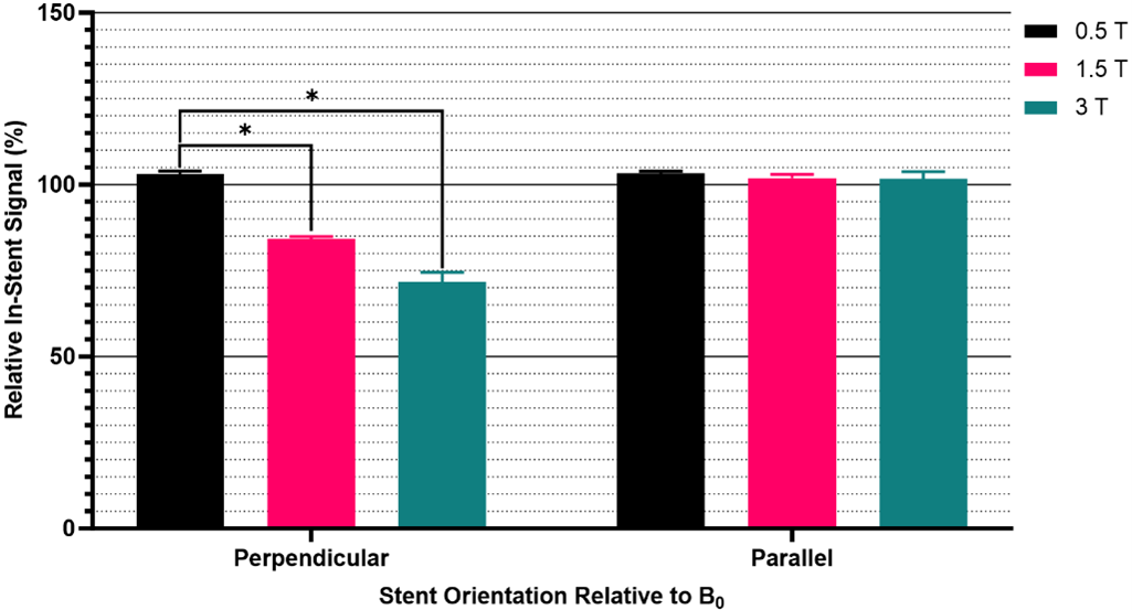
The results of the RF shielding assessment are provided in Figure 4. Device orientation and field strength significantly influenced RF shielding effects. RF shielding was negligible with the stent oriented parallel to the main field, but the perpendicular orientation reduced relative in-stent signal intensity to 72% at 3 T and 84% at 1.5 T.
Conclusion
Results from TRM bSSFP confirmed the scaling of susceptibility-induced field distortions with field strength. Clinical protocolling at 1.5 T and 3 T mitigated the effects of increased field distortions. The most substantial evidence is provided by bSSFP at 3 T, where artifact width was reduced relative to 1.5 T and comparable to 0.5 T. Protocolling trade-offs that decrease sensitivity to magnetic field distortions are frequently costly in terms of increased scan time or reduced signal-to-noise ratio. Alternatively, low-field MRI protocols require fewer of these protocolling compromises, suggesting an opportunity for improved clinical imaging around implanted devices and in regions with substantial magnetic susceptibility distortions.Acknowledgements
Funding for this research was provided by grants from the NSERC Discovery program and INOVAIT Focus Fund, and by scholarships from Dalhousie's Faculty of Engineering Graduate Award and the Bruce & Dorothy Rosetti Engineering Research Scholarship.References
1. Hargreaves BA, Worters PW, Pauly KB, Pauly JM, Koch KM, Gold GE. Metal-Induced Artifacts in MRI. American Journal of Roentgenology. 2011;197(3):547-555. doi:10.2214/AJR.11.73642. Schenck JF. The role of magnetic susceptibility in magnetic resonance imaging: MRI magnetic compatibility of the first and second kinds. Med Phys. 1996;23(6):815-850. doi:10.1118/1.597854
3. ASTM Committee F04 on Medical and Surgical Materials and Devices. F2119-07(2013) Standard Test Method for Evaluation of MR Image Artifacts from Passive Implants. West Conshohocken, PA, United States: ASTM International doi:10.1520/F2119-07R13
4. U.S. Food & Drug Administration. Recognized Consensus Standards: Medical Devices - ASTM F2119-07. September 2023. https://www.accessdata.fda.gov/scripts/cdrh/cfdocs/cfstandards/detail.cfm?standard__identification_no=31055.
5. Choi JW, Roh HG, Moon WJ, Chun YI, Kang CH. Optimization of MR Parameters of 3D TOF-MRA for Various Intracranial Stents at 3.0T MRI. Neurointervention. 2011;6(2):71-77. doi:10.5469/neuroint.2011.6.2.71
6. Schindelin J, Arganda-Carreras I, Frise E, et al. Fiji: an open-source platform for biological-image analysis. Nat Methods. 2012;9(7):676-682. doi:10.1038/nmeth.2019
7. Benjamini Y, Krieger AM, Yekutieli D. Adaptive linear step-up procedures that control the false discovery rate. Biometrika. 2006;93(3):491-507. doi:10.1093/biomet/93.3.491
Figures
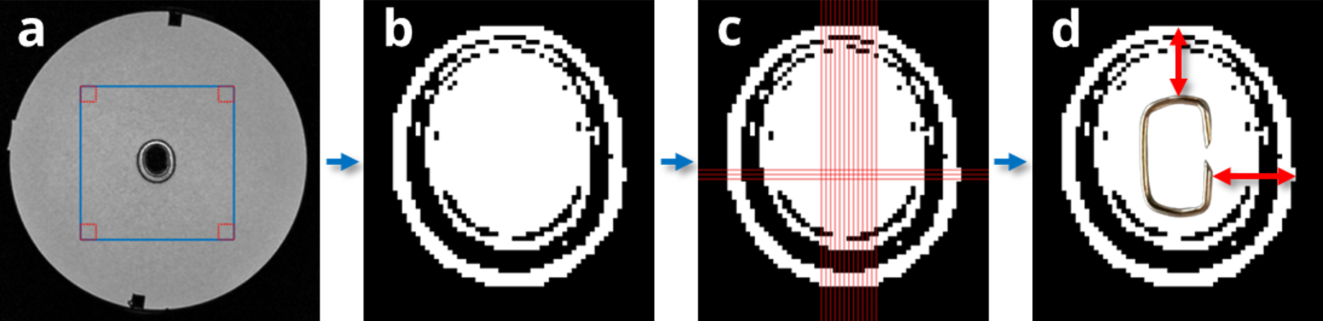
Figure 1: Maximum in-plane artifact width measurement pipeline. The region of interest (blue box) and four background regions (red boxes) are automatically positioned on each image in (a). Signal is sampled from background regions in (a) and used to threshold the region of interest to ±30% of mean background signal intensity in (b). The largest discontinuous distance is measured in (c) from which the device dimension is subtracted and divided by two. This process is completed in the vertical and horizontal directions to produce the artifact width measurements represented in (d).
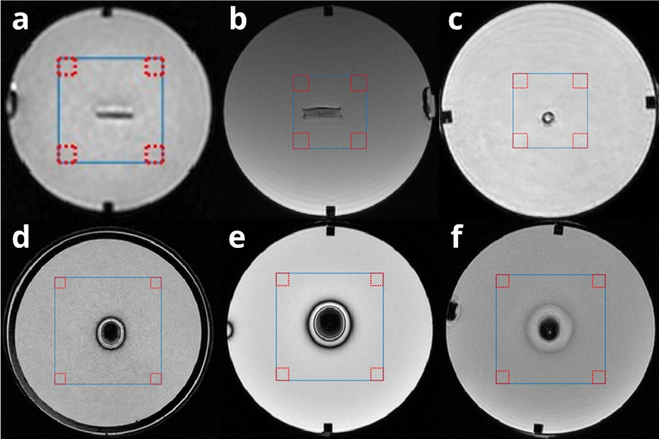
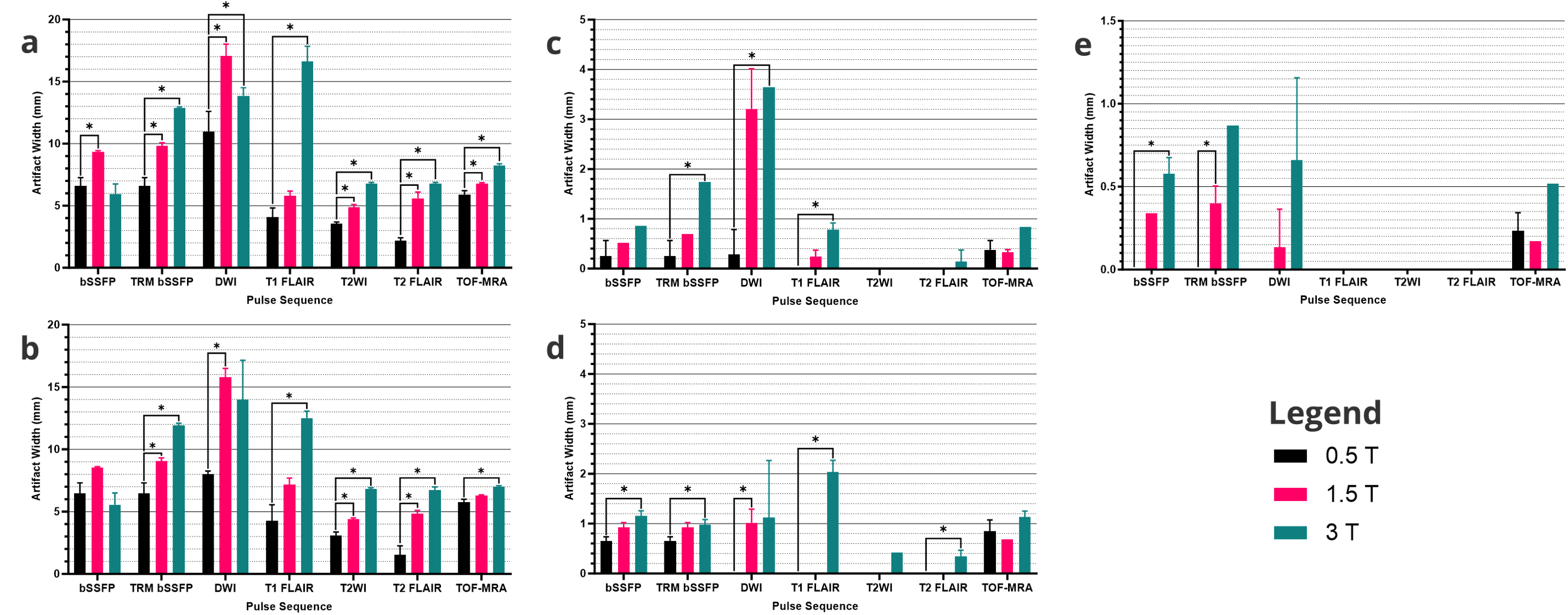
Figure 3: Average maximum in-plane artifact width measurements. Plots are grouped by device and orientation of the longest device axis relative to the main magnetic field as follows: (a) staple, perpendicular, (b) staple, parallel, (c) stent, perpendicular, (d) stent, parallel, and (e) coil, perpendicular.