2842
Rapid Zero-shot Image Denoising for Quantitative Imaging on a Point-Of-Care 46-mT-MRI System1C.J. Gorter MRI Center, Department of Radiology, LUMC, Leiden, Netherlands, 2Division of Image Processing, Department of Radiology, LUMC, Leiden, Netherlands, 3Wellcome Centre for Integrative Neuroimaging, FMRIB, Nuffield Department of Clinical Neurosciences, University of Oxford, Oxford, United Kingdom, 4Philips Research Hamburg, Hamburg, Germany
Synopsis
Keywords: Low-Field MRI, Low-Field MRI, denoising
Motivation: Low-field MRI holds the promise of expanding access to healthcare. The low signal-to-noise ratio (SNR) poses a significant challenge to acquiring diagnostically useful information in a reasonable scanning time.
Goal(s): To overcome the challenge of low SNR in low-field MRI, achieving fast, self-supervised denoising.
Approach: A rapid 4D-denoising method utilizing the Zero-Shot-Noise2Noise framework is proposed, without the need for intensive network training.
Results: This method provides fast denoising in just 10-20 seconds per case and significantly boosts SNR efficiency, reducing the number of measrued TIs and TEs needed for precise, high-quality T1/T2 mapping.
Impact: This study's fast 4D-denoising approach revolutionizes low-field MRI by enhancing SNR without extensive training datasets, enabling faster, more efficient imaging and broadening diagnostic accessibility in resource-limited settings.
Introduction
Low-field MRI (<0.1T) has the potential to expand healthcare access, particularly in low- and middle-income regions1,2. However, its inherently low signal-to-noise ratio (SNR) presents a challenge in terms of scan time and spatial resolution, affecting detailed pathology detection and diagnosis. Recently, deep learning-based denoising methods have received increasing attention, and have been applied to low-field MRI data sets3,4. However, supervised learning-based denoising suffers from the lack of high-quality low-field training data, while transfer learning from networks trained on high-field MRI data5 requires careful tuning to account for differences in contrast and noise distribution. In this work, we adapted the zero-shot noise2noise (ZS-N2N)6 concept for low-field MRI and added an additional “relaxation/contrast” dimension (T1,T2) for a self-supervised, fast 4D-denoising in about 10-20 seconds per subject, potentially allowing fewer TI/TE points required for T1/T2 fitting.Methods
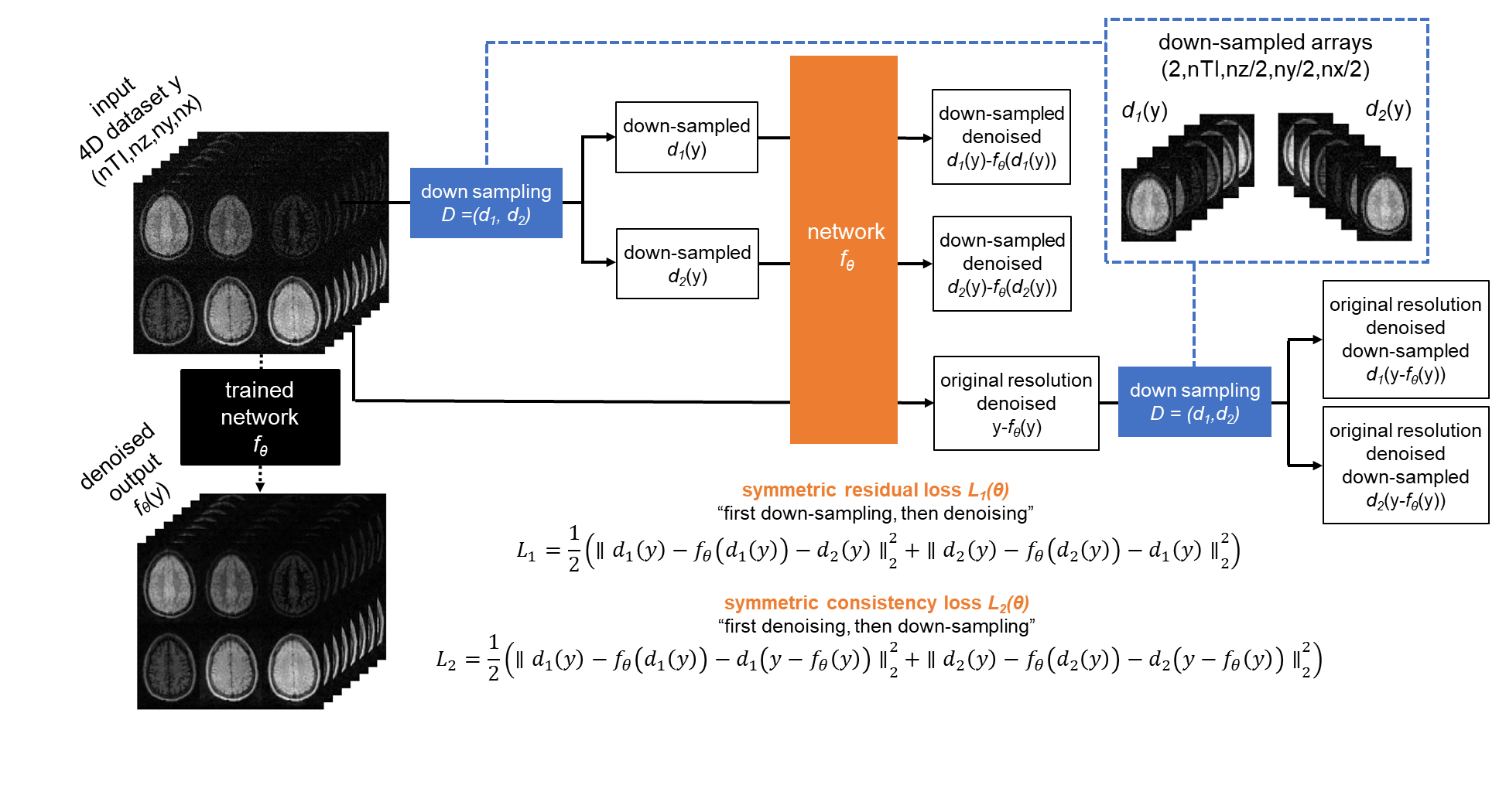
Noise2noise7 has demonstrated that networks trained on noisy image pairs can achieve similar quality as conventional supervised learning. In ZS-N2N, a single source image can be down-sampled into two sub-images, whose noise shares the same distribution while remaining statistically independent. In this study, given a quantitative MR-parameter mapping dataset (source images) with dimensions (nt,nx,ny,nz), two low-resolution down-sampled images (nt,nx/2,ny/2,nz/2) can be formed by applying two 3D convolutions on the source images, with stride of two and “symmetric” kernels: $$$k_1=\left[\left[\begin{array}{cc}0.25&0\\0&0.25\end{array}\right],\left[\begin{array}{cc}0&0.25\\0.25&0\end{array}\right]\right]$$$ and $$$k_2=\left[\left[\begin{array}{cc}0&0.25\\0.25&0\end{array}\right],\left[\begin{array}{cc}0.25&0\\0&0.25\end{array}\right]\right]$$$, as $$$d_1(y)=y\circledast{k_1}$$$ and $$$d_2(y)=y\circledast{k_2}$$$. In the loss calculation for a given network $$$\hat{x}=f_{\hat{\theta}}(y)$$$, the network parameters are fitted initially with a symmetric residual-learning loss calculated as:$$L_1=\frac{1}{2}\left(\left\|d_1(y)-f_\theta\left(d_1(y)\right)-d_2(y)\right\|_2^2+\left\|d_2(y)-f_\theta\left(d_2(y)\right)-d_1(y)\right\|_2^2\right).$$Here the network is optimized between two down-sampled images $$$d_1(y)$$$,$$$d_2(y)$$$from the same source image. Secondly, the data-consistency loss is calculated by first denoising the source image and then applying down sampling:$$L_2=\frac{1}{2}\left(\left\|d_1(y)-f_\theta\left(d_1(y)\right)-d_1\left(y-f_\theta(y)\right)\right\|_2^2+\left\|d_2(y)-f_\theta\left(d_2(y)\right)-d_2\left(y-f_\theta(y)\right)\right\|_2^2\right).$$This constrains the network output to be consistent with the source image thereby avoiding overfitting to the down-sampled images. The total loss for the optimization would be $$$L_{\text{total }}=L_1+L_2$$$.To enable a short processing time and avoid overfitting, a simplistic 3D convolutional neural network $$$f_\theta$$$ is used. It incorporates a 3D convolutional neural network structure with three convolutional layers, employing a LeakyReLU activation with a negative slope of 0.2, performing convolutions with kernel sizes of 3x3x3 in the first two layers and 1x1x1 in the third layer. The denoising pipeline is illustrated in Fig.1. For a standard inversion-recovery T1 mapping dataset with 6 TIs and a matrix size of (6,32,94,70), the network with its modest 70k parameters completes denoising of the entire dataset in less than 20 seconds on an NVIDIA RTX6000 GPU over 2000 epochs.
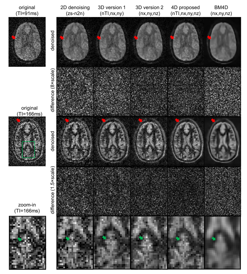
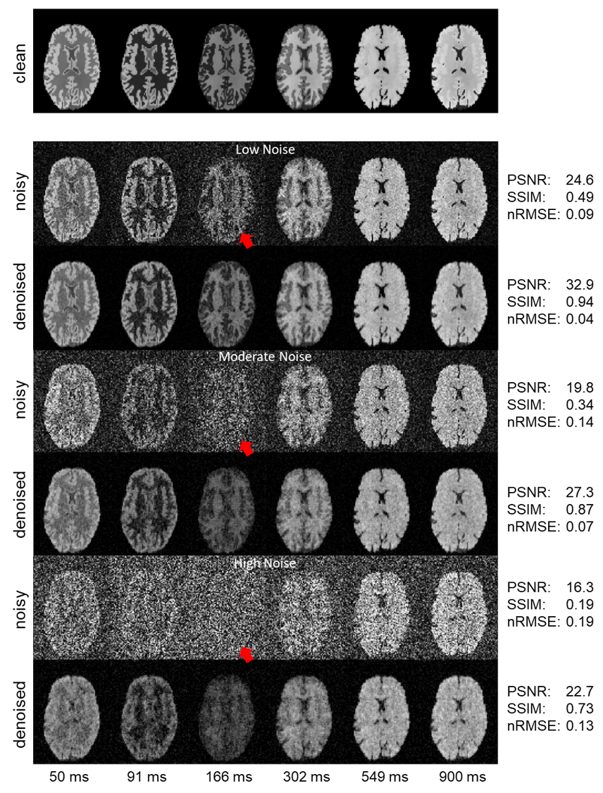
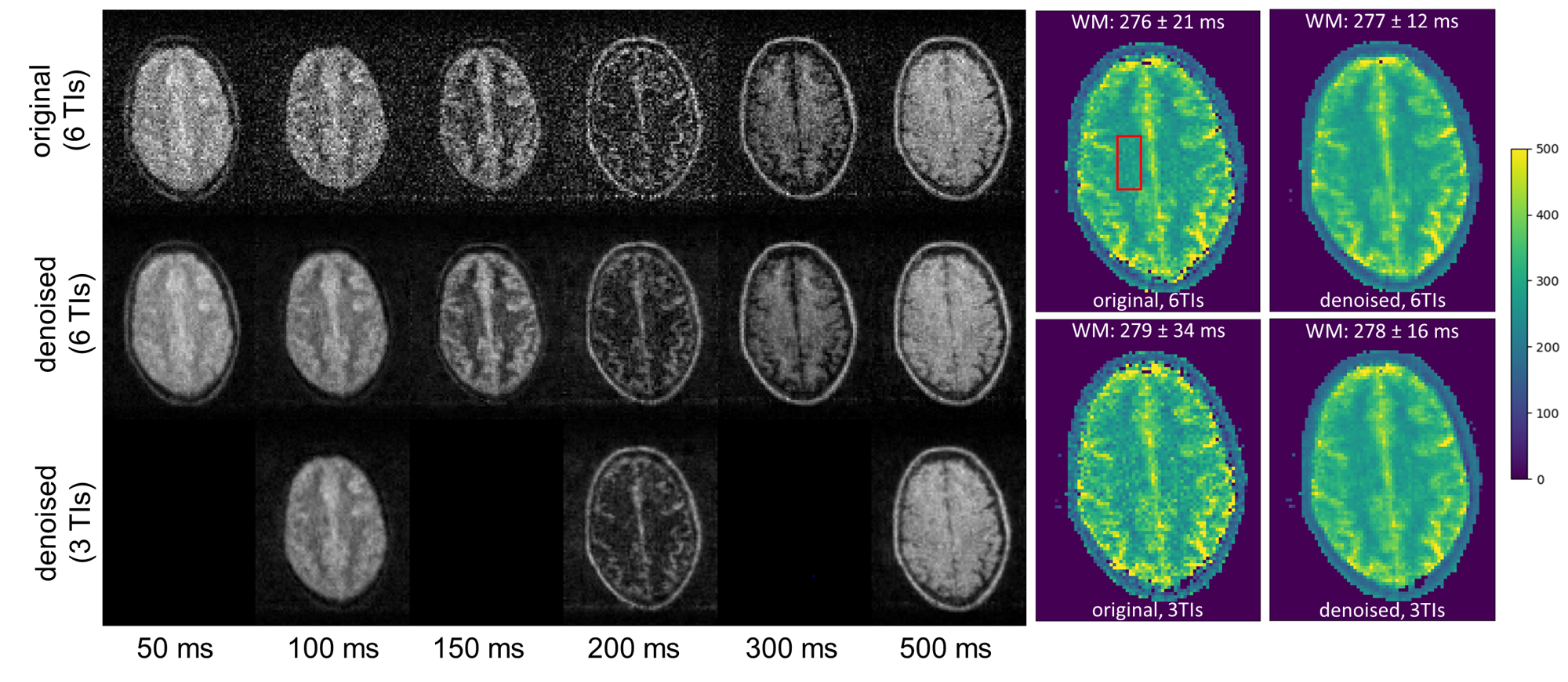
Four volunteer datasets were acquired on a 46 mT Halbach low-field system1 for testing using a 3D TSE sequence. Two datasets8 were acquired to estimate T2 of lipid and muscle in the calf muscle with 10 TEs and an echo-spacing of 11 ms, resolution 2.5x2.5x5mm3. Two T1 mapping8 datasets were acquired in brain (2.5x2.5x5mm3), with slightly differing parameters to achieve different contrasts: 1) TR=1250ms and 6 inversion times (TIs): 50,100,150,200,300,500 ms; 2) TR=1200ms and 6 TIs: 50,91,166,302,549,900 ms. The proposed method (4D-dataset, 3D-CNN) was compared to the original ZS-N2N (2D-CNN), two extended 3D versions either by adding the inversion time dimension (version 1) or the z-direction (version 2), and BM4D9. A simulated phantom T1 mapping (50,91,166,302,549,900 ms) study was added to test the performance of the algorithm with 3 noise levels (low,middle,high). To enable zero-mean Gaussian noise distribution of each image, a simple phase correction was performed before denoising to rotate the magnetization to the real-axis10.
Results
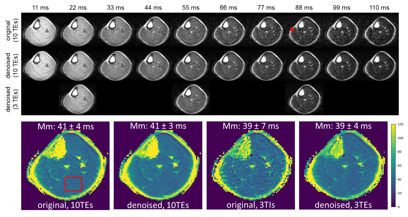
Figure 2 compares different denoising methods, highlighting the finer structures captured with the proposed 4D-denosing approach. Figure 3 evaluates the proposed method over three noise levels using various metrics in simulation, demonstrating increased PSNR/SSIM and reduced nRMSE with respect to the noise-free ground-truth images after denoising. Figure 4 shows T1 mapping results, contrasting the effects of using different numbers of TIs and the impacts of denoising. Figure 5 shows T2 mapping, highlighting the consistency and efficiency of our method to reduce scan time.Discussion and conclusion
In this work, we have proposed a zero-shot denoising approach for low-field quantitative MRI, without training on any other sources. It has demonstrated its potential to allow for less fitting points in T1,T2 mapping. This is essential since, e.g., it took roughly 36 minutes8 to acquire these 6 TIs datasets, where denoising can really improve the scan-efficiency to reach the same SNR of T1,T2 mapping with reduced number of fitting points. However, the optimized choice of TIs/TEs has not been investigated yet, remaining a topic for the future. To further explore the denoising performance of a multi-contrast protocol (e.g. T1w,T2w,Flair/DWI) at low-field may also be interesting.Acknowledgements
No acknowledgement found.References
1. O’Reilly T, Teeuwisse WM, de Gans D, Koolstra K, Webb AG. In vivo 3D brain and extremity MRI at 50 mT using a permanent magnet Halbach array. Magn Reson Med. 2021;85(1).
2. Kimberly WT, Sorby-Adams AJ, Webb AG, et al. Brain imaging with portable low-field MRI. Nature Reviews Bioengineering. 2023;1(9).
3. Koonjoo N, Zhu B, Bagnall GC, Bhutto D, Rosen MS. Boosting the signal-to-noise of low-field MRI with deep learning image reconstruction. Scientific Reports 2021 11:1. 2021;11(1):1-16.
4. Le DBT, Sadinski M, Nacev A, Narayanan R, Kumar D. Deep Learning-based Method for Denoising and Image Enhancement in Low-Field MRI. IST 2021 - IEEE International Conference on Imaging Systems and Techniques, Proceedings. Published online 2021.
5. Man C, Lau V, Su S, et al. Deep learning enabled fast 3D brain MRI at 0.055 tesla. Sci Adv. 2023;9(38).
6. Mansour Y, Heckel R. Zero-Shot Noise2Noise: Efficient Image Denoising without any Data. In: ; 2023.
7. Lehtinen J, Munkberg J, Hasselgren J, et al. Noise2Noise: Learning image restoration without clean data. In: 35th International Conference on Machine Learning, ICML 2018. Vol 7. ; 2018.
8. O’Reilly T, Webb AG. In vivo T1 and T2 relaxation time maps of brain tissue, skeletal muscle, and lipid measured in healthy volunteers at 50 mT. Magn Reson Med. 2022;87(2).
9. Dabov K, Foi A, Egiazarian K. Video denoising by sparse 3D transform-domain collaborative filtering. European Signal Processing Conference. 2007;16(8):145-149.
10. Prah DE, Paulson ES, Nencka AS, Schmainda KM. A simple method for rectified noise floor suppression: Phase-corrected real data reconstruction with application to diffusion-weighted imaging. Magn Reson Med. 2010;64(2).
11. Van Der Walt S, Schönberger JL, Nunez-Iglesias J, et al. Scikit-image: Image processing in python. PeerJ. 2014;2014(1).
Figures