2838
Beyond Boundaries – A versatile Console for Advanced Low-Field MRI1Physikalisch-Technische Bundesanstalt (PTB), Braunschweig and Berlin, Germany, 2Department of Radiology, Leiden University Medical Center (LUMC), C.J.Gorter MRI Center, Leiden, Netherlands
Synopsis
Keywords: Low-Field MRI, Low-Field MRI, open-source, console, acquisition
Motivation: We challenge proprietary barriers in low-field MRI to enhance methodological integration. Our focus is on improving system versatility for advanced imaging.
Goal(s): To create a versatile, MRI console driven by open-source software, capable of integrating sophisticated low-field imaging techniques. This involves for instance additional sensors or real-time adaptions.
Approach: We implemented Spectrum-Instrumentation measurement cards with a high-performance reconstruction system. The open-source Python software, incorporating a Pulseq interpreter, allows to streamline flexible, fast, and transparent low-field imaging applications.
Results: Successful implementation evidenced by high-fidelity Pulseq sequence execution to image 3D printed brain phantoms on a system capable of in-vivo applications.
Impact: The open and versatile design of our proposed console paves the way for advanced techniques in low-field MRI, enabling widespread adoption in research facilities and fostering innovative MRI applications in resource-limited settings.
Introduction
The emergence of low-field MRI presents a cost-effective and portable solution in magnetic resonance imaging1-4. Especially for portable imaging applications, the employment of advanced approaches, such as EMI suppression5 and elaborate reconstruction models for B0 inhomogeneity corrections, has yielded substantial improvements in image fidelity6-8. The domain is currently experiencing swift progress, particularly in the integration of auxiliary sensors9 and the dynamic optimization of imaging parameters through feedback loop analytics10. Nonetheless, most available MRI consoles are not inherently designed to facilitate these complex techniques11 or their proprietary nature poses limitations on customizability (e.g. limited on-board computing power, extension modules, or number of analog input/output channels). Our novel approach aims to bridge this gap and by providing a versatile, yet powerful console that enables a simple implementation of sophisticated methodologies and is easy to customize. It serves not only as a console but also as a high-performance reconstruction system enabling real-time data processing steps. It is tailored for advanced techniques and has the potential to significantly enhance the capabilities of low-field MRI systems, propelling the imaging performance beyond its current limits.Methods
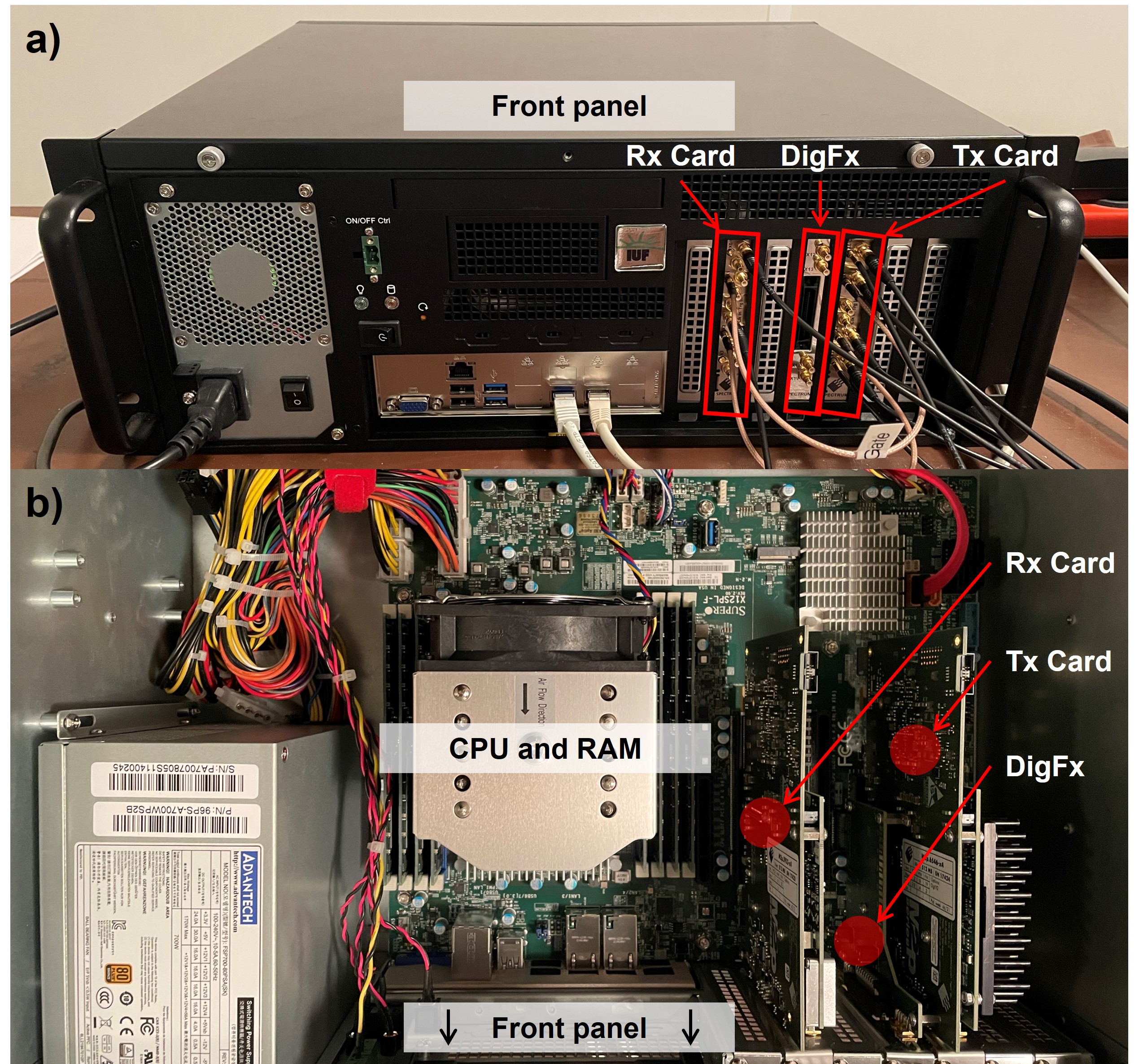
HardwareSpectrum-Instrumentation measurement cards served as the foundational core component of the console. As arbitrary waveform generator the M2p.6546-x4 with additional 16 GPIO ports (FX2 connector) and as digitizer the M2p.5933-x4 were chosen. This allows for a total of 4 analog (16-bit) transmit channels with 40 MS/s. For reception 8 single-ended or 4 differential analog input channels can be used with up to 40 MS/s at 16-bit resolution. Both cards accommodate a 512 MS (1 GB) memory. The measurement-cards were installed via the PCI express slots of the console main board. The console is equipped with a 24 core Intel CPU (6312U) and 256 GB RAM. The configuration is shown in Figure 1.
Software
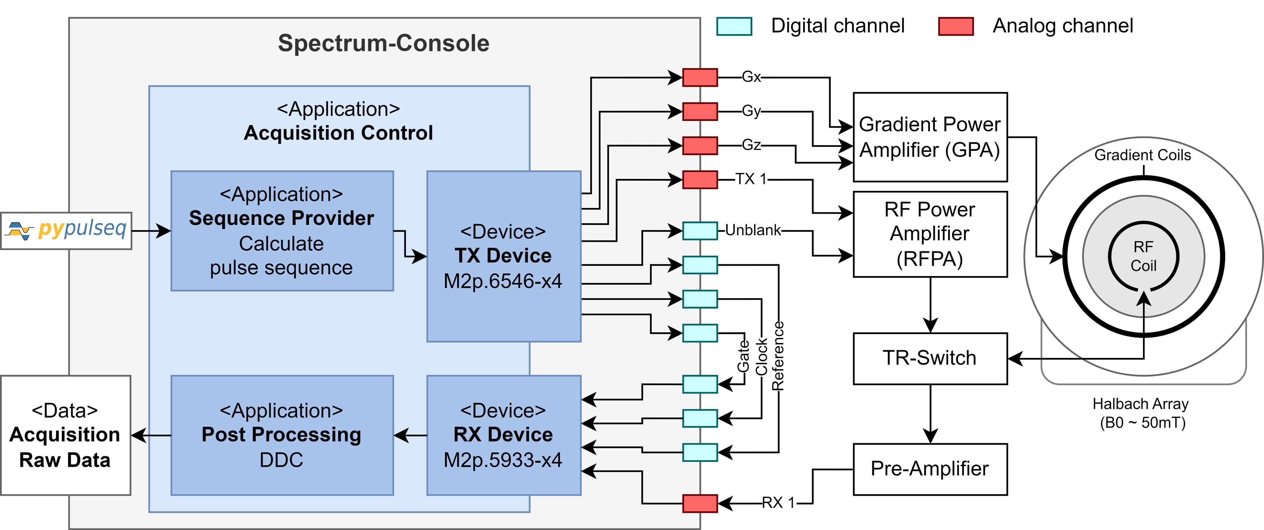
The console is controlled by a Python-based open-source software, which is publicly accessible at https://github.com/schote/spectrum-console. The application implements an interpreter for Pulseq12 to convert pulse sequences defined in the open-access Pulseq format into the necessary RF and gradient waveforms, and digital control signals (Figure 2).
Experiments
The performance of the console was assessed by conducting a series of experiments on 3D printed brain phantoms on a ~50mT low-field MRI scanner1. Our initial tests included frequency calibration, transmit adjustments, timing corrections, phase stability assessment, and 2D spin echo imaging (TE/TR: 20/300 ms, 128x128 pixel). The post-processing workflow, entirely realized in Python, entails sample rate conversion, which is achieved by employing a combination of averaging, band-pass filtering, and finite impulse response (FIR) decimation, complemented by intensity correction.
Results
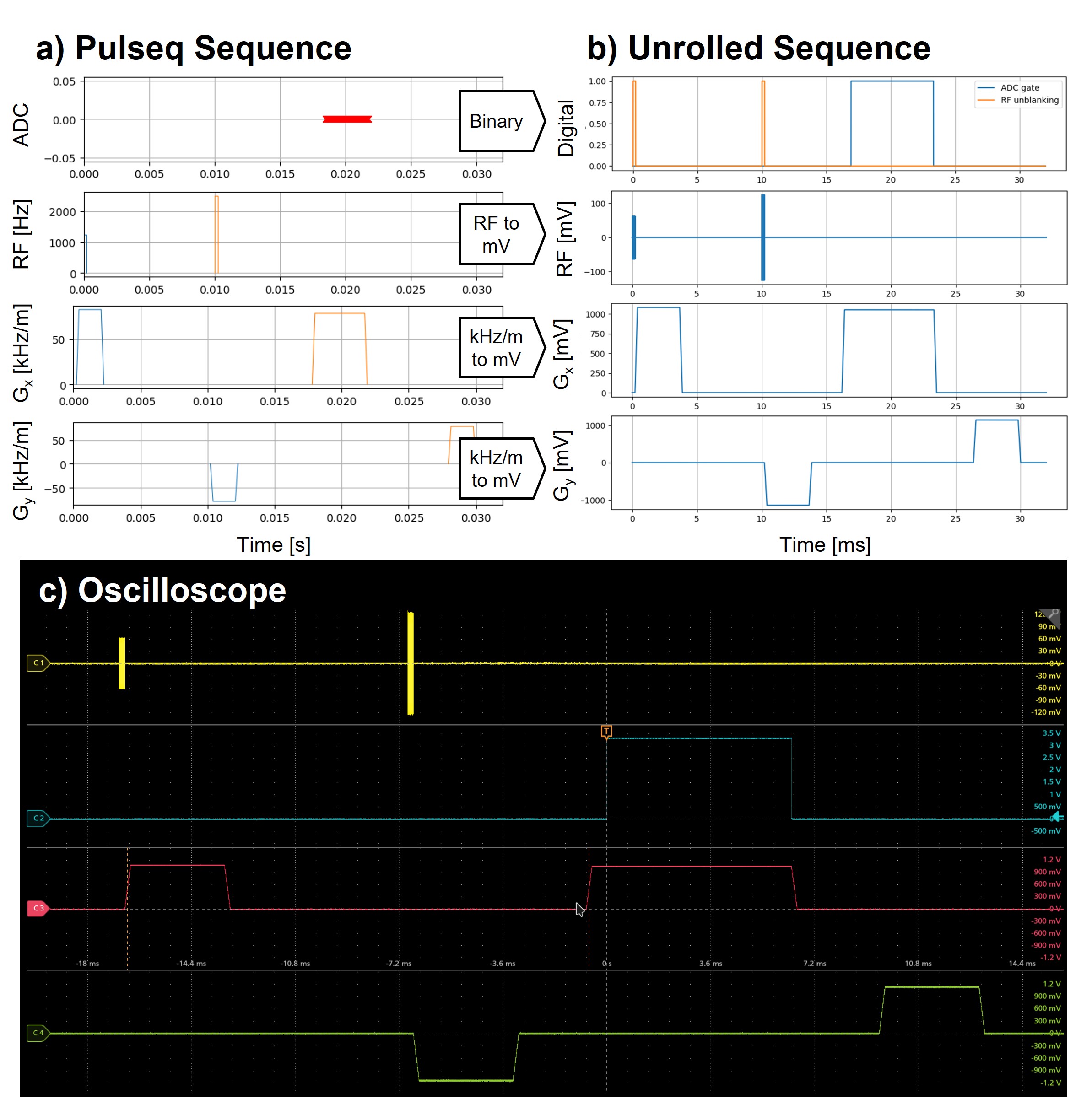
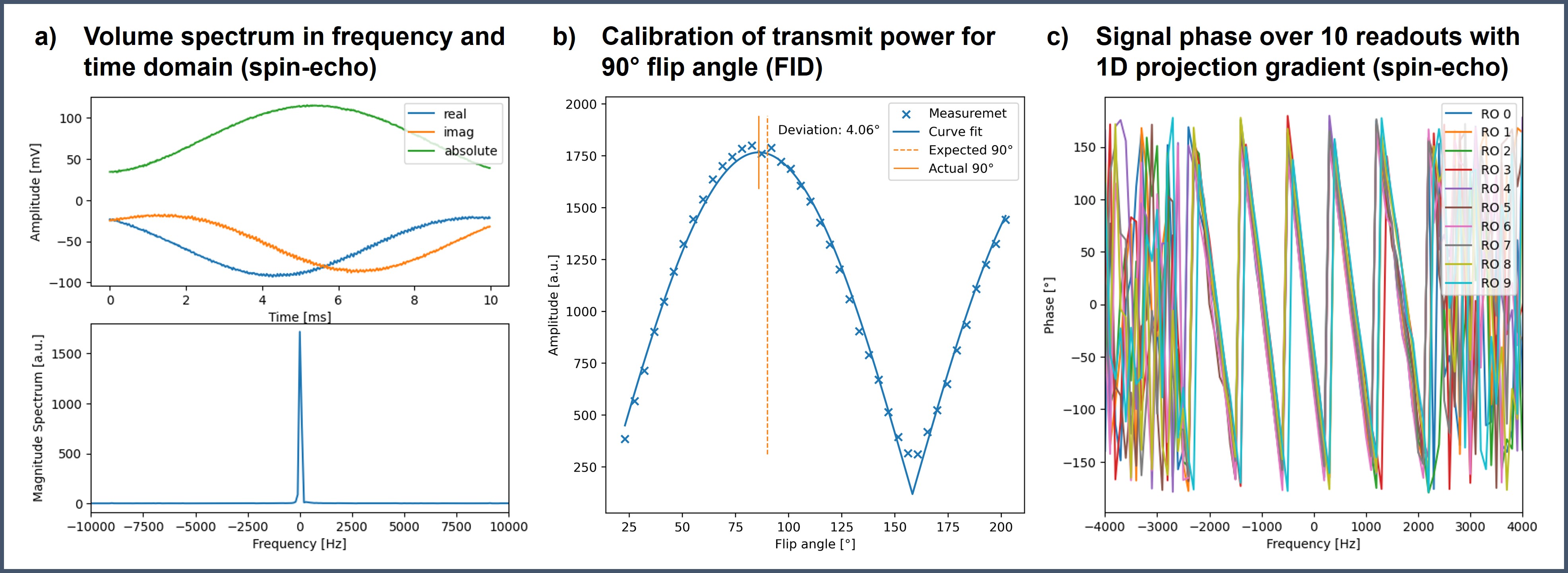
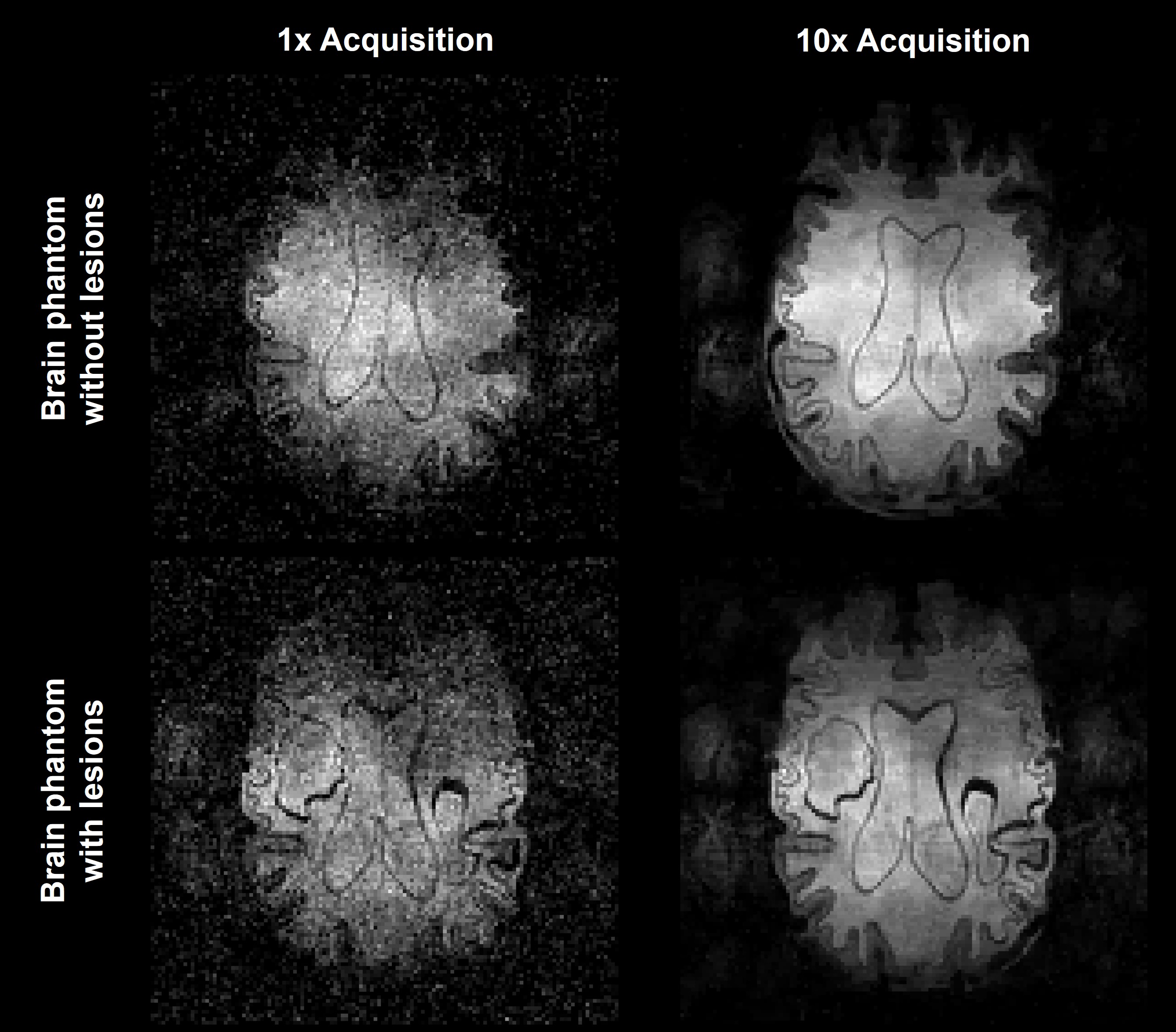
Real-time computed (~10.19 s for 2D spin-echo sequence (41.18 s execution duration), 128×128 pixels) Pulseq sequences demonstrated high fidelity compared to the measured waveforms (Figure 3). Frequency spectrum, flip angle calibration curve and the receive phase stability are shown in Figure 4a-b. Two subsequent 2D spin-echo imaging experiments of brain phantoms are shown in Figure 5a-b, showcasing the imaging capability of the MRI console. Intensity corrections were applied to these images based on noise measurements to correct for sensitivity variations from using a high Q-factor RF coil13.Discussion
The presented MR console was successfully implemented and tested on a low-field MRI scanner. The Python-based open-source implementation of the console software, along with the integration of Pulseq compatibility is publicly available and facilitates broad and straightforward adoption across the community for a multitude of applications. In the current setup, gradient waveforms are replayed through the analog transmission channels capable of driving the GPA directly. Nonetheless, it is also feasible to dispatch these waveforms through the synchronous GPIO channels what would liberate three analog transmission channels. Moreover, the system's ability to synchronize up to eight measurement cards simplifies the expansion to even more transmit and receive channels as necessitated by more demanding applications, like B0-field supervision, noise-cancelling or GIRF based trajectory corrections. The utilized cards enable direct data streaming to GPU memory, which paves the way for rapid AI-driven image reconstruction techniques and the potential to implement real-time adaptive feedback systems. This could significantly optimize both the sequence execution and the performance of external hardware components, which potentially enhances the image quality and the diagnostic value. Furthermore, the implementation facilitates effortless integration with the web-based acquisition control platform, ScanHub14.Conclusion
Our proposed console, combining commercial hardware with open-source software, brings forth a blend of flexibility, transparency, and customizability, simplifying the application of advance low-field MRI techniques. Preliminary imaging trials on brain phantoms have affirmed its potential, setting the stage for future advancements in the domain.Acknowledgements
This work is part of the Metrology for Artificial Intelligence for Medicine (M4AIM) project that is funded by the Federal Ministry for Economic Affairs and Energy (BMWi) in the frame of the QI-Digital initiative.
The project (21NRM05 and 22HLT02 A4IM) has received funding from the European Partnership on Metrology, co-financed by the European Union’s Horizon Europe Research and Innovation Program and by the Participating States.
References
- T. O’Reilly, W. M. Teeuwisse, D. Gans, K. Koolstra, und A. G. Webb, „In vivo 3D brain and extremity MRI at 50 mT using a permanent magnet Halbach array“, Magn. Reson. Med., Bd. 85, Nr. 1, S. 495–505, Jan. 2021, doi: 10.1002/mrm.28396.
- C. Z. Cooley et al., “Design and implementation of a low-cost, tabletop MRI scanner for education and research prototyping,” Journal of Magnetic Resonance, vol. 310, p. 106625, Jan. 2020, doi: 10.1016/j.jmr.2019.106625.
- L. L. Wald, P. C. McDaniel, T. Witzel, J. P. Stockmann, and C. Z. Cooley, „Low‐cost and portable MRI“, J Magn Reson Imaging, Bd. 52, Nr. 3, S. 686–696, Sep. 2020, doi: 10.1002/jmri.26942.
- T. Guallart‐Naval et al., „Benchmarking the performance of a low‐cost magnetic resonance control system at multiple sites in the open MaRCoS community“, NMR in Biomedicine, Bd. 36, Nr. 1, S. e4825, Jan. 2023, doi: 10.1002/nbm.4825.
- Y. Liuet al., „A low-cost and shielding-free ultra-low-field brain MRI scanner“, Nature Communications, Bd. 12, Nr. 1, S. 7238, Dez. 2021, doi: 10.1038/s41467-021-27317-1.
- D. Schote et al., „Physics-Informed Deep Learning for Image Distortion Correction from B0-inhomogeneities in Low-Field MRI“, in Proc. Intl. Soc. Mag. Reson. Med., London, 2022, S. 1816.
- D. Schote, L. Winter, C. Kolbitsch, and A. Kofler, „Reliable Low-Field B0-Maps by Deep Learning with Physical Constraints“, in Proc. Intl. Soc. Mag. Reson. Med., Toronto, 2023, S. 4344.
- K. Koolstra, T. O’Reilly, P. Börnert, and A. Webb, „Image distortion correction for MRI in low field permanent magnet systems with strong B0 inhomogeneity and gradient field nonlinearities“, Magn Reson Mater Phy, Jan. 2021, doi: 10.1007/s10334-021-00907-2.
- S. A. Srinivas et al., „External Dynamic InTerference Estimation and Removal (EDITER) for low field MRI“, 2023.
- A. Loktyushin et al., „MRzero -- Fully automated discovery of MRI sequences using supervised learning“, Magnetic Resonance in Med, Bd. 86, Nr. 2, S. 709–724, Aug. 2021, doi: 10.1002/mrm.28727.
- V. Negnevitskyet al., „MaRCoS, an open-source electronic control system for low-field MRI“, Journal of Magnetic Resonance, Bd. 350, S. 107424, Mai 2023, doi: 10.1016/j.jmr.2023.107424.
- K. J. Layton et al., „Pulseq: A rapid and hardware-independent pulse sequence prototyping framework: Rapid Hardware-Independent Pulse Sequence Prototyping“, Magn. Reson. Med., Bd. 77, Nr. 4, S. 1544–1552, Apr. 2017, doi: 10.1002/mrm.26235.
- A. Webb and T. O’Reilly, „Tackling SNR at low-field: a review of hardware approaches for point-of-care systems“, MAGMA, Bd. 36, Nr. 3, S. 375–393, Juli 2023, doi: 10.1007/s10334-023-01100-3.
- D. Schote, J. Behrens, L. Winter, C. Kolbitsch, and C. Dinh, „ScanHub: Open-Source Platform for MR Scanner Control, Acquisitions and Postprocessing“, in Proc. Intl. Soc. Mag. Reson. Med., Toronto, Kanada, 2023, S. 2391.
- A. Chambolle and T. Pock, “A First-Order Primal-Dual Algorithm for Convex Problems with Applications to Imaging,” J Math Imaging Vis, vol. 40, no. 1, pp. 120–145, May 2011, doi: 10.1007/s10851-010-0251-1.
Figures