2837
Design and comissioning of a new field-cycling imaging scanner for clinical applications1Aberdeen Biomedical Imaging Centre, University of Aberdeen, Aberdeen, United Kingdom
Synopsis
Keywords: Low-Field MRI, Low-Field MRI, Field-Cycling
Motivation: Field-cycling imaging is a new imaging technique that allows access to new imaging biomarkers. To allow investigation of clinical applications a high performance and patient acceptable scanner is needed.
Goal(s): We have constructed a new clinical-grade resistive field-cycling imaging system operating at 0.2 T with a 60 cm bore and high performance gradient and magnet amplifiers, with a custom made outer covering.
Approach: The system is in the late stage of commissioning. The field homogeneity and temporal stability have been assessed.
Results: The final system is visually attractive and has < 1 ppm temporal stability and < 8 ppm field homogeneity.
Impact: This system will allow us to investigate the clinical utility of field-cycling and low-field imaging in a much larger patient population. The high performance gradient amplifiers will enable much faster imaging, which will enable new possibilities for new sequence development.
Introduction
Field-cycling imaging is a novel low-field MRI technique that allows the variation of the spin-lattice relaxation time T1 with magnetic field, known as T1 dispersion, to be exploited as a new image contrast mechanism. T1 dispersion, particularly at low field, is a rich source of information on molecular dynamics and can provide access to unique features such as 1H-14N cross-relaxation effects which are completely hidden to conventional MRI.Our research group have previously presented results from several pilot studies, including stroke1 and osteoarthritis2, using a home-built 0.2 T prototype field-cycling imaging scanner3 that highlight the potential clinical utility of field-cycling. As a technology demonstrator, this prototype scanner had several limitations including a narrow bore (50 cm), weak imaging gradients and relatively poor temporal and spatial homogeneity. These limitations, along with the visual profile of the scanner, made clinical studies with larger cohorts challenging as the scanner is not well tolerated by claustrophobic or anxious participants.
In this work we present our progress on a next generation field-cycling imaging scanner, sited in Aberdeen Biomedical Imaging Centre in Aberdeen Royal Infirmary, which sets out to address these limitations in order to allow new studies into potential clinical applications of this new imaging technique.
Methods
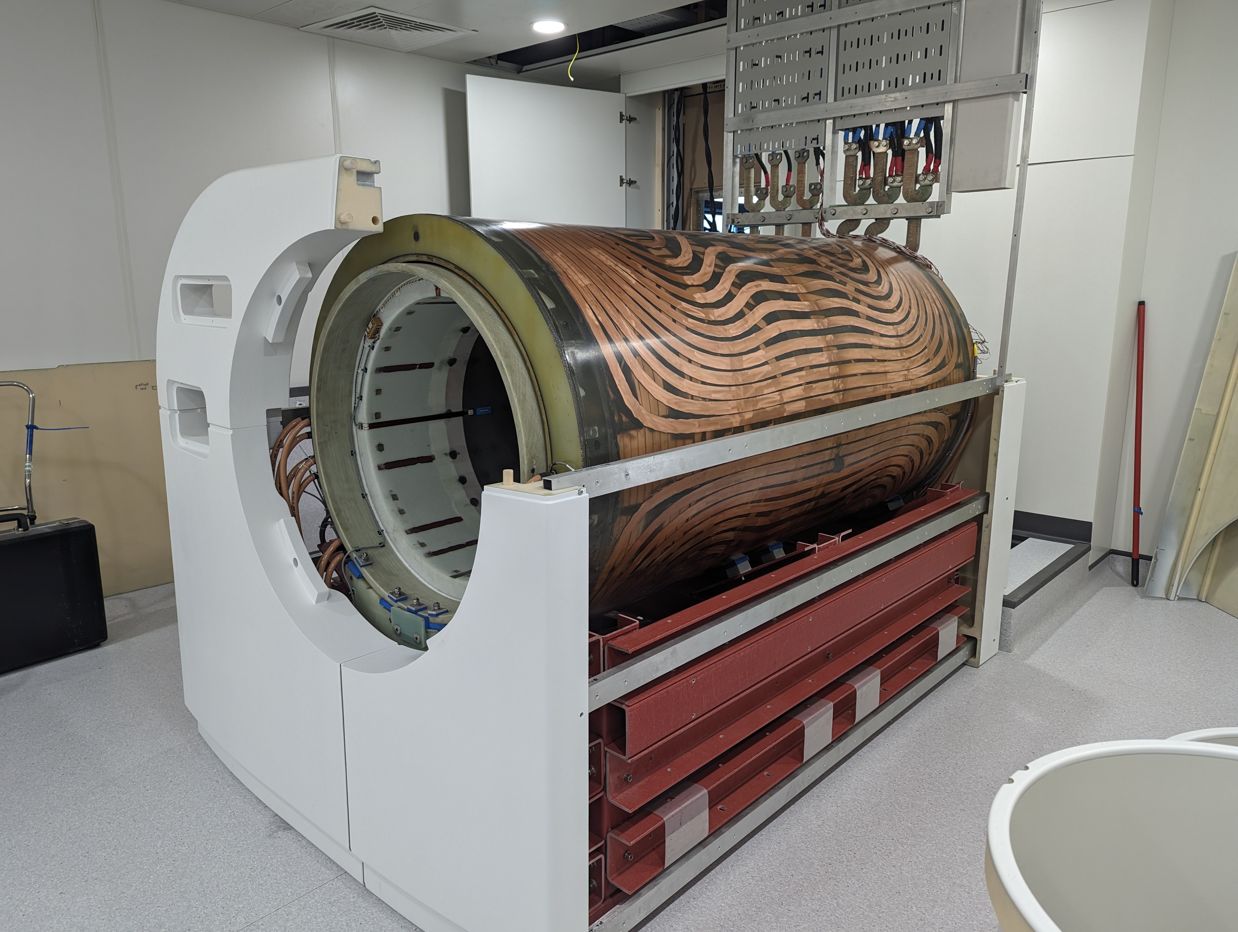
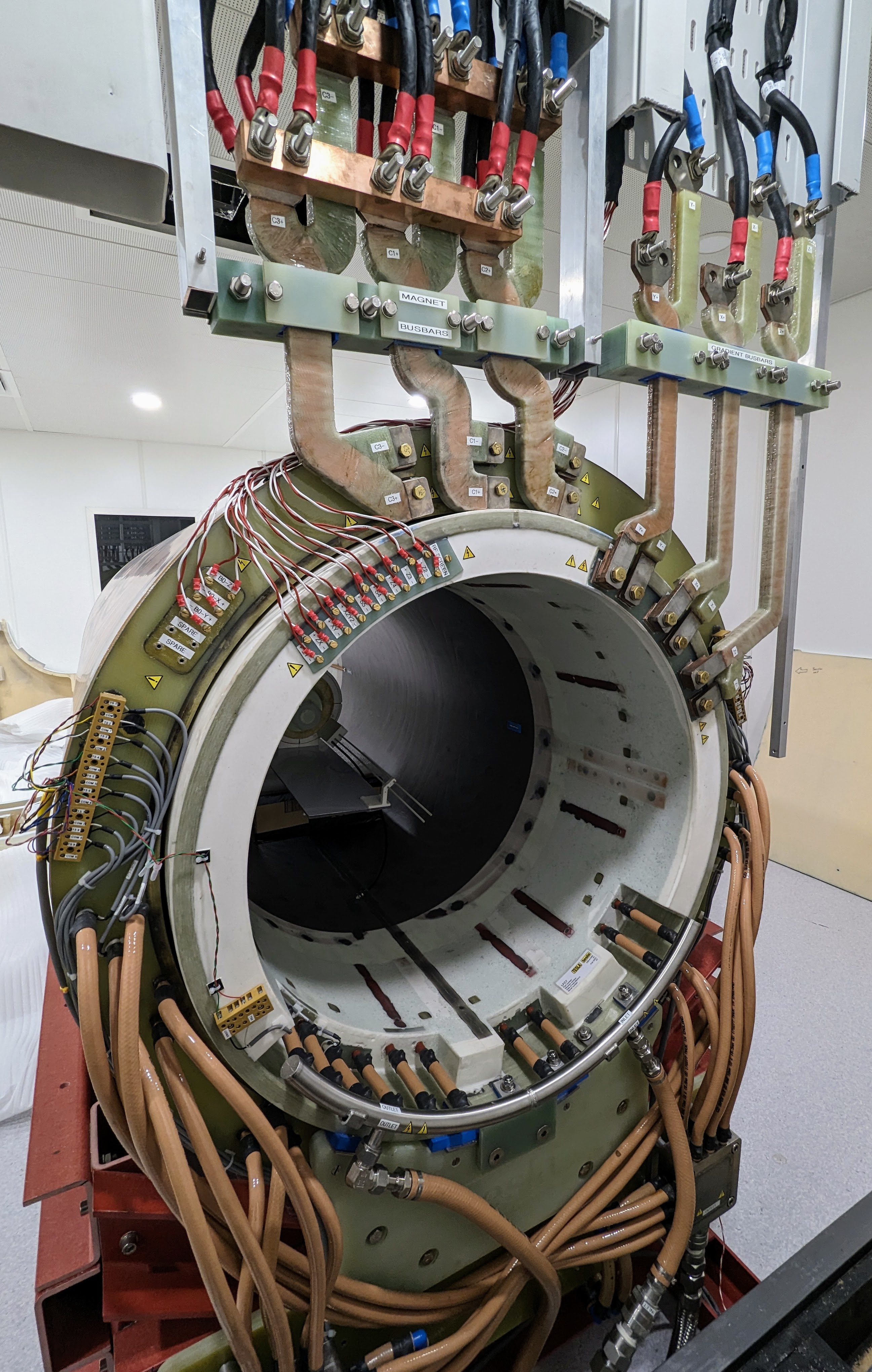
Our new field-cycling imaging magnet (Figure 1) is comprised of three independent resistive solenoid segments coaxially wound in a glass-expoxy (Tesla Engineering Ltd, Storrington, UK). We have elected for this design in order to lower the overall inductance of the system to allow faster switching of B0. Each magnet segment has inductance of approximately 87 mH, room temperature resistance of 72 mΩ and are each driven by a modified gradient amplifier (model QDCM2100D, Performance Controls Inc, Mongromeryville, US). Both the magnet and amplifiers are water-cooled (Figure 2) such that the system can be operated continuously at a peak current of 675 A per magnet segment. The maximum B0 switching rate is 20T/s (corresponding to a field ramp of 10 ms) and the field range of the system is between 0 and 0.2 T. To reach field strengths below the Earth's field, the magnet assembly has three-axis Earth's field cancellation coils (visible in Figure 1), controlled as additional shim channels.The system is equipped with gradient and shim coils up to 4th order. The gradient coils are driven by a three-axis gradient amplifier (model DA1500, Performance Controls Inc) while the shim coils are driven by a set of dedicated shim amplifiers (model SA2-50, Performance Controls Inc). The maximum gradient strength achievable is 38 mT/m.
The scanner has an in-house designed birdcage body-coil, driven by a 4 kW RF power amplifier (model BT04000-AlphaS, Tomco Technologies, Stepney, Australia) and a 16-channel anterior-posterior receive-only array coil based on an in-house design and constructed by Wideblue Ltd (Glasgow, UK).
The scanner is controlled using a commercial console (RS2D, Mundolsheim, France), modified to allow B0 to be controlled as a "4th gradient" in the pulse sequence environment.
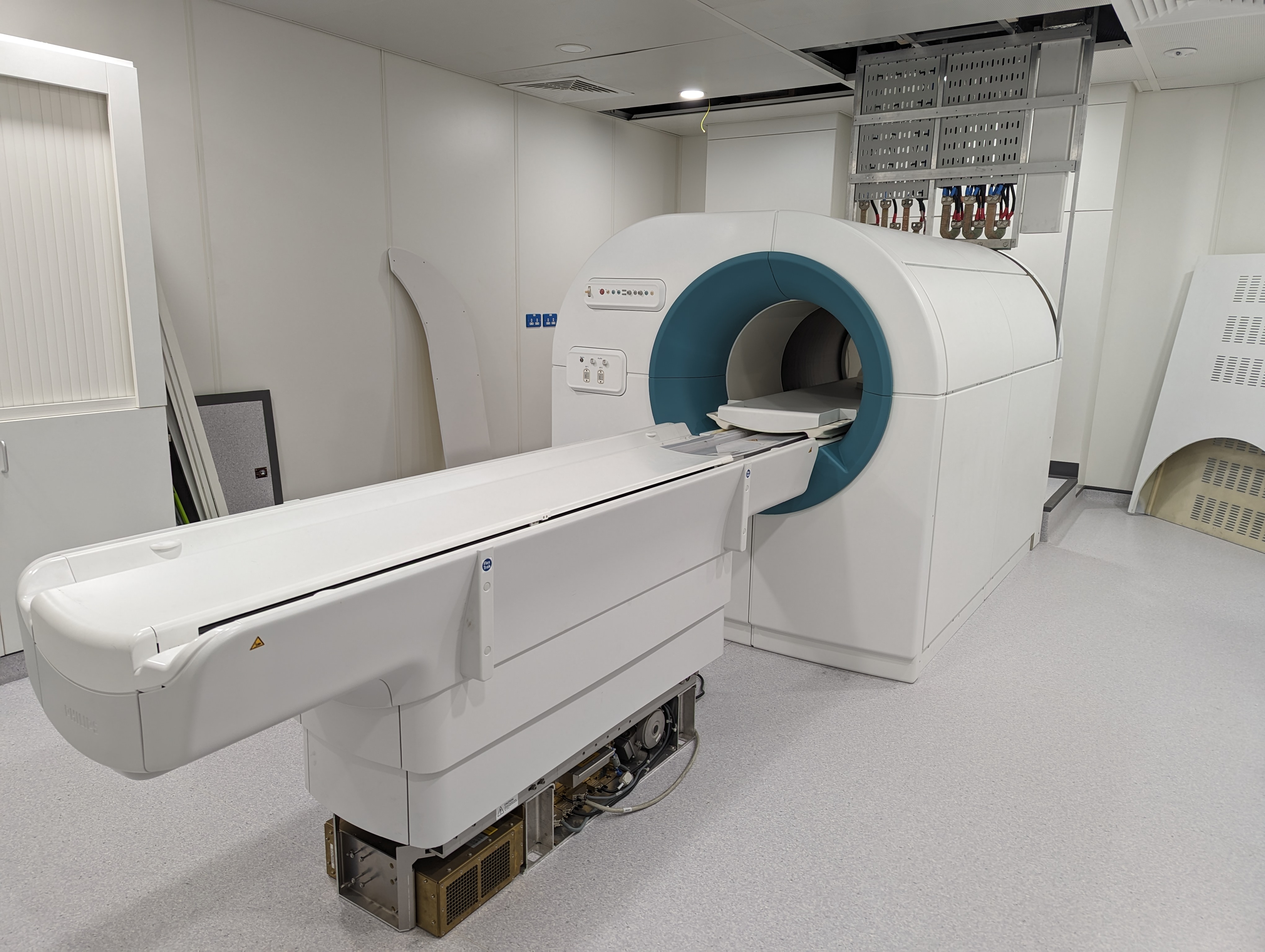
The magnet cowling (Figure 3) is constructed from solid ABS plastic. The design and construction was performed by Wideblue Ltd.
The spatial and temporal stability of the magnet was assessed using a manufacturer supplied in-bore goniometer and an NMR-based magnetometer (model PT2026, Metrolab, Geneva, Switzerland)
Results
To assess the thermal stability of the magnet, the system was cold-started and ramped to 0.2T, with the magnetometer located at isocentre. We observed that the thermal stabilisation time is on the order of 20 minutes, during which the measured field strength drifted by several mT. Following thermalisation the field temporal stability reaches approximately 0.6 ppm, or approximately 5 Hz.Spatial homogeneity was assessed using the goniometer and magnetometer to measure the field strength at 156 points describing a 24 cm diameter sphere at isocentre from which corrective shim currents were derived. Following an iterative shimming process a field homogeneity of < 8 ppm (64 Hz) was achieved over this sphere.
Real world gradient and magnet coupling was assessed by observing the drive voltage error across the main magnet while a series of short ramp, high amplitude gradient pulses were applied at 50 Hz. No voltage deviation was observed at any stage, indicating a high degree of decoupling between the magnet and gradients.
Conclusions
We have successfully commissioned a new field-cycling imaging scanner which is equipped with high performance magnet and gradient amplifiers. The measured temporal stability and field homogeneity of the system is excellent, and the scanner is visually appealing. This will enable us to move towards larger trials aimed at assessing the clinical utility of field cycling imaging on clinical populations.Acknowledgements
No acknowledgement found.References
1. Bödenler, M, Maier, O, Stollberger, R, et al. Joint multi-field T1 quantification for fast field-cycling MRI. Magn Reson Med. 86: 2049–2063 (2021)
2. Ross, P.J et al. A New Method for Investigating Osteoarthritis using Fast Field Cycling Nuclear Magnetic Resonance. Physical Medica 88, pp. 142-147 (2021)
3. Broche, L.M., Ross, P.J., Davies, G.R. et al. A whole-body Fast Field-Cycling scanner for clinical molecular imaging studies. Sci Rep 9, 10402 (2019)
Figures